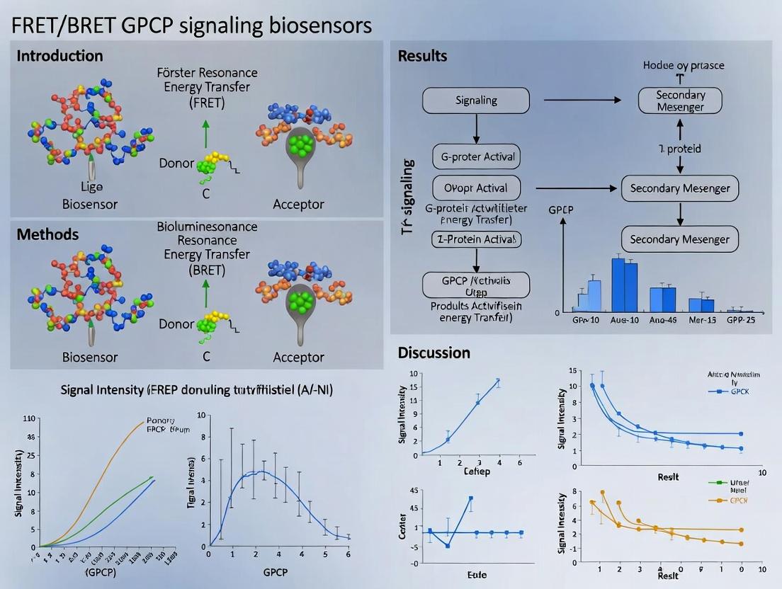
FRET vs BRET GPCR Biosensors: A Complete Guide for Drug Discovery and Signaling Research
This comprehensive guide explores FRET and BRET biosensors for monitoring GPCR signaling dynamics in live cells.
FRET vs BRET GPCR Biosensors: A Complete Guide for Drug Discovery and Signaling Research
Abstract
This comprehensive guide explores FRET and BRET biosensors for monitoring GPCR signaling dynamics in live cells. We cover the foundational principles of these resonance energy transfer technologies, detailing methodological approaches for real-time measurement of second messengers, conformational changes, and protein-protein interactions. The article provides practical troubleshooting advice, optimization strategies for signal-to-noise ratio and specificity, and a comparative analysis of sensor validation techniques. Designed for researchers and drug development professionals, this resource synthesizes current best practices to enable robust, quantitative analysis of GPCR pharmacology and accelerate therapeutic discovery.
Understanding FRET and BRET Biosensors: Core Principles for GPCR Signaling
G protein-coupled receptors (GPCRs) represent the largest family of cell surface receptors, encoded by over 800 genes in humans. They transduce extracellular signals (hormones, neurotransmitters, light, odors) into intracellular responses, regulating virtually every physiological process. Consequently, GPCR dysfunction is implicated in a vast array of diseases, making them the target of approximately 35% of all FDA-approved drugs.
The advent of real-time biosensors based on Förster/ Bioluminescence Resonance Energy Transfer (FRET/BRET) has revolutionized our ability to monitor GPCR signaling dynamics with high spatial and temporal resolution directly in living cells. This application note details key protocols and reagents within the context of ongoing FRET/BRET biosensor research.
Key GPCR Signaling Pathways & Biosensor Targets
The complexity of GPCR signaling arises from their ability to engage multiple transducers. The table below summarizes core pathways and measurable biosensor outputs.
Table 1: Core GPCR Signaling Pathways and Biosensor Readouts
| Pathway/Effector | Primary G Protein | Key Second Messenger/Biosensor Target | Example Physiological Role | Disease Link |
|---|---|---|---|---|
| Gαs | Gs | ↑ cAMP (e.g., EPAC-based FRET sensor) | Heart rate, gluconeogenesis | Asthma, heart failure |
| Gαi/o | Gi/o | ↓ cAMP, ↑ ERK activation | Neurotransmission, immune cell migration | Depression, cancer metastasis |
| Gαq/11 | Gq/11 | ↑ IP3, DAG, Ca²⁺ (e.g., Cameleon sensors) | Vasoconstriction, exocrine secretion | Hypertension, metabolic disorders |
| Gβγ | All classes | Activation of GIRK channels, PI3Kγ, GRKs | Neuronal inhibition, chemotaxis | Pain, inflammation |
| β-arrestin | N/A (recruited post-activation) | Receptor internalization & scaffolding (e.g., BRET between GPCR and β-arrestin) | Signal desensitization, MAPK signaling | Chronic opioid tolerance |
Experimental Protocols
Protocol: Live-Cell FRET Assay for Gαq-Mediated Ca²⁺ Release
Objective: To measure real-time GPCR-Gq activation using a Cameléon-type FRET biosensor (YC3.60).
Materials:
- HEK293T cells
- Plasmid: GPCR of interest
- Plasmid: YC3.60 (Cameleon Ca²⁺ biosensor)
- Appropriate cell culture medium and transfection reagent
- FRET-compatible microplate reader or fluorescence microscope
- Agonist/antagonist compounds
- Hanks' Balanced Salt Solution (HBSS) with 20 mM HEPES, pH 7.4
Procedure:
- Cell Seeding & Transfection: Seed HEK293T cells in a poly-D-lysine-coated 96-well black-walled microplate at 50,000 cells/well. 24h later, co-transfect with GPCR and YC3.60 plasmid DNA (1:1 ratio, 100 ng total/well) using your preferred transfection reagent.
- Incubation: Incubate transfected cells for 24-48h at 37°C, 5% CO₂.
- Dye Loading/Preparation (Optional): Not required for Cameleon. Replace medium with 80 µL/well of HBSS/HEPES imaging buffer.
- FRET Measurement (Plate Reader):
- Pre-equilibrate plate to 37°C in the reader.
- Configure reader for ratiometric FRET: Excite CFP at 433-455 nm, collect emissions at 475-495 nm (CFP channel) and 520-540 nm (FRET/YFP channel) simultaneously.
- Establish a 60-second baseline reading.
- Automatically inject 20 µL of 5x concentrated agonist solution.
- Monitor the 520/475 nm emission ratio over time (typically 5-10 minutes).
- Data Analysis: Normalize the emission ratio (R) to the initial baseline ratio (R₀). Plot R/R₀ vs. time. Calculate EC₅₀/IC₅₀ from dose-response curves.
Protocol: BRET² Assay for GPCR-β-Arrestin Interaction
Objective: To quantify GPCR-β-arrestin proximity using BRET² with GFP² and Rluc8.
Materials:
- HEK293T cells
- Plasmid: GPCR-Rluc8 (C-terminally tagged)
- Plasmid: β-Arrestin2-GFP² (C-terminally tagged)
- Cell culture medium and transfection reagent
- Microplate reader capable of detecting BRET (e.g., PHERAstar, TriStar²)
- Coelenterazine 400a (DeepBlueC) substrate
- Assay buffer: PBS with 0.1% glucose and 0.5 mM MgCl₂.
Procedure:
- Cell Preparation: Seed and co-transfect cells as in Protocol 3.1, using a 1:5 ratio of GPCR-Rluc8 to β-Arrestin2-GFP² DNA (optimize per receptor).
- Harvesting: 48h post-transfection, wash cells with PBS, detach gently, and resuspend in assay buffer. Adjust cell density to ~1 x 10⁶ cells/mL.
- BRET Measurement:
- Distribute 95 µL of cell suspension per well in a white 96-well microplate.
- Add agonist/antagonist in a 5 µL volume. Incubate 15-30 min at 37°C.
- Add 10 µL of 25 µM Coelenterazine 400a (final conc. 2.5 µM) to each well.
- Immediately measure luminescence (Rluc8 donor) at 410 nm and fluorescence (GFP² acceptor) at 515 nm sequentially (integration time 0.5-1s).
- Data Analysis:
- Calculate BRET ratio = (Emission at 515 nm) / (Emission at 410 nm).
- Subtract the BRET ratio from cells expressing GPCR-Rluc8 alone (background).
- Plot net BRET ratio vs. ligand concentration.
The Scientist's Toolkit: Essential Research Reagents
Table 2: Key Reagent Solutions for FRET/BRET GPCR Biosensing
| Reagent / Material | Function & Description | Example Product / Note |
|---|---|---|
| FRET-based cAMP Biosensor (e.g., EPAC-camps) | Recombinant protein or plasmid encoding a cAMP sensor. Changes FRET upon cAMP binding. | "pGLO-20F" EPAC-based plasmid; commercially available kits (Cisbio cAMP Gs Dynamic Kit). |
| Cameléon-type Ca²⁺ Biosensor (e.g., YC3.60) | Genetically encoded Ca²⁺ indicator (Cameleon). FRET between CFP and YFP changes with Ca²⁺ binding. | Addgene plasmid #; also available as stable cell lines. |
| GPCR-Rluc8 Fusion Construct | Donor for BRET assays. Rluc8 is a bright, stable Renilla luciferase mutant fused to GPCR C-terminus. | Custom cloned or from cDNA repositories (cDNAs.org). |
| β-Arrestin-GFP² Fusion Construct | Acceptor for BRET² assays. GFP² is a green fluorescent protein variant with optimized emission for BRET². | Available from academic sources or commercial vendors (e.g., PerkinElmer). |
| Coelenterazine 400a (DeepBlueC) | Substrate for Rluc8 in BRET². Emits blue light (~400 nm) to excite GFP². | Available from Nanolight, GoldBio, etc. Critical for low background BRET². |
| Stable Cell Lines Expressing Biosensor | Cell lines (e.g., HEK293, CHO) stably expressing a GPCR pathway biosensor for HTS. | Available through commercial partners (e.g., DiscoverX PathHunter, or generated in-house via lentiviral transduction). |
| FRET-Compatible Microplate Reader | Instrument for high-throughput, live-cell kinetic FRET/BRET measurements. | Devices like BMG Labtech PHERAstar, CLARIOstar; or PerkinElmer EnVision. |
| Confocal Microscopy System with FRET Capability | For spatially resolved, single-cell FRET imaging (e.g., acceptor photobleaching method). | Zeiss LSM, Nikon A1, or Olympus FV3000 systems with appropriate lasers/filters. |
Table 3: Quantitative Advantages of Real-Time Biosensor Assays
| Parameter | Traditional Endpoint Assay (e.g., ELISA) | FRET/BRET Real-Time Biosensor Assay | Improvement/Advantage |
|---|---|---|---|
| Temporal Resolution | Minutes to hours (single timepoint) | Sub-second to second (continuous) | Enables kinetic profiling (kon/koff, signaling waves) |
| Information Content | Single pathway node (e.g., total cAMP) | Multiple nodes (activation, translocation, complex formation) | Pathway deconvolution & bias analysis |
| Assay Format | Typically lysate-based | Live-cell, functional | Preserves cellular context & compartmentalization |
| HTS Compatibility | Moderate to High | High (with optimized stable lines) | Suitable for primary drug screening |
| Artifact Potential | Higher (lysis, fixation artifacts) | Lower (non-invasive, minimal perturbation) | More physiologically relevant data |
| Key Output Metrics | IC₅₀, EC₅₀, potency | EC₅₀, kinetics, efficacy, bias factor | Comprehensive pharmacological profiling |
Real-time FRET/BRET biosensors have moved GPCR research from static, endpoint measurements to dynamic, mechanistic dissection of signaling in living systems. This capability is crucial for understanding the nuanced role of GPCRs in physiology and complex diseases, and for driving the discovery of next-generation therapeutics with improved efficacy and reduced side-effect profiles, such as biased agonists. The protocols and tools outlined here provide a foundation for implementing these powerful techniques in both basic research and drug discovery pipelines.
This application note details the fundamental photophysics of Förster Resonance Energy Transfer (FRET) and Bioluminescence Resonance Energy Transfer (BRET), providing essential context and protocols for their application in developing GPCR signaling biosensors.
Photophysical Principles
Förster Resonance Energy Transfer (FRET)
FRET is a non-radiative energy transfer process between two light-sensitive molecules (chromophores). A donor chromophore in an excited state transfers energy to an acceptor chromophore through dipole-dipole interactions.
Key Quantitative Parameters:
- Förster Distance (R₀): The distance at which energy transfer efficiency is 50%. Typically 2-8 nm.
- Transfer Efficiency (E):
E = 1 / [1 + (r/R₀)^6], whereris the donor-acceptor distance. - Spectral Overlap Integral (J): Measures the overlap between donor emission and acceptor absorption spectra.
Bioluminescence Resonance Energy Transfer (BRET)
BRET is a natural phenomenon involving non-radiative energy transfer from a bioluminescent donor enzyme (e.g., Renilla luciferase) to a fluorescent acceptor protein. It does not require external light excitation, eliminating autofluorescence and photobleaching.
Key Quantitative Parameters:
- BRET Ratio: The ratio of acceptor emission intensity to donor emission intensity.
- BRET Signal (mBU): MillBRET Units, a standardized measure.
- Background Signal: Inherently lower than FRET due to lack of excitation light.
Table 1: Comparison of FRET and BRET Core Principles
| Parameter | FRET | BRET |
|---|---|---|
| Energy Donor | Fluorescent Protein/Dye (e.g., CFP, GFP) | Bioluminescent Enzyme (e.g., RLuc, Nluc) |
| Energy Acceptor | Fluorescent Protein/Dye (e.g., YFP, mCherry) | Fluorescent Protein (e.g., GFP, YFP) |
| Excitation Source | External Light | Enzyme-Substrate Reaction |
| Key Dependency | Spectral Overlap, Distance, Orientation | Spectral Overlap, Distance, Orientation |
| Typical R₀ | 2-8 nm | 4-8 nm |
| Autofluorescence | Possible | Minimal |
| Photobleaching | Yes | No |
Application to GPCR Signaling Biosensors
GPCRs are dynamic membrane proteins. FRET/BRET biosensors monitor conformational changes or protein-protein interactions in real-time within live cells. Common designs include:
- Intramolecular: Donor and acceptor flank a sensor domain (e.g., ligand-binding domain). Conformational change alters distance/orientation.
- Intermolecular: Donor and acceptor are fused to separate interacting proteins (e.g., GPCR and β-arrestin). Interaction brings them into proximity.
Experimental Protocols
Protocol: Generation of an Intramolecular FRET GPCR Biosensor
Objective: To create a cell-based biosensor for monitoring GPCR activation via intramolecular FRET.
Materials:
- cDNA encoding your GPCR of interest.
- Donor (e.g., mTurquoise2) and acceptor (e.g., cpVenus) FP vectors.
- Appropriate cell line (e.g., HEK293).
- Transfection reagent.
- FRET-compatible microplate reader or fluorescence microscope.
- Phosphate-Buffered Saline (PBS).
- Ligand/Agonist for the target GPCR.
Procedure:
- Molecular Cloning: Using standard molecular biology techniques, generate a fusion construct in the order: Donor FP - GPCR - Acceptor FP. Ensure linkers (e.g., (GGGGS)₃) are included between domains for flexibility.
- Cell Culture and Transfection: Seed HEK293 cells in a poly-D-lysine coated 96-well black-walled, clear-bottom plate. At 60-80% confluency, transiently transfect with the biosensor construct using a suitable transfection reagent. Include untransfected cells as a control.
- Incubation: Culture transfected cells for 24-48 hours at 37°C, 5% CO₂ to allow for expression.
- FRET Measurement (Microplate Reader): a. Gently replace medium with pre-warmed PBS or assay buffer. b. For a single-timepoint dose-response: Add varying concentrations of ligand to wells. Incubate for the optimized time (e.g., 5 min). c. Measure fluorescence intensities using appropriate filters: * Donor Excitation / Donor Emission (IDD) * Donor Excitation / Acceptor Emission (IDA - the FRET signal) * (Optional) Acceptor Excitation / Acceptor Emission (IAA) to check acceptor expression.
- Data Calculation:
a. Subtract background signals from control wells.
b. Calculate the FRET Ratio:
I<sub>DA</sub> / I<sub>DD</sub>. c. Normalize data as % change from baseline (unstimulated) or as a BRET/FRET ratio. d. Plot FRET Ratio vs. ligand concentration to generate a dose-response curve.
Protocol: β-Arrestin Recruitment Assay using BRET²
Objective: To monitor ligand-induced interaction between a GPCR and β-arrestin using enhanced BRET (BRET² with GFP²/Rluc).
Materials:
- GPCR tagged with Renilla luciferase (Rluc8) at its C-terminus.
- β-Arrestin2 tagged with GFP² (a variant of GFP).
- HEK293T cells.
- Coelenterazine 400a (DeepBlueC) substrate.
- BRET-compatible microplate reader (capable of detecting 400 nm and 510 nm).
- White-walled, opaque 96-well assay plates.
Procedure:
- Cell Culture and Transfection: Co-transfect HEK293T cells in a 10 cm dish with constant amounts of GPCR-Rluc8 and varying amounts of β-arrestin2-GFP² constructs to optimize the donor/acceptor expression ratio.
- Cell Seeding: 24h post-transfection, detach cells and seed into a white 96-well plate.
- Assay Execution: 48h post-transfection, replace medium with PBS. Add Coelenterazine 400a to a final concentration of 5 µM.
- Kinetic Reading: Immediately measure luminescence/fluorescence sequentially using filters for:
- Donor Emission: 370-450 nm (Rluc8 signal).
- Acceptor Emission: 500-550 nm (GFP² signal via BRET).
- Take readings every 1-2 minutes for 10-15 minutes to identify peak signal.
- Stimulation: For endpoint assays, after adding substrate, incubate for the optimized time (e.g., 5 min), then add ligand or vehicle control. Measure luminescence immediately after.
- Data Analysis:
a. Calculate the BRET Ratio for each well:
(Emission at 510 nm) / (Emission at 410 nm). b. Calculate the Net BRET by subtracting the BRET ratio from cells expressing the donor construct alone (GPCR-Rluc8). c. Plot Net BRET vs. ligand concentration or time.
Diagrams
Diagram 1: FRET Energy Transfer Mechanism
Diagram 2: BRET Energy Transfer Mechanism
Diagram 3: Intramolecular FRET GPCR Biosensor
The Scientist's Toolkit
Table 2: Essential Research Reagents & Materials for FRET/BRET GPCR Biosensing
| Item | Function & Rationale | Example Products/Types |
|---|---|---|
| Fluorescent Proteins (Donors) | Genetically-encoded FRET donors. High quantum yield, photostability. | mTurquoise2, Cerulean, CFP, ECFP. |
| Fluorescent Proteins (Acceptors) | Genetically-encoded FRET acceptors. Good spectral overlap with donor. | cpVenus, YFP, mCitrine, mCherry (for red-shifted pairs). |
| Bioluminescent Donors | Enzymatic donors for BRET; no excitation light needed. | Rluc8 (bright, stable), Nanoluc (Nluc, ultra-bright). |
| Luciferase Substrates | Enzyme cofactor for bioluminescent donors. | Coelenterazine-h (for Rluc), Coelenterazine 400a (for BRET²), Furimazine (for Nluc). |
| Specialized Cell Lines | Provide consistent, relevant background for GPCR studies. | HEK293, CHO-K1, or engineered lines lacking specific GPCRs. |
| Transfection Reagents | Introduce biosensor DNA constructs into cells. | PEI, Lipofectamine 3000, electroporation systems. |
| Assay Plates | Optimized for optical readouts. | Black-walled, clear-bottom (FRET); white-walled, opaque (BRET). |
| Ligands/Agonists | Pharmacological tools to activate the target GPCR. | Endogenous ligands (e.g., adrenaline), synthetic agonists. |
| Microplate Reader | Instrument for sensitive, wavelength-specific detection. | Multi-mode readers with dual-emission filters (e.g., PHERAstar, CLARIOstar). |
| Live-Cell Imaging System | For kinetic, single-cell FRET imaging. | Inverted microscopes with sensitive cameras (e.g., sCMOS), LED light sources. |
G Protein-Coupled Receptor (GPCR) biosensors based on Förster Resonance Energy Transfer (FRET) or Bioluminescence Resonance Energy Transfer (BRET) are fundamental tools for studying real-time signaling dynamics in living cells. These biosensors enable the quantification of conformational changes, protein-protein interactions, and second messenger levels downstream of receptor activation. Their design hinges on three core components: the Donor, the Acceptor, and the Sensing Element. This document, framed within a thesis on FRET/BRET GPCR signaling biosensors, provides detailed application notes and protocols for researchers and drug development professionals.
Core Components: Definitions and Roles
The Donor
The donor is a fluorophore (for FRET) or a luciferase (for BRET) that emits light upon excitation by an external light source (FRET) or upon reaction with its substrate (BRET). This emitted light serves as the energy source for the acceptor if they are in close proximity (<10 nm).
The Acceptor
The acceptor is a fluorophore that absorbs the energy emitted by the donor and re-emits it at a longer wavelength. In FRET, it must be excitable by the donor's emission. In BRET, it is typically a fluorescent protein that is not directly excited by the luciferase substrate.
The Sensing Element
This is the biologically active module that undergoes a conformational change or mediates a specific interaction in response to the target signaling event (e.g., GPCR activation, G protein dissociation, cAMP production). It is strategically positioned between or adjacent to the donor and acceptor to modulate the efficiency of energy transfer (E).
The relationship is defined by the Förster equation: E = 1 / [1 + (R/R₀)⁶], where R is the distance between donor and acceptor, and R₀ is the Förster distance at which energy transfer is 50% efficient.
Quantitative Comparison of Common Donor/Acceptor Pairs
Table 1: Common FRET/BRET Pairs for GPCR Biosensors
| Pair Name | Type | Donor (Ex/Emp) | Acceptor (Ex/Emp) | R₀ (Å) | Key Application |
|---|---|---|---|---|---|
| CFP/YFP | FRET | ~433/475 nm | ~516/529 nm | ~49-52 | Canonical pair for cAMP, PKC, & GPCR conformational sensors. |
| mTurquoise2/sYFP2 | FRET | ~434/474 nm | ~515/527 nm | ~57 | Improved brightness & photostability over CFP/YFP. |
| Nluc/Venus | BRET | Substrate: Furimazine (Em: ~460 nm) | ~515/528 nm | ~48-50 | Excellent for low-background, high dynamic range assays. |
| Nluc/mNeonGreen | BRET | Substrate: Furimazine (Em: ~460 nm) | ~506/517 nm | ~45 | Higher acceptor brightness than Venus. |
| mCerulean/mCitrine | FRET | ~433/475 nm | ~516/529 nm | ~52 | Early improved variant with reduced pH sensitivity. |
Table 2: Example Sensing Elements in GPCR Biosensors
| Biosensor Target | Sensing Element | Donor-Acceptor Placement | Signal Change Upon Activation |
|---|---|---|---|
| GPCR Conformation | Full GPCR (e.g., β2AR) | Donor & Acceptor in intracellular loops 3 & 4. | FRET/BRET Increase or Decrease (conformation-dependent). |
| Gα Protein Activation | Gα subunit (e.g., Gαs) | Donor on Gα, Acceptor on Gγ; or intramolecular within Gα. | FRET/BRET Decrease (upon Gαβγ dissociation). |
| cAMP Level | EPAC cAMP-binding domain | Donor & Acceptor flanking the domain. | FRET/BRET Increase (cAMP binding induces conformational change). |
| β-arrestin Recruitment | β-arrestin2 protein | Donor on GPCR C-terminus, Acceptor on β-arrestin2. | BRET/FRET Increase (upon recruitment). |
Detailed Protocol: Measuring GPCR Activation via an Intramolecular BRET Biosensor
This protocol details the use of a GPCR fused to a NanoLuc (Nluc) donor and a circularly permuted Venus (cpVenus) acceptor within its third intracellular loop (ICL3), a common design for monitoring conformational changes.
Materials & Reagents
Research Reagent Solutions Table
| Item | Function/Brief Explanation |
|---|---|
| HEK293T Cells | Commonly used mammalian cell line with high transfection efficiency. |
| PEI Max Transfection Reagent | Polyethylenimine-based polymer for plasmid DNA delivery into cells. |
| Serum-Free DMEM | Medium for diluting DNA/PEI complexes during transfection. |
| GPCR-Nluc-cpVenus Plasmid | Intramolecular BRET biosensor construct. |
| Furimazine Substrate | Cell-permeable luciferase substrate for Nluc (e.g., from Nano-Glo kit). |
| PBS, 1X (without Ca2+/Mg2+) | For washing cells and reagent dilution. |
| White 96-well or 384-well Plates | Optically clear plates for luminescence/fluorescence detection. |
| Microplate Reader | Capable of sequential luminescence (BRET donor) and fluorescence (BRET acceptor) detection. |
| Receptor Agonist/Antagonist | Pharmacological agents to stimulate or inhibit the target GPCR. |
Protocol Steps
Day 1: Cell Seeding
- Seed HEK293T cells in a white 96-well plate at a density of 4-6 x 10⁴ cells/well in complete growth medium (e.g., DMEM + 10% FBS). Incubate overnight at 37°C, 5% CO₂ to achieve ~80% confluence at transfection.
Day 2: Transfection
- Prepare transfection mix for one well: Dilute 100 ng of GPCR BRET biosensor plasmid DNA in 25 µL of serum-free DMEM.
- Dilute PEI Max reagent at a 3:1 ratio (PEI:DNA, w/w) in 25 µL of serum-free DMEM. Incubate for 5 minutes at RT.
- Combine the diluted PEI with the diluted DNA, mix gently, and incubate for 15-20 minutes at RT to form complexes.
- Add the 50 µL DNA-PEI complex dropwise to the well containing cells and medium. Gently swirl the plate. Return to incubator for 24-48 hours.
Day 4: BRET Measurement
- Prepare a working solution of Furimazine substrate in PBS (e.g., 1:1000 dilution from stock).
- Carefully aspirate the medium from transfected cells and wash once gently with 100 µL PBS.
- Add 80 µL of PBS containing Furimazine substrate to each well. Incubate for 3-5 minutes at RT or 37°C to allow substrate penetration and signal stabilization.
- Using a microplate reader, perform sequential integration:
- Donor Emission (Nluc): Measure luminescence through a 460 nm bandpass filter (or 450-470 nm).
- Acceptor Emission (cpVenus): Immediately measure luminescence through a 535 nm bandpass filter (or 525-555 nm).
- Calculate the BRET Ratio: BRET Ratio = (Acceptor Emission @535nm) / (Donor Emission @460nm). A baseline ratio is established in untreated cells. The change (ΔBRET Ratio) upon agonist addition is the key metric.
Day 4 (Alternative): Kinetic Agonist Response
- After step 7, add only 60 µL of PBS/Furimazine to wells.
- Initiate baseline readings in the plate reader (2-3 cycles).
- Pause the reader, automatically inject 20 µL of 5X concentrated agonist (or vehicle control) prepared in PBS/Furimazine into selected wells, and immediately resume kinetic reading for 5-15 minutes. Plot BRET Ratio vs. Time.
Diagram: Intramolecular GPCR BRET Biosensor Workflow
Diagram 1: GPCR BRET biosensor workflow and output.
Diagram: GPCR-cAMP-PKA Signaling Pathway
Diagram 2: GPCR-cAMP pathway and biosensor integration.
Within the context of a broader thesis on developing GPCR signaling biosensors, selecting the optimal resonance energy transfer (RET) technique is a critical first step. Förster (Fluorescence) Resonance Energy Transfer (FRET) and Bioluminescence Resonance Energy Transfer (BRET) are the two principal methodologies. This application note provides a detailed comparison of their excitation mechanisms, spectral requirements, and instrumentation, along with protocols for their application in live-cell GPCR assays.
FRET (Förster Resonance Energy Transfer)
FRET involves non-radiative energy transfer from a photo-excited donor fluorophore to an acceptor fluorophore. The donor is excited by an external light source at a specific wavelength. Efficient transfer requires close proximity (1-10 nm) and dipole-dipole coupling.
Key Experiment: Live-Cell FRET Assay for GPCR Activation.
- Objective: Measure conformational changes in a GPCR or interactions between GPCR subunits (e.g., Gα and Gβγ) using FRET-based biosensors.
- Protocol:
- Biosensor Transfection: Plate cells (e.g., HEK293) and transiently transfect with plasmid(s) encoding the FRET biosensor. Common pairs include CFP/YFP (e.g., Epac-based cAMP sensor) or the newer mTurquoise2/sYFP2.
- Preparation: 24-48 hours post-transfection, wash cells with assay buffer (e.g., HBSS with 20 mM HEPES).
- Instrument Setup: Use a plate reader or microscope equipped with:
- Donor excitation filter (e.g., 430-450 nm for CFP).
- A dichroic mirror to separate emission.
- Two emission filters: donor channel (e.g., 460-500 nm for CFP) and acceptor channel (e.g., 520-550 nm for YFP).
- Baseline Reading: Measure donor and acceptor emission intensities for 2-5 minutes to establish baseline FRET ratio (Acceptor emission / Donor emission).
- Ligand Stimulation: Automatically add agonist ligand to wells.
- Kinetic Measurement: Continuously record dual-emission intensities for 15-30 minutes.
- Data Analysis: Calculate the normalized FRET ratio (R/R0). Correct for background, bleed-through (crosstalk), and direct acceptor excitation.
BRET (Bioluminescence Resonance Energy Transfer)
BRET relies on energy transfer from a bioluminescent donor enzyme (typically a luciferase) to an acceptor fluorophore. The donor is excited chemically via the oxidation of its substrate (e.g., coelenterazine). No external light source is required for excitation, eliminating photobleaching and autofluorescence.
Key Experiment: BRET Assay for GPCR-β-arrestin Interaction.
- Objective: Quantify ligand-induced recruitment of β-arrestin to an activated GPCR, a key step in desensitization.
- Protocol:
- Construct Design & Transfection: Co-transfect cells with two constructs: the GPCR of interest tagged with a luciferase (e.g., Renilla Luciferase, Rluc8) and β-arrestin tagged with an acceptor fluorophore (e.g., GFP10, Venus, or a far-red fluorescent protein).
- Preparation: 24-48 hours post-transfection, detach cells and resuspend in substrate-containing buffer. For Rluc8, use 5µM coelenterazine h.
- Equilibration: Incubate cell suspension for 5-10 minutes at 37°C to allow substrate penetration and signal stabilization.
- Instrument Setup: Use a plate reader capable of sequential filter-based or spectrometer-based detection.
- Measurement:
- Baseline: Dispense cell suspension into a white-walled microplate. Measure donor emission (e.g., 475 nm, +/- 20 nm) and acceptor emission (e.g., 535 nm, +/- 20 nm). Calculate baseline BRET ratio.
- Ligand Addition: Inject vehicle (control) or agonist directly into wells.
- Kinetic Recording: Immediately begin sequential dual-emission readings every 1-2 minutes for 30-60 minutes.
- Data Analysis: Calculate the BRET ratio as (Acceptor Emission / Donor Emission). Subtract the BRET ratio from cells expressing the donor construct alone (background BRET). Report as net BRET or milliBRET units (mBU = net BRET ratio * 1000).
Quantitative Comparison: Spectral Overlap & Instrumentation
| Property | FRET | BRET |
|---|---|---|
| Donor Excitation | External light source (laser, lamp) | Chemical (enzyme-substrate reaction) |
| Common Donor | CFP, mTurquoise2, mCerulean | Renilla Luciferase (Rluc, Rluc8), NanoLuc (Nluc) |
| Common Acceptor | YFP, mVenus, mCitrine, mRuby3 | GFP10, Venus, YFP, TagRFP, dTomato |
| Typical Donor Emiss. Peak | ~475 nm (CFP) | ~480 nm (Rluc-coelenterazine h) |
| Typical Acceptor Emiss. Peak | ~530 nm (YFP) | ~530 nm (Venus) |
| Spectral Overlap (J) | Must be high between donor emission & acceptor excitation spectra. | Must be high between donor bioluminescence & acceptor excitation spectra. |
| Critical Parameter | Donor Acceptor distance/orientation, bleed-through correction. | Donor/Acceptor ratio, substrate kinetics, signal-to-noise. |
| Excitation Light Artifacts | Photobleaching, autofluorescence, direct acceptor excitation. | None. |
Table 2: Comparison of Instrumentation Needs
| Requirement | FRET | BRET |
|---|---|---|
| Light Source | Required (Xenon lamp, LED, laser). | Not required for excitation. |
| Excitation Filters | Required (specific to donor). | Not applicable. |
| Emission Filters | Required (for donor and acceptor). | Required (for donor and acceptor luminescence). |
| Detector Sensitivity | High-sensitivity PMT or CCD camera. | Very high-sensitivity PMT (due to lower photon output vs fluorescence). |
| Primary Instrument | Fluorescence plate reader, microscope, flow cytometer. | Luminescence plate reader, microscope with luminescence module. |
| Key Consideration | Need for precise optical filtering to minimize crosstalk. | Need for highly sensitive detection and injectors for kinetic assays. |
Visualizing GPCR RET Biosensor Pathways & Workflows
The Scientist's Toolkit: Research Reagent Solutions
Table 3: Essential Reagents for FRET/BRET GPCR Biosensor Research
| Reagent / Material | Function in Experiment | Example(s) / Notes |
|---|---|---|
| FRET Donor Plasmids | Encodes the donor fluorophore protein, fused to one component of the biosensor (e.g., GPCR, Gα subunit). | pmTurquoise2-C1, pcDNA3.1-CFP, Epac-camps (CFP-YFP cAMP sensor). |
| FRET Acceptor Plasmids | Encodes the acceptor fluorophore protein, fused to the interacting partner (e.g., Gγ subunit, β-arrestin). | pmVenus-C1, pcDNA3.1-YFP. |
| BRET Donor Plasmids | Encodes the luciferase enzyme, fused to the bait protein (e.g., GPCR at C-terminus). | pRLuc-N1 (Renilla), pNluc-C1 (NanoLuc, brighter). Rluc8 mutants offer enhanced stability. |
| BRET Acceptor Plasmids | Encodes the acceptor fluorophore, fused to the prey protein (e.g., β-arrestin2, G protein subunit). | pGFP10-N1, pVenus-N1, pTagRFP-N1. |
| Luciferase Substrate | Chemical fuel for bioluminescent donor excitation. | Coelenterazine h (for Rluc), Furimazine (for NanoLuc). Aliquot and store in dark at -80°C. |
| Cell Line | Expression system for biosensors. Should have low background and appropriate GPCR signaling machinery. | HEK293, HeLa, CHO-K1. Stable lines reduce transfection variability. |
| Transfection Reagent | Introduces plasmid DNA into mammalian cells. | Polyethylenimine (PEI), Lipofectamine 3000. Choice depends on cell line and efficiency needed. |
| Assay Buffer | Physiological buffer for live-cell measurements during ligand stimulation. | Hanks' Balanced Salt Solution (HBSS) with 20 mM HEPES, pH 7.4. |
| White/Clear Bottom Plates | Microplates optimized for luminescence/fluorescence detection. | White plates for BRET (reflect light); clear bottom for microscopy FRET. |
| Reference Agonist/Antagonist | Pharmacological controls to validate biosensor function. | Known full agonist and inverse antagonist for the GPCR under study. |
This document, framed within a thesis on FRET/BRET GPCR signaling biosensors, provides application notes and detailed protocols for the development and implementation of these critical research tools.
Application Notes: Generational Classification and Performance Metrics
Biosensors for monitoring GPCR activity have evolved significantly, enhancing sensitivity, dynamic range, and cellular context.
Table 1: Evolution of FRET/BRET GPCR Biosensor Generations
| Generation | Core Design Principle | Typical Dynamic Range (ΔF/F or ΔR/R %) | Key Advantages | Primary Limitations |
|---|---|---|---|---|
| First-Gen | Single-chain, intramolecular FRET/BRET (e.g., CAMYEL). | 10-25% | Simple ratiometric measurement; reduced variability from expression levels. | Low dynamic range; limited subcellular targeting; often measures total [cAMP] vs. microdomains. |
| Second-Gen | Targeted, intermolecular FRET/BRET pairs (e.g., Epac-based cAMP sensors). | 25-50% | Improved targeting (plasma membrane, organelles); higher specificity for local signaling events. | Larger construct size may perturb native biology; requires careful donor/acceptor co-expression. |
| Third-Gen (Current) | Circularly permutated or single-fluorophore designs (e.g., cpGFP-based, FLIM applications). | 50-100%+ | Very high dynamic range; compatibility with multiplexing; enables high-throughput screening (HTS). | Can be more photolabile; requires specialized equipment (e.g., for FLIM). |
| Next-Gen Constructs | Nanobody-tethered, optogenetic actuators, or CRISPR-integrated reporters. | Variable, often >100% | Unprecedented specificity for active receptor conformations (e.g., Nluc-tagged nanobodies); spatiotemporal control. | Complex molecular biology; potential immunogenicity; validation in native systems is critical. |
Table 2: Quantitative Comparison of Representative GPCR Biosensors
| Biosensor Name | Type (FRET/BRET) | Target Readout | Dynamic Range (Reported) | Reference (Example) |
|---|---|---|---|---|
| CAMYEL | BRET (RLuc8/YPet) | Bulk cAMP | ~15-20% ΔBRET | Jiang et al., 2007 |
| Epac1-camps | FRET (CFP/YFP) | cAMP | ~25-30% ΔR/R | Nikolaev et al., 2004 |
| GRK-based β-arrestin recruitment | BRET (Nluc/Venus) | β-arrestin-2 recruitment | ~50-80% ΔBRET | Olsen et al., 2020 |
| dLight1.1 | Single FP (cpGFP) | Dopamine (D1R) | ~340% ΔF/F | Patriarchi et al., 2018 |
| nanoBRET-targeted Nluc-nanobody | BRET (Nluc/HaloTag) | Active-state GPCR conformation | ~200% ΔBRET | Stoeber et al., 2018 |
Detailed Protocols
Protocol 2.1: Transient Transfection and Live-Cell BRET Assay for a Next-Gen Nanobody-Based GPCR Sensor
Objective: To measure ligand-induced, real-time conformational changes in a GPCR using a Nluc-tagged nanobody and a cell-surface targeted HaloTag acceptor.
Research Reagent Solutions & Essential Materials
| Item | Function/Description |
|---|---|
| HEK293T cells | Standard mammalian cell line with high transfection efficiency. |
| Plasmid: GPCR-of-interest (untagged) | The target GPCR expressed in its native form. |
| Plasmid: Nluc-tagged anti-GPCR nanobody | Emits 475nm light upon furimazine addition; binds active GPCR. |
| Plasmid: HaloTag- transmembrane anchor (SNAP-Tag alternative) | Cell-surface localized BRET acceptor; labeled with membrane-impermeable HaloTag Janelia Fluor 646 (acceptor dye). |
| FuGENE HD Transfection Reagent | Low-toxicity, high-efficiency transfection reagent. |
| Furimazine (NanoBRET Substrate) | Cell-permeable luciferase substrate for Nluc. |
| Live-cell imaging medium (e.g., FluoroBrite DMEM) | Low autofluorescence medium for optimal signal detection. |
| 96-well white, clear-bottom microplate | For cell culture and luminescence/fluorescence detection. |
| Plate reader with dual luminescence filters (e.g., 475nm & 650nm) | Must be capable of sequential integration for donor and acceptor emission. |
Methodology:
- Day 1: Cell Seeding: Seed HEK293T cells at 80,000 cells/well in a 96-well plate in complete growth medium.
- Day 2: Transfection: For each well, prepare a DNA mix containing: 50 ng GPCR plasmid, 50 ng Nluc-nanobody plasmid, and 100 ng HaloTag-anchor plasmid. Complex with 0.5 µL FuGENE HD in 20 µL serum-free medium. After 15 min incubation, add dropwise to cells.
- Day 3: Acceptor Labeling: 24h post-transfection, replace medium with 80 µL/well FluoroBrite medium containing 100 nM Janelia Fluor 646 HaloTag ligand. Incubate for 30 min at 37°C. Wash cells 3x with FluoroBrite medium.
- Day 3: BRET Measurement: Add 80 µL fresh FluoroBrite medium. Equilibrate plate in reader at 37°C. Add furimazine substrate to a final concentration of 5 µM. Immediately measure luminescence sequentially through the donor filter (475/30 nm) and the acceptor filter (650 LP or 640/30 nm). This establishes the baseline BRET ratio (Acceptor Emission / Donor Emission).
- Day 3: Agonist Stimulation: Add vehicle or agonist (in 20 µL medium) at desired concentrations. Continuously or intermittently measure the BRET ratio for 5-15 minutes.
- Data Analysis: Calculate BRET ratio = (Acceptor Emission) / (Donor Emission). Normalize data as ΔBRET = (BRET sample - BRET vehicle control) or as % change from baseline.
Protocol 2.2: Validation of Sensor Specificity Using CRISPR-Cas9 Knock-in
Objective: To integrate a FRET biosensor (e.g., a cAMP sensor) into a safe-harbor locus (AAVS1) for endogenous, stable expression.
Research Reagent Solutions & Essential Materials
| Item | Function/Description |
|---|---|
| sgRNA targeting human AAVS1 locus | Guides Cas9 to a defined, transcriptionally active genomic site. |
| Donor plasmid: FRET sensor (e.g., Epac-SH187) flanked by homology arms | Template for homology-directed repair (HDR) at the cut site. |
| SpCas9 expression plasmid or RNP complex | Creates a double-strand break at the target locus. |
| Electroporator (e.g., Neon System) or lipid-based transfection (for difficult cells) | For efficient delivery of CRISPR components. |
| Puromycin or other selection antibiotic | Selects for cells that have integrated the donor plasmid. |
| Cloning cylinders | For isolation of single-cell clones. |
| PCR primers flanking integration site & internal to sensor | For genotypic validation of correct integration. |
| Confocal microscope with FRET capability | For functional validation of the clonal cell line. |
Methodology:
- Design & Preparation: Clone the FRET biosensor into an AAVS1 donor vector containing ~800bp left and right homology arms and a puromycin resistance gene. Formulate sgRNA and SpCas9 as plasmid or ribonucleoprotein (RNP) complex.
- Delivery: Co-deliver the donor plasmid and CRISPR components (sgRNA + Cas9) into the target cell line (e.g., HEK293 or a relevant immortalized cell line) via electroporation (recommended for RNP) or lipid transfection.
- Selection & Cloning: 48 hours post-delivery, begin selection with puromycin (e.g., 1-2 µg/mL). Maintain selection for 7-10 days until distinct colonies form. Use cloning cylinders to isolate 20-30 single colonies and expand them in 24-well plates.
- Genotypic Validation: Perform genomic PCR on clonal lines using: a) an external forward primer (upstream of left homology arm) and an internal reverse primer (within the sensor) to confirm 5' integration, and b) an internal forward primer and an external reverse primer to confirm 3' integration. Sequence the PCR products.
- Functional Validation: Image validated clones on a confocal microscope. Measure baseline FRET ratio (e.g., YFP/CFP). Stimulate with forskolin (10 µM) and a GPCR agonist to confirm the sensor responds appropriately to cAMP elevation. Select the clone with the highest dynamic range and normal morphology for future experiments.
Diagrams
Title: Evolution of GPCR Biosensor Design Generations
Title: Next-Gen NanoBRET GPCR Conformational Assay Workflow
Title: CRISPR-Cas9 Knock-in Strategy for Endogenous Biosensor Expression
Implementing FRET/BRET Biosensors: Protocols and Applications in Drug Screening
Within the context of FRET/BRET GPCR signaling biosensor research, the precise delivery and expression of genetically-encoded biosensors in live cells is foundational. This protocol details a robust workflow for transient transfection, expression optimization, and preparation for live-cell imaging experiments. The goal is to achieve consistent, high signal-to-noise biosensor expression for reliably monitoring dynamic GPCR-mediated events such as cAMP production, IP3 accumulation, or ERK activation.
Table 1: Common Transfection Reagents for Biosensor Delivery
| Transfection Reagent | Typical Efficiency in HEK293 (%) | Cytotoxicity | Optimal DNA (µg) per 35mm dish | Serum Condition During Transfection |
|---|---|---|---|---|
| Linear Polyethylenimine (PEI) | 80-95 | Low | 1.0 - 2.0 | Serum-free |
| Lipofectamine 3000 | 70-90 | Moderate | 1.0 - 1.5 | With serum (Opti-MEM mix) |
| Calcium Phosphate | 50-80 | High | 2.0 - 5.0 | With serum |
| Electroporation (Neon System) | >90 | Moderate-High | 2.0 - 5.0 | N/A |
Table 2: Critical Live-Cell Imaging Parameters
| Parameter | Recommended Setting/Range | Rationale |
|---|---|---|
| Temperature | 37°C (± 0.5°C) | Maintain physiological signaling |
| CO₂ Level | 5% | Maintain pH for media (7.2-7.4) |
| Imaging Medium | Phenol-red free, with HEPES | Minimize autofluorescence, stabilize pH outside incubator |
| Objective Magnification/NA | 40x / 1.3 NA or 60x / 1.4 NA | Optimal balance of resolution, light collection, and field of view |
| Bin Size | 2x2 (for speed) or 1x1 (for resolution) | Trade-off between signal and spatiotemporal resolution |
Experimental Protocols
Protocol 3.1: Transfection of FRET/BRET Biosensor Plasmids using Linear PEI
Materials:
- HEK293 cells (or other adherent cell line)
- Complete growth medium (e.g., DMEM + 10% FBS)
- Sterile, plasmid-grade H₂O
- Linear PEI (1 mg/mL stock in H₂O, pH adjusted to 7.0)
- Opti-MEM or serum-free DMEM
- FRET/BRET biosensor plasmid DNA (purified via endotoxin-free kit)
Method:
- Day 0: Cell Seeding. Seed cells onto poly-D-lysine-coated 35mm glass-bottom imaging dishes at 30-50% confluence (e.g., 1.5 x 10⁵ cells/dish) in 2 mL complete medium. Aim for 70-80% confluence at imaging.
- Day 1: Transfection Complex Preparation (for one dish). a. Dilute 1.0 µg of biosensor plasmid DNA in 50 µL of Opti-MEM. Mix gently. b. Dilute 3.0 µL of PEI stock (1 mg/mL) in 50 µL of Opti-MEM (3:1 PEI:DNA ratio). Mix gently. c. Incubate both solutions at room temperature for 5 minutes. d. Combine the diluted PEI with the diluted DNA. Mix immediately by vortexing for 10 seconds. e. Incubate the DNA-PEI mixture at room temperature for 15-20 minutes to allow complex formation.
- Transfection. Add the 100 µL transfection complex dropwise to the dish containing cells in complete medium. Gently swirl the dish.
- Expression. Return cells to the 37°C, 5% CO₂ incubator.
- Medium Change (Optional). 4-6 hours post-transfection, replace the medium with 2 mL of fresh, pre-warmed complete medium to reduce toxicity.
Protocol 3.2: Expression Optimization and Live-Cell Preparation
Materials:
- Live-cell imaging medium (e.g., Fluorobrite DMEM + 2% FBS + 20mM HEPES)
- Microscope stage-top incubator with CO₂ and temperature control
Method:
- Expression Timing. For most FRET biosensors (e.g., Epac-based cAMP sensors), optimal imaging occurs 18-36 hours post-transfection. Monitor expression using the donor fluorescence channel (e.g., CFP for CFP/YFP FRET pair). Expression that is too high can lead to buffering artifacts and mislocalization.
- Serum Starvation (Context-Dependent). For assays measuring GPCR activation by ligands, serum starve cells for 1-2 hours prior to imaging in serum-free imaging medium to reduce basal pathway activity.
- Dish Preparation for Imaging. a. Gently aspirate the growth medium from the transfected cells. b. Wash cells once with 1 mL of pre-warmed live-cell imaging medium. c. Add 1.5 mL of fresh, pre-warmed live-cell imaging medium. d. Place the dish lid loosely to allow gas exchange or use a lid with a gas-permeable membrane.
- Microscope Setup. Pre-warm the stage-top incubator and objective heater to 37°C. Allow the environment to stabilize for at least 30 minutes before placing samples. Set the CO₂ to 5%.
Protocol 3.3: Basic Live-Cell FRET Imaging Acquisition Setup
Method:
- Locate Expressed Cells. Using low-intensity epifluorescence illumination in the donor channel (e.g., CFP excitation), quickly identify 5-10 fields of view with healthy, moderately expressing cells.
- Define Acquisition Settings. a. Exposure Times: Set exposure times for donor and acceptor channels (e.g., CFP and YFP) to utilize 50-80% of the camera's dynamic range without saturating pixels. Keep exposure times identical across experiments. b. FRET Channel: Acquire the FRET signal (e.g., YFP emission with CFP excitation). This channel contains both the FRET signal and direct acceptor excitation/bleed-through. c. Timelapse Interval: Set based on the biological process (e.g., 30 seconds for cAMP dynamics, 5 minutes for ERK translocation). d. Focus Stabilization: Engage the hardware autofocus system (if available) to compensate for drift.
- Initiate Acquisition. Start the timelapse experiment, allowing 3-5 initial timepoints to establish a stable baseline before adding pharmacological stimuli via a pipette or perfusion system.
Diagrams
GPCR Biosensor Signaling Pathway
Biosensor Transfection & Imaging Workflow
The Scientist's Toolkit
Table 3: Key Research Reagent Solutions
| Item | Function/Application in GPCR Biosensor Research |
|---|---|
| Linear Polyethylenimine (PEI) | Cationic polymer for high-efficiency, low-cost transient transfection of plasmid DNA into many mammalian cell lines. |
| Poly-D-Lysine Coated Dishes | Enhances cell adherence, crucial for reducing focal plane drift during extended timelapse imaging. |
| Phenol-Red Free Imaging Medium | Reduces background autofluorescence, increasing the signal-to-noise ratio for sensitive FRET/BRET measurements. |
| HEPES Buffer | Maintains physiological pH in imaging medium when outside a controlled CO₂ environment. |
| FRET Biosensor Plasmid | Genetically-encoded construct (e.g., Epac-camps, AKAR) that changes fluorescence properties upon binding a specific signaling molecule. |
| Stage-Top Incubator | Maintains cells at 37°C and 5% CO₂ on the microscope stage for physiological live-cell imaging. |
| High NA Oil-Immersion Objective | Collects maximum emitted light, essential for dim, dynamic fluorescence signals. |
Within the broader thesis on FRET/BRET GPCR signaling biosensors, the real-time quantification of second messengers—cyclic adenosine monophosphate (cAMP), calcium ions (Ca²⁺), and inositol 1,4,5-trisphosphate (IP₃)—is paramount. These molecules are critical downstream effectors of G protein-coupled receptor (GPCR) activation, governing cellular responses. Genetically encoded biosensors based on Förster/ Bioluminescence Resonance Energy Transfer (FRET/BRET) have revolutionized our ability to monitor these key pathways with high spatiotemporal resolution in live cells, providing invaluable insights for basic research and drug discovery.
Key Second Messenger Pathways & Biosensor Design
Second messenger pathways are initiated by GPCR activation. Gₛ proteins stimulate adenylyl cyclase (AC) to produce cAMP. Gᵢ proteins inhibit AC, reducing cAMP. Gq proteins activate phospholipase Cβ (PLCβ), which hydrolyzes phosphatidylinositol 4,5-bisphosphate (PIP₂) into diacylglycerol (DAG) and IP₃; IP₃ then triggers Ca²⁺ release from the endoplasmic reticulum (ER). Biosensors typically consist of a sensing domain that binds the second messenger, flanked by a donor and acceptor fluorophore/luciferase. Conformational change upon binding alters the FRET/BRET efficiency.
Diagram Title: GPCR-Triggered Second Messenger Pathways
Comparison of Key FRET/BRET Biosensors
Table 1: Features of Representative Biosensors for cAMP, Ca²⁺, and IP₃/PLC Activity
| Second Messenger | Biosensor Name | Sensor Type | Donor | Acceptor | Dynamic Range (ΔR/R %) | Key Application |
|---|---|---|---|---|---|---|
| cAMP | Epac1-camps (FRET) | Single-chain, full Epac1 | CFP | YFP | ~30-40% | General cAMP signaling in cytosol |
| cAMP | H187 (BRET) | PKA-based, split-luciferase | Nanoluc | Fluorescent protein | ~200% (BRET ratio) | High-throughput screening (HTS) |
| Ca²⁺ | D3cpv (FRET) | Calmodulin/M13 peptide | CFP | cpVenus | ~400% (YFP/CFP) | Slow, sustained cytosolic Ca²⁺ |
| Ca²⁺ | YC3.6 (FRET) | Calmodulin/M13 peptide | CFP | cpCitrine | ~500% (FRET ratio) | Fast, high-sensitivity Ca²⁺ imaging |
| IP₃/PLC | LIBRA (FRET) | IP₃ receptor ligand-binding domain | CFP | YFP | ~10-15% | Direct IP₃ concentration measurement |
| IP₃/PLC | nIR-IP₃R (BRET) | IP₃R domain, membrane-tethered | Nanoluc | HaloTag-JF646 | ~80% (BRET ratio) | Membrane-localized, near-infrared imaging |
Experimental Protocols
Protocol 4.1: Transfection and Live-Cell FRET Imaging for cAMP using Epac1-camps
Objective: To measure GPCR-mediated cAMP production in HEK293 cells. Key Reagents: See "The Scientist's Toolkit" below. Procedure:
- Cell Culture & Transfection: Seed HEK293 cells on poly-D-lysine-coated 35mm glass-bottom dishes. At 60-70% confluency, transfect with 1 µg of Epac1-camps plasmid DNA using a cationic lipid reagent. Incubate for 24-48h.
- Preparation & Imaging Buffer: Prepare Hanks' Balanced Salt Solution (HBSS) with 20mM HEPES, pH 7.4. Pre-warm to 37°C.
- Microscope Setup: Use an inverted epifluorescence or confocal microscope with environmental control (37°C, 5% CO₂). Configure filters: CFP excitation (430/24 nm), CFP emission (470/24 nm), and FRET (YFP) emission (535/30 nm). Use a 440nm dichroic mirror.
- Dual-Emission Rationetric Imaging: Replace culture medium with imaging buffer. Select cells expressing moderate sensor levels. Acquire time-lapse images: capture CFP and FRET channels simultaneously or sequentially every 10-30 seconds.
- Stimulation & Calibration: Acquire baseline for 1-2 minutes. Add GPCR agonist (e.g., 10 µM Isoproterenol for β₂-adrenergic receptor) directly to dish. Image for 10-15 minutes. At endpoint, add 10 µM Forskolin (AC activator) and 100 µM IBMX (phosphodiesterase inhibitor) for maximum cAMP response (Rmax). Then add 100 µM of a specific PKA inhibitor (e.g., Rp-8-Br-cAMPS) to obtain minimum response (Rmin).
- Data Analysis: For each time point, calculate the emission ratio R = FRET channel intensity / CFP channel intensity. Normalize data as (R - Rmin) / (Rmax - Rmin) or as ΔR/R₀ (%).
Diagram Title: Live-Cell FRET Imaging Workflow
Protocol 4.2: BRET Assay for IP₃ Dynamics using nIR-IP₃R Sensor
Objective: To measure Gq-coupled receptor-induced IP₃ production via BRET in a microplate reader format. Key Reagents: See "The Scientist's Toolkit". Procedure:
- Cell Seeding & Transfection: Seed HEK293T cells in white-walled, clear-bottom 96-well plates. Co-transfect with plasmids encoding the Nanoluc-IP₃R donor and HaloTag-JF646 acceptor fused to membrane anchor. Include a donor-only control.
- Labeling: 24h post-transfection, add HaloTag ligand JF646 to culture medium (final ~100 nM). Incubate for 30-60 min at 37°C. Wash cells gently 3x with assay buffer (e.g., Dulbecco's PBS with Ca²⁺/Mg²⁺ and 0.1% glucose).
- Substrate Addition & Plate Reader Setup: Add Nanoluc furimazine substrate at recommended dilution directly before reading. Use a plate reader capable of sequential luminescence (e.g., 460 nm filter) and near-infrared fluorescence (e.g., 670 nm filter) detection.
- Kinetic BRET Measurement: Initiate reading to establish a stable baseline luminescence and acceptor signal for 2-5 minutes. Pause the reader, automatically inject a pre-loaded GPCR agonist (e.g., 100 µM Carbachol for muscarinic receptors), and immediately resume reading for 15-20 minutes.
- Data Processing: Calculate the BRET ratio for each well as (emission at 670 nm) / (emission at 460 nm). Subtract the average BRET ratio from donor-only wells to obtain net BRET. Plot net BRET vs. time.
The Scientist's Toolkit
Table 2: Essential Research Reagent Solutions
| Reagent/Material | Function/Description | Example Product/Catalog |
|---|---|---|
| Epac1-camps Plasmid | FRET-based cAMP biosensor expression vector. | Addgene plasmid # 61586 |
| nIR-IP₃R BRET Kit | Includes plasmids & protocols for IP₃ sensing. | Often assembled from parts: Nluc-IP₃R (Addgene), HaloTag-JF646 ligand (Promega). |
| Poly-D-Lysine | Coats glass-bottom dishes for improved cell adhesion. | Millipore Sigma, A-003-E |
| Cationic Lipid Transfection Reagent | Delivers plasmid DNA into mammalian cells. | Lipofectamine 3000 (Thermo) |
| Nanoluc Furimazine Substrate | High-intensity, stable luciferase substrate for BRET. | Nano-Glo Luciferase Assay System (Promega) |
| HaloTag JF646 Ligand | Cell-permeable fluorescent ligand to label BRET acceptor. | Promega, GA1120 |
| Forskolin & IBMX | AC activator and PDE inhibitor for cAMP assay calibration. | Tocris, 1099 & 2845 |
| White 96-well Assay Plates | Optically ideal plates for luminescence/BRET readings. | Corning, 3917 |
| Live-Cell Imaging Buffer (HBSS/HEPES) | Maintains pH and health of cells during imaging. | Gibco, 14025092 |
| Inverted Fluorescence Microscope | Equipped with controlled environment, sensitive camera, and appropriate filter sets. | Systems from Nikon, Zeiss, or Olympus. |
Application Notes
Within the broader thesis on FRET/BRET GPCR biosensor research, monitoring the real-time conformational dynamics of G Protein-Coupled Receptors (GPCRs) is paramount for elucidating signaling mechanisms and profiling novel therapeutics. Modern sensors leverage intramolecular Förster Resonance Energy Transfer (FRET) or Bioluminescence Resonance Energy Transfer (BRET) between donor and acceptor probes inserted into a single GPCR. The central principle is that agonist-induced conformational changes alter the distance/orientation between the probes, producing a quantifiable change in the FRET/BRET ratio. These sensors enable the detection of ligand efficacy (full, partial, inverse agonist), kinetic profiling, and allosteric modulator effects in live cells, providing a direct readout of receptor state versus downstream signaling events.
Key advancements include the development of cpGFP/cpYFP-based FRET sensors (e.g., β2AR-FP) and NanoLuc-based BRET sensors (e.g., Nluc inserted at receptor intracellular loop 3, with a fluorescent protein acceptor at the C-terminus). BRET sensors, requiring only a single genetic construct and no external illumination, are particularly advantageous for high-throughput screening (HTS) in drug discovery. Furthermore, “TRUPATH”-like designs incorporate G protein subunits tagged with BRET pairs, offering an indirect but highly sensitive readout of activation via G protein engagement. Recent trends focus on creating pathway-selective and bias-factor sensors by targeting specific transducer proteins (Gs, Gi, Gq, β-arrestin).
Table 1: Performance Metrics of Representative GPCR Conformational Biosensors
| Sensor Name (Receptor) | Technology | Dynamic Range (ΔFRET/ΔBRET %) | Reference Agonist (EC₅₀) | Key Application | Reference (Example) |
|---|---|---|---|---|---|
| β2AR-FP | Intramolecular FRET (CFP/YFP) | ~10-15% FRET increase | Isoproterenol (nM range) | Real-time kinetics of activation | Lohse et al., 2012 |
| Rho-FP | Intramolecular FRET (CFP/YFP) | ~5% FRET decrease | Light (full agonist) | Study of purified receptor | Hofmann et al., 2005 |
| Nluc-β2AR-mVenus | Intramolecular BRET (Nluc/mVenus) | ~50-100 mBRET units | Isoproterenol (low nM) | HTS, plate reader assays | Stoddart et al., 2015 |
| D2R-Nluc-Gγ9 | TRUPATH (Receptor/G protein BRET) | ~80-120 mBRET units | Quinpirole (nM range) | G protein subtype selectivity profiling | Olsen et al., 2020 |
| AT1R-SNAP-/CLIP-βarr2 | Intramolecular BRET (Nluc/FlAsH) | ~200% BRET increase | Angiotensin II (nM range) | Arrestin conformation & bias | Charest et al., 2020 |
Table 2: Comparison of FRET vs. BRET Methodologies for GPCR Conformational Sensing
| Parameter | Intramolecular FRET | Intramolecular BRET (NanoLuc-based) |
|---|---|---|
| Donor | CFP, cpGFP (requires external light) | NanoLuc (substrate: furimazine) |
| Acceptor | YFP, cpYFP, mVenus | Fluorescent protein (e.g., mVenus, HaloTag) |
| Excitation Light | Required (can cause photobleaching) | Not required (reduces autofluorescence) |
| Signal-to-Noise Ratio | Moderate (background autofluorescence) | High (low background) |
| Throughput | Medium (microscopy, specialized plate readers) | High (standard plate readers) |
| Primary Use | Kinetic imaging in single cells | Population assays, HTS, kinetic plate reads |
| Key Limitation | Photobleaching, spectral overlap | Substrate cost, potential donor saturation |
Experimental Protocols
Protocol 1: Live-Cell BRET Assay for GPCR Conformational Activation Using an Intramolecular Sensor
Objective: To measure real-time conformational changes of a GPCR (e.g., β2AR) in response to ligand stimulation using a BRET2 pair (NanoLuc-mVenus) in a 96-well plate format.
Materials: See "The Scientist's Toolkit" below.
Method:
- Cell Seeding & Transfection:
- Seed HEK293T cells in poly-D-lysine-coated white 96-well assay plates at 80,000 cells/well in DMEM+/+.
- After 24 hours, transfect cells with the plasmid encoding the intramolecular BRET sensor (e.g., β2AR-Nluc-mVenus) using a suitable transfection reagent (e.g., PEI, Lipofectamine 3000). Use 100 ng DNA per well.
- Incubate transfected cells for 24-48 hours at 37°C, 5% CO₂ to allow for expression.
BRET Measurement Preparation:
- Gently aspirate the culture medium.
- Wash cells once with 100 µL/well of pre-warmed, clear HBSS (pH 7.4) supplemented with 0.1% BSA (HBSS/BSA).
- Add 90 µL/well of HBSS/BSA.
Substrate Addition & Baseline Reading:
- Dilute the NanoLuc substrate, furimazine, in HBSS/BSA to a 100X final working concentration (typically 1:1000 from stock).
- Add 10 µL of the diluted furimazine to each well for a 1X final concentration. Mix gently.
- Incubate the plate for 3-5 minutes at room temperature to allow for signal stabilization.
- Using a multi-mode plate reader capable of sequential luminescence/fluorescence filtering, take a baseline BRET reading. First, integrate the luminescence signal from the donor (NanoLuc) using a 460/40 nm bandpass filter. Immediately after, measure the acceptor emission (mVenus) using a 535/30 nm bandpass filter.
Ligand Addition & Kinetic Measurement:
- Prepare 10X concentrated ligand solutions (agonist, antagonist, etc.) in HBSS/BSA.
- Add 10 µL of the 10X ligand solution to the appropriate wells using a multichannel pipette. For vehicle control, add 10 µL of HBSS/BSA.
- Immediately after ligand addition, initiate a kinetic BRET measurement cycle. Record donor and acceptor emissions every 10-30 seconds for a period of 5-15 minutes.
Data Analysis:
- For each well and time point, calculate the BRET ratio as: BRET = (Acceptor Emission @535nm) / (Donor Emission @460nm).
- Normalize data as needed. Common analyses include:
- ΔmBRET: Subtract the average vehicle baseline BRET ratio from ligand-treated ratios.
- Dose-response: Plot ΔmBRET at a fixed time point (e.g., 5 minutes post-agonist) against log[ligand] to determine EC₅₀ values using a 4-parameter logistic fit.
Protocol 2: Validation of Sensor Specificity & Pharmacology
Objective: To confirm that the observed BRET/FRET signal change is specific to receptor activation and to perform basic pharmacological characterization.
Method:
- Antagonist/Inverse Agonist Inhibition:
- Pre-incubate cells expressing the sensor with a range of concentrations of a known antagonist/inverse agonist (e.g., ICI 118,551 for β2AR) in HBSS/BSA for 15-30 minutes.
- Add the reference agonist at its approximate EC₈₀ concentration (from preliminary data) and measure BRET as in Protocol 1.
- Plot the inhibition of the agonist-induced ΔBRET response against log[antagonist] to determine an IC₅₀.
Ligand Efficacy Profiling:
- Test a panel of ligands (full agonists, partial agonists, inverse agonists) across a full concentration range using Protocol 1.
- Compare the maximal ΔBRET response (Emax) and potency (EC₅₀) relative to the reference full agonist. Partial agonists will show a reduced Emax.
Control for Expression Artifacts:
- Perform a parallel experiment using a non-functional sensor with a point mutation (e.g., DRY motif mutant) that disrupts activation. A lack of BRET response confirms the signal is activation-dependent.
- Titrate the amount of transfected DNA to ensure the response is not saturated by receptor over-expression.
Diagrams
Title: GPCR Activation State Change Detected by Intramolecular BRET
Title: Live-Cell GPCR Conformational BRET Assay Workflow
The Scientist's Toolkit: Key Research Reagent Solutions
Table 3: Essential Materials for Intramolecular BRET GPCR Assays
| Item | Function & Description | Example Product/Catalog # |
|---|---|---|
| Intramolecular BRET Sensor Plasmid | Single construct encoding the GPCR with donor (NanoLuc) and acceptor (mVenus) inserted at specific loops (ICL3, C-tail). Critical for measuring conformational change. | Custom clone from academic labs (e.g., pNluc-β2AR-mVenus). |
| HEK293T Cells | Robust, easily transfected mammalian cell line with low endogenous GPCR expression, standard for biosensor studies. | ATCC CRL-3216. |
| Poly-D-Lysine | Coating reagent to improve cell adherence to assay plates during washing steps. | Sigma-Aldrich, P6407. |
| White Opaque 96-well Assay Plates | Optically optimized plates for luminescence/BRET readings, minimizing cross-talk between wells. | Corning, #3912. |
| NanoLuc Luciferase Substrate (Furimazine) | High-efficiency, low background substrate for the NanoLuc donor. Supplied as a stabilized solution. | Promega, Nano-Glo Live Cell Assay System (N2013). |
| HBSS, with Ca²⁺/Mg²⁺ | Physiological salt solution for maintaining cell health during live-cell assays. | Gibco, 14025092. |
| Fatty Acid-Free BSA | Added to HBSS (0.01-0.1%) to reduce non-specific ligand/receptor binding to plastic and cells. | Sigma-Aldrich, A8806. |
| Reference Agonist/Antagonist | Pharmacological tools for assay validation and as controls (e.g., Isoproterenol, ICI 118,551 for β2AR). | Tocris Bioscience. |
| Multi-Mode Microplate Reader | Instrument capable of sequential or simultaneous luminescence/fluorescence detection with injectors for kinetic reads. | BMG Labtech CLARIOstar Plus, Tecan Spark. |
| Data Analysis Software | For curve fitting (EC₅₀/IC₅₀) and kinetic analysis. | GraphPad Prism, BRET data analysis modules. |
Monitoring β-Arrestin Recruitment and Protein-Protein Interactions
Application Notes
Within the broader context of FRET/BRET GPCR signaling biosensor research, monitoring β-arrestin recruitment is pivotal for understanding biased agonism, receptor internalization, and G protein-independent signaling pathways. Current methodologies leverage genetically encoded biosensors, primarily based on Bioluminescence Resonance Energy Transfer (BRET) and Fluorescence Resonance Energy Transfer (FRET), to quantify dynamic, real-time interactions in living cells. These techniques are essential for drug discovery, enabling the high-throughput screening of compounds that selectively modulate β-arrestin-mediated signaling.
Recent advancements include the development of intramolecular biosensors (e.g., β-arrestin2 conformational sensors) and intermolecular recruitment assays. The former detects conformational changes within β-arrestin upon receptor engagement, while the latter directly measures proximity between a GPCR and β-arrestin. Nanoluciferase (Nluc)-based BRET systems have become the gold standard for recruitment assays due to their superior signal-to-noise ratio and dynamic range. Furthermore, triple BRET/FRET systems now allow simultaneous monitoring of G protein and β-arrestin pathways, providing a comprehensive view of ligand bias. Key quantitative metrics include BRET/FRET ratios, Emax, EC50, and Z'-factors for assay robustness.
Protocols
Protocol 1: Intermolecular β-Arrestin Recruitment Assay using Nanoluciferase BRET2
This protocol details a standard assay to monitor the interaction between a GPCR of interest and β-arrestin in HEK293T cells.
Research Reagent Solutions
| Item | Function |
|---|---|
| HEK293T Cells | A robust, easily transfected mammalian cell line for heterologous protein expression. |
| GPCR-Nluc Fusion Plasmid | GPCR C-terminally tagged with Nanoluciferase (donor). |
| β-Arrestin2-GFP10 Fusion Plasmid | β-Arrestin2 tagged with GFP10 acceptor for BRET2. |
| Furimazine Substrate | Cell-permeable substrate for Nluc, produces 460nm light. |
| Polyethylenimine (PEI) | Transfection reagent for plasmid DNA delivery. |
| HBSS Buffer | Hanks' Balanced Salt Solution for assay execution in a physiological environment. |
| White 96-well Microplates | Optically clear plates for luminescence/fluorescence detection. |
| Microplate Reader | Capable of sequential luminescence (460nm) and fluorescence (510nm) detection. |
Detailed Methodology:
- Cell Seeding & Transfection: Seed HEK293T cells in a white 96-well plate at 80,000 cells/well. After 24 hours, co-transfect with 50ng GPCR-Nluc and 150ng β-Arrestin2-GFP10 plasmid DNA per well using PEI.
- Incubation: Culture transfected cells for 48 hours at 37°C, 5% CO2 to allow protein expression.
- Assay Preparation: Gently replace medium with 80µL of pre-warmed HBSS.
- Substrate Addition: Add 20µL of furimazine (diluted in HBSS to a final concentration of 5µM) to each well.
- Agonist Stimulation: Immediately after substrate addition, add 10µL of agonist/drug or vehicle control. Incubate plate at room temperature for 2-5 minutes.
- Signal Detection: Read the plate using a compatible microplate reader. First, measure Nluc donor luminescence (filter: 460nm, bandwidth 25nm). Immediately after, measure GFP10 acceptor emission (filter: 510nm, bandwidth 25nm).
- Data Calculation: Calculate the BRET ratio as (Acceptor Emission at 510 nm) / (Donor Luminescence at 460 nm). Correct by subtracting the BRET ratio from cells expressing the donor construct alone.
Protocol 2: Intramolecular β-Arrestin Conformational FRET Biosensor Assay
This protocol uses a single-construct biosensor (e.g., β-arrestin2-cpVenus-mVenus) to detect ligand-induced conformational changes via FRET.
Research Reagent Solutions
| Item | Function |
|---|---|
| β-Arrestin2 FRET Biosensor Plasmid | Intramolecular sensor with donor (mTFP/CFP) and acceptor (cpVenus/YFP) fluorophores. |
| HEK293 Cells stably expressing GPCR | Provides a consistent receptor background for biosensor recruitment. |
| PBS (Phosphate Buffered Saline) | Assay buffer for maintaining cell integrity during readings. |
| Fluorescence Microplate Reader | Equipped with dual monochromators for FRET pair excitation/emission. |
Detailed Methodology:
- Cell Preparation: Seed HEK293 cells stably expressing the target GPCR in a black-walled, clear-bottom 96-well plate.
- Transfection: Transfect cells with the β-arrestin2 FRET biosensor plasmid using a suitable transfection reagent. Incubate for 24-48 hours.
- Assay Execution: Wash cells once with PBS and add 100µL of PBS per well.
- Baseline Reading: Place plate in a pre-warmed (37°C) microplate reader. Take three baseline readings of donor and FRET channel emissions.
- Agonist Addition: Automatically inject 25µL of 5X concentrated agonist solution. Mix gently.
- Kinetic Reading: Monitor donor (e.g., Excitation 433nm/Emission 475nm) and FRET acceptor (e.g., Excitation 433nm/Emission 527nm) fluorescence every 30 seconds for 15-30 minutes.
- Data Analysis: Calculate the FRET ratio as (FRET channel emission) / (Donor channel emission). Normalize data as ΔFRET Ratio = (FRET Ratio / Baseline FRET Ratio). Plot ΔFRET Ratio over time.
Table 1: Comparative Performance of BRET-based β-Arrestin Recruitment Assays
| Biosensor Configuration | Dynamic Range (ΔBRET) | Signal-to-Noise Ratio (SNR) | Typical EC50 for Model Agonist (e.g., Angiotensin II at AT1R) | Assay Window (Z'-Factor) | Reference (Example) |
|---|---|---|---|---|---|
| AT1R-Nluc / Arr2-GFP10 | 0.15 - 0.25 | 8:1 - 15:1 | 0.5 - 2.0 nM | 0.5 - 0.7 | Inoue et al., 2019 |
| Nluc-βarr2 / GFP10-GPCR | 0.10 - 0.20 | 5:1 - 10:1 | Comparable to standard | 0.4 - 0.6 | Namkung et al., 2016 |
| Triple BRET (Gq + βarr2) | 0.08 (Gq), 0.18 (βarr) | 6:1 - 12:1 | Varies by pathway | >0.5 | Avet et al., 2022 |
Table 2: Key Parameters for FRET-based β-Arrestin Conformational Sensors
| Biosensor Name | Donor | Acceptor | Response (ΔFRET%) to Balanced Agonist | Response Direction | Utility for Biased Ligand Screening |
|---|---|---|---|---|---|
| βarr2-cpVenus-mVenus | mTFP1 | cpVenus | +15% to +25% | Increase | High |
| βarr2-CCP-Venus | CFP | Venus | -8% to -15% | Decrease | Moderate |
| Rho-βarr2 (Visual Arrestin) | ECFP | Venus | +20% to +30% | Increase | For Rhodopsin studies |
Visualization
Title: BRET Assay Workflow for β-Arrestin Recruitment
Title: GPCR Signaling Pathways & Biosensor Monitoring Points
Within the broader thesis investigating FRET/BRET GPCR signaling biosensors, high-throughput plate reader assays are indispensable tools. They enable the rapid, quantitative profiling of compound libraries against G Protein-Coupled Receptors (GPCRs), whose signaling dynamics are precisely monitored by these genetically encoded biosensors. This application note details protocols for utilizing plate readers in conjunction with FRET/BRET biosensors to screen for agonists, antagonists, and allosteric modulators, thereby accelerating the drug discovery pipeline.
Key Signaling Pathways & Biosensor Principles
FRET (Förster Resonance Energy Transfer) and BRET (Bioluminescence Resonance Energy Transfer) biosensors allow real-time monitoring of GPCR activation and downstream signaling events in live cells.
Diagram Title: GPCR Activation Leading to Biosensor Readout
Research Reagent Solutions Toolkit
| Item | Function in FRET/BRET GPCR Assays |
|---|---|
| Stable Cell Line | Cells expressing the GPCR of interest and the FRET/BRET biosensor (e.g., cAMP, Ca2+, β-arrestin). Provides assay consistency. |
| FRET Pair (e.g., CFP/YFP) | Donor and acceptor fluorescent proteins in a single biosensor construct. Conformational change alters energy transfer efficiency. |
| BRET Pair (e.g., NLuc/mVenus) | Bioluminescent donor (Luciferase) and fluorescent acceptor. Eliminates excitation light, reducing autofluorescence. |
| Compound Library | Small molecules or biologics for screening. Typically dispensed in 384- or 1536-well plate format. |
| Assay-Ready Plates | Microplates (white, opaque-walled) optimized for luminescence/fluorescence with minimal signal crosstalk. |
| Cell Culture Medium | Phenol-red free medium to minimize background fluorescence during plate reading. |
| Luciferase Substrate | For BRET: coelenterazine-h or furimazine. Essential for generating the bioluminescent donor signal. |
| Reference Agonist/Antagonist | Pharmacological controls (e.g., Isoproterenol for β-AR, Caffeine for cAMP) to validate assay performance and Z'-factor. |
| Kinase/Pathway Inhibitors | Tools to probe specific downstream signaling nodes (e.g., PKI for PKA, UBO-QIC for Gαq). |
Quantitative Performance Metrics for HTS
Key metrics validate the robustness of a plate reader assay for high-throughput screening (HTS).
| Metric | Formula / Description | Target Value for HTS | Example Data (cAMP BRET Assay) |
|---|---|---|---|
| Z'-Factor | 1 - [(3σc+ + 3σc-)/|μc+ - μc-|] | ≥ 0.5 | 0.72 |
| Signal-to-Noise (S/N) | (μsignal - μbackground) / σ_background | > 10 | 18.5 |
| Signal-to-Background (S/B) | μsignal / μbackground | > 5 | 8.2 |
| Coeff. of Variation (CV) | (σ / μ) * 100% | < 10% | 6.5% |
| Dynamic Range | Max Ratio - Min Ratio (e.g., ΔmBRET) | Maximize | 180 mBRET units |
| EC50/IC50 of Control | Potency of reference compound | Consistent with literature | ISO EC50 = 1.2 nM |
Protocol 1: BRET-based cAMP Biosensor Assay for GPCR Profiling
This protocol details a live-cell, kinetic assay for profiling compounds acting on Gαs- or Gαi-coupled GPCRs using a CAMYEL-type biosensor.
Materials:
- Cell line expressing target GPCR and CAMYEL (cAMP sensor using RLuc8 and YFP).
- White, tissue-culture treated 384-well microplates.
- CO2-independent, phenol-red free imaging medium.
- Coelenterazine-h substrate (in anhydrous EtOH).
- Plate reader with dual PMTs (YFP filter: 535/30 nm, RLuc filter: 475/30 nm).
Procedure:
- Cell Preparation: Harvest cells in log growth phase. Seed in 384-well plate at 20,000 cells/well in 40 µL complete medium. Culture for 24h (or as required for adherence/confluence).
- Compound Addition: Using an acoustic dispenser or pin tool, transfer 100 nL of test compound from DMSO stock library to each well. Include controls: DMSO vehicle (basal), reference full agonist (max response), and 10 µM Forskolin (max cAMP).
- Equilibration: Incubate plate at 37°C for 15-30 minutes.
- Substrate Addition: Dilute coelenterazine-h to 5 µM in pre-warmed imaging medium. Using a reagent dispenser, add 20 µL to each well (final [coelenterazine-h] = 2 µM).
- Kinetic BRET Reading: Immediately place plate in pre-warmed (37°C) plate reader. Read luminescence sequentially through the 475 nm (Donor, RLuc8) and 535 nm (Acceptor, YFP) filters every 60-120 seconds for 10-20 minutes.
- Data Processing: Calculate the BRET ratio as (Acceptor emission / Donor emission). Normalize data as % Forskolin response = [(Ratiocompound - Ratiobasal) / (RatioForskolin - Ratiobasal)] * 100.
Protocol 2: FRET-based Ca2+ Mobilization Assay for Gαq-Coupled GPCRs
This protocol uses the biosensor GCAMP or a Cameleon-type FRET sensor for high-throughput Ca2+ flux measurements.
Materials:
- Cell line expressing target GPCR and a FRET-based Ca2+ biosensor (e.g., TN-XXL).
- Black-walled, clear-bottom 384-well plates.
- HEPES-buffered saline (HBSS) with or without Ca2+/Mg2+.
- Plate reader equipped with a dual-emission fluorescence optic module or fast monochromators.
Procedure:
- Cell Seeding: Seed cells as in Protocol 1. Load cells with 1-2 µM of the FRET acceptor dye (e.g., a cell-permeable YFP enhancer) if required, 1 hour prior to assay.
- Plate Preparation: Gently replace medium with 20 µL of pre-warmed HBSS.
- Baseline Reading: Place plate in reader (37°C). Set excitation for CFP (~433 nm). Read emissions at 475 nm (CFP) and 527 nm (YFP) for 5 cycles (10s interval) to establish baseline FRET ratio.
- Compound Addition: Pause reader. Using the onboard injectors, add 20 µL of 2X concentrated test compound prepared in HBSS. Final DMSO concentration ≤0.5%.
- Kinetic FRET Measurement: Resume reading immediately. Collect the CFP and YFP signals every 2-5 seconds for 2-3 minutes.
- Data Analysis: Calculate the F527/F475 ratio over time. Quantify peak amplitude (ΔRatio) or area under the curve (AUC) for each well. Generate concentration-response curves for hit compounds.
Diagram Title: HTS Workflow for GPCR Biosensor Assays
Integrated into a research thesis on FRET/BRET GPCR biosensors, these plate reader protocols provide a robust framework for high-throughput compound screening and detailed pharmacological profiling. The quantitative data generated bridges molecular signaling events with functional drug discovery outcomes.
Optimizing FRET/BRET Signals: Troubleshooting Poor SNR and Common Pitfalls
Within FRET/BRET GPCR biosensor research, a low signal-to-noise ratio (SNR) is a critical bottleneck that compromises the detection of dynamic signaling events. This application note focuses on two primary, interrelated culprits: donor/acceptor spectral or physical mismatch and suboptimal expression levels. Accurate diagnosis and correction of these issues are essential for robust biosensor performance in live-cell assays for drug discovery.
Table 1: Common FRET Pairs and Their Spectral Properties Affecting SNR
| Donor | Acceptor | Donor Ex (nm) | Donor Em (nm) | Acceptor Ex (nm) | Acceptor Em (nm) | Förster Radius (R0 in Å) | Common SNR Pitfall |
|---|---|---|---|---|---|---|---|
| CFP | YFP | 433 | 475/503 | 514 | 527 | ~49 | Bleed-through; Acceptor direct excitation |
| GFP | mCherry | 488 | 510 | 587 | 610 | ~51 | Lower spectral overlap vs. CFP/YFP |
| GFP | RFP | 488 | 510 | 558 | 583 | ~44 | Potential for significant spectral crosstalk |
| NanoLuc | GFP2/HiBiT | N/A (BRET) | 460 (max) | N/A | 510 | N/A (BRET pair) | Donor emission spill into acceptor channel |
| mLuc7 | mNeonGreen | N/A (BRET) | 460 (max) | N/A | 517 | N/A (BRET pair) | Requires very high expression for detection |
Table 2: Expression Level Effects on Biosensor SNR
| Expression Level | FRET/BRET Ratio | Background Noise | Resultant SNR | Primary Issue |
|---|---|---|---|---|
| Very Low | Low | Low | Very Low | Insufficient signal above detector noise |
| Optimal | High | Moderate | High | Ideal balance |
| Very High | High (or Artifactually Low) | Very High | Low | Scattering, aggregation, cellular toxicity |
| Unequal (D ≠ A) | Artificially Low/High | Variable | Low | Incomplete complex formation; excess unpaired fluorophore |
Experimental Protocols
Protocol 1: Diagnosing Spectral Mismatch & Bleed-Through
Objective: Quantify and correct for spectral crosstalk and direct acceptor excitation. Materials: Cells expressing donor-only, acceptor-only, and full biosensor constructs.
- Image Acquisition:
- Acquire images in three channels using appropriate filter sets:
- Donor Channel (IDD): Exc: Donor Ex, Em: Donor Em.
- Acceptor Channel (IAA): Exc: Acceptor Ex, Em: Acceptor Em.
- FRET Channel (I_DA): Exc: Donor Ex, Em: Acceptor Em.
- Acquire images in three channels using appropriate filter sets:
- Calculate Correction Factors:
- From donor-only cells:
Bleed-Through (a) = mean(I_DA) / mean(I_DD). - From acceptor-only cells:
Direct Excitation (b) = mean(I_DA) / mean(I_AA). - From acceptor-only cells:
Cross-excitation (c) = mean(I_DD) / mean(I_AA).
- From donor-only cells:
- Apply Corrected FRET Calculation (e.g., corrected FRET, Fc):
Fc = I_DA - (a * I_DD) - (b * I_AA).- Normalize
Fcto donor (Fc / I_DD) or acceptor as required.
Protocol 2: Titrating Expression for Optimal SNR
Objective: Determine the optimal plasmid transfection range for maximal SNR. Materials: Biosensor plasmid, transfection reagent, live-cell imaging medium.
- Transfection Titration:
- Seed HEK293 cells in a 24-well plate at equal density.
- Transfect with a gradient of biosensor plasmid DNA (e.g., 100 ng, 250 ng, 500 ng, 1000 ng, 2000 ng per well) using a constant amount of transfection reagent.
- Include untransfected control.
- Analysis:
- 24-48h post-transfection, image live cells expressing the biosensor.
- For each transfection condition (n>20 cells):
- Measure mean FRET/BRET ratio (signal).
- Measure standard deviation of ratio in unstimulated cells or from background ROI (noise).
- Calculate
SNR = (Mean Signal) / (Standard Deviation of Noise).
- Plot SNR vs. Transfection DNA amount and SNR vs. Expression Level (fluorescence/luminescence intensity). The peak identifies the optimal expression window.
Visualization
Title: Diagnostic Workflow for Low SNR in FRET/BRET Biosensors
Title: Integrated GPCR Biosensor Structure & Reporting Mechanism
The Scientist's Toolkit
Table 3: Key Research Reagent Solutions for SNR Optimization
| Item | Function in Diagnosis/Optimization |
|---|---|
| Donor-Only & Acceptor-Only Constructs | Essential controls for calculating spectral bleed-through correction factors. |
| Validated FRET/BRET Pair Vectors (e.g., mTurquoise2-sYFP2, NanoLuc-GFP2) | Pre-optimized pairs with high quantum yield, photostability, and Förster radius. |
| Low-Autofluorescence Imaging Medium | Reduces cellular background noise, improving SNR in live-cell assays. |
| Transfection Grade Plasmid Kits | Ensure high-purity DNA for consistent, reproducible expression levels across titrations. |
| Fluorescent Protein Spectral Viewer Tools (Online databases) | Allow visualization of spectral overlap to pre-empt mismatch issues during biosensor design. |
| Stable Cell Line Generation System (e.g., Flp-In T-REx) | Enables consistent, tunable biosensor expression, eliminating transfection variability. |
| Sensitive Detectors (e.g., EM-CCD, sCMOS cameras) | Critical for detecting low-intensity signals without introducing read noise. |
Within the context of FRET/BRET GPCR signaling biosensor research, accurate quantification of dynamic protein-protein interactions and conformational changes is paramount. These measurements are fundamentally compromised by spectral bleed-through (crosstalk), direct acceptor excitation, donor emission in the acceptor channel, and the irreversible loss of signal due to photobleaching. This document provides essential application notes and detailed protocols for implementing the correction controls necessary to derive quantitative, reliable FRET/BRET data, which is critical for both basic research and high-content drug screening campaigns.
Core Concepts and Correction Formulae
Spectral Bleed-Through (SBT) / Crosstalk
This refers to the detection of donor or acceptor signal in the "wrong" detection channel. It must be experimentally measured using cells expressing the donor-only or acceptor-only constructs under identical imaging conditions.
Photobleaching
The irreversible destruction of a fluorophore by light exposure. It leads to a time-dependent decay in signal and can preferentially affect the donor or acceptor, artificially altering the FRET ratio. Corrections involve monitoring signal decay in control samples and limiting exposure.
Standard Correction Methodology for Sensitized Emission FRET
The corrected FRET signal (FRETC) is derived from the raw FRET channel signal (FRETRaw) by subtracting contributions from donor bleed-through (BTD) and acceptor direct excitation (BTA).
General Formula:
FRET_C = FRET_Raw - (a * Donor_Channel) - (b * Acceptor_Channel)
Where a and b are bleed-through coefficients determined from control samples.
Table 1: Typical Bleed-Through Coefficients for Common FRET Pairs (Microscopy)
| FRET Pair (Donor -> Acceptor) | Donor Bleed-Through (a) | Acceptor Direct Excitation (b) | Recommended Donor Channel Filter Set | Recommended FRET/Acceptor Filter Set |
|---|---|---|---|---|
| CFP -> YFP (e.g., Venus) | 0.35 - 0.55 | 0.05 - 0.15 | Ex: 430/24, Em: 470/24 | Ex: 430/24, Em: 535/30 |
| GFP -> RFP (e.g., mCherry) | 0.05 - 0.15 | 0.20 - 0.40 | Ex: 470/40, Em: 525/50 | Ex: 470/40, Em: 610/75 |
| Cerulean -> Venus | 0.40 - 0.60 | 0.01 - 0.08 | Ex: 433/25, Em: 475/35 | Ex: 433/25, Em: 542/27 |
| mTurquoise2 -> sfGFP | 0.30 - 0.45 | 0.01 - 0.05 | Ex: 434/17, Em: 474/28 | Ex: 434/17, Em: 527/45 |
| BRET Pair | Background RLuc Signal (465 nm) | Cross-Talk (GFP2 -> Rluc8) | Donor Emission Filter | Acceptor Emission Filter |
| Rluc8 -> GFP2 | N/A | Typically < 0.5% | 465 nm (±20 nm) | 510-530 nm |
Note: Coefficients are instrument-specific and must be determined empirically.
Table 2: Impact of Uncorrected Errors on Apparent FRET Efficiency
| Error Source | Direction of Effect on Apparent FRET | Typical Magnitude of Error (Uncorrected) | Primary Control Experiment |
|---|---|---|---|
| Donor Bleed-Through | False Increase | +10% to +50% of raw signal | Donor-only cells |
| Acceptor Direct Excitation | False Increase | +5% to +30% of raw signal | Acceptor-only cells |
| Donor Photobleaching | False Increase or Decrease* | Up to ±100% over time | Donor-only time series |
| Acceptor Photobleaching | False Decrease | Up to -100% over time | Acceptor-only time series |
| Background Autofluorescence | Variable | Highly sample-dependent | Untransfected cells |
Donor photobleaching can artificially increase FRET ratio if acceptor is also excited, but generally decreases net FRET signal.
Detailed Experimental Protocols
Protocol 1: Determining Bleed-Through Coefficients for Sensitized Emission FRET (Microscopy)
Objective: To empirically determine coefficients a (donor bleed-through) and b (acceptor direct excitation) for use in the correction formula.
Materials: See "The Scientist's Toolkit" below.
Procedure:
- Cell Preparation:
- Plate cells (e.g., HEK293T) onto appropriate imaging dishes (e.g., µ-Slide 8-well chambered coverslips).
- Transfert three separate samples:
- Donor-only control: Express the donor fluorophore (e.g., CFP) linked to the biosensor backbone.
- Acceptor-only control: Express the acceptor fluorophore (e.g., YFP) linked to the biosensor backbone.
- FRET biosensor sample: Express the full biosensor (e.g., CFP-GPCR-YFP).
- Culture for 24-48 hours to achieve optimal expression.
Image Acquisition:
- Use a widefield or confocal microscope with stable light source and appropriate filter sets (Table 1).
- For each sample, acquire three images sequentially using the following settings:
- Donor Channel: Donor excitation / donor emission filters.
- FRET Channel: Donor excitation / acceptor emission filters.
- Acceptor Channel: Acceptor excitation / acceptor emission filters.
- Maintain identical exposure times, gain, laser power, and chamber environment (CO2, temperature) for all samples.
- Focus on cells with moderate expression levels to avoid saturation.
Image Analysis & Coefficient Calculation:
- Using analysis software (e.g., ImageJ/FIJI, MetaMorph), define identical Regions of Interest (ROIs) around cells in each channel.
- Measure mean fluorescence intensity (Background Subtracted) for each ROI.
- Calculate coefficient
a:- From the Donor-only sample, for each cell/ROI, plot the intensity in the FRET channel (IFRET(D-only)) against the intensity in the Donor channel (IDonor(D-only)).
- Perform linear regression. The slope of the best-fit line (forced through zero) is the bleed-through coefficient
a.
- Calculate coefficient
b:- From the Acceptor-only sample, for each cell/ROI, plot the intensity in the FRET channel (IFRET(A-only)) against the intensity in the Acceptor channel (IAcceptor(A-only)).
- Perform linear regression. The slope of the best-fit line is the direct excitation coefficient
b.
Apply Correction:
- For each cell expressing the full FRET biosensor, apply the formula:
I_FRET_Corrected = I_FRET_Raw - (a * I_Donor) - (b * I_Acceptor) - The corrected FRET efficiency can then be calculated as:
E = I_FRET_Corrected / (I_FRET_Corrected + G * I_Donor), where G is an instrument-specific gamma factor.
- For each cell expressing the full FRET biosensor, apply the formula:
Protocol 2: Acceptor Photobleaching FRET Control Protocol
Objective: To validate FRET occurrence and calculate FRET efficiency by selectively destroying the acceptor fluorophore.
Materials: As in Protocol 1, requires a microscope capable of region-of-interest (ROI) photobleaching.
Procedure:
- Prepare cells expressing the full FRET biosensor.
- Pre-bleach Acquisition: Acquire a high-quality image pair: Donor channel and Acceptor channel. Use low light to minimize pre-bleaching.
- Acceptor Bleaching: Define an ROI covering a portion of a cell. Expose this ROI to intense light at the acceptor's excitation wavelength (e.g., 514 nm laser for YFP) for a defined period (e.g., 30-100 iterations) until the acceptor signal is reduced by >70%.
- Post-bleach Acquisition: Immediately acquire a post-bleach image pair (Donor and Acceptor channels) using the same settings as step 2.
- Analysis:
- Measure donor intensity (ID) inside the bleached ROI before (IDpre) and after (IDpost) bleaching.
- Measure acceptor intensity to confirm successful bleaching (IApost << IApre).
- Calculate apparent FRET efficiency:
E_app = 1 - (I_D_pre / I_D_post). - A significant increase in donor fluorescence after acceptor bleaching confirms FRET.
Protocol 3: Monitoring and Correcting for Photobleaching in Time-Lapse Experiments
Objective: To account for signal loss during kinetic measurements of GPCR activation.
Procedure:
- Control Time Series: Perform a time-lapse experiment on donor-only and acceptor-only control cells using the exact same acquisition settings (interval, exposure, illumination) as the main FRET biosensor experiment.
- Data Fitting: For each control, plot the fluorescence intensity over time. Fit the decay to an exponential decay model:
I(t) = I0 * exp(-k*t), wherekis the bleaching rate constant. - Correction Application: For each cell in the experimental (FRET biosensor) time-lapse, the intensities in the donor (ID) and acceptor (IA) channels can be corrected frame-by-frame:
I_D_corrected(t) = I_D(t) / exp(-k_D * t)I_A_corrected(t) = I_A(t) / exp(-k_A * t)- These corrected intensities are then used in the bleed-through correction formula (Protocol 1) and subsequent FRET ratio calculations.
Visualizations
Diagram 1: Essential Controls Workflow for FRET/BRET Biosensor Data
Diagram 2: FRET/BRET GPCR Biosensor Signaling Pathway
The Scientist's Toolkit
Table 3: Essential Research Reagent Solutions for FRET/BRET Controls
| Item | Function in Control Experiments | Example Product/Catalog # (Representative) |
|---|---|---|
| Donor-Only Plasmid | Expresses donor fluorophore (CFP, Rluc8) fused to the biosensor scaffold. Serves as the critical control for measuring spectral bleed-through. | pCMV-CFP-GPCR (backbone specific), pcDNA3.1-Rluc8. |
| Acceptor-Only Plasmid | Expresses acceptor fluorophore (YFP, GFP2) fused to the biosensor scaffold. Serves as the critical control for measuring direct excitation. | pCMV-GPCR-YFP, pcDNA3.1-GFP2. |
| Low-Autofluorescence Cell Line | Minimizes background noise, improving signal-to-noise ratio for accurate bleed-through and photobleaching measurements. | HEK293T GripTite, CHO-K1. |
| Phenol-Red Free Imaging Medium | Reduces background fluorescence in the visible spectrum during live-cell microscopy. | Gibco FluoroBrite DMEM. |
| Live-Cell Imaging Optimized Chamber | Provides stable environment (gas, temperature) for time-lapse control experiments. | Ibidi µ-Slide 8 Well, Lab-Tek II Chambered Coverglass. |
| Validated FRET Positive Control Plasmid | Construct with known high FRET efficiency (e.g., tandem dimer) to validate microscope setup and correction protocols. | mCerulean3-linker-mVenus (e.g., Addgene #74286). |
| Validated FRET Negative Control Plasmid | Construct with known low/no FRET (e.g., distant fluorophores) to establish baseline. | CFP-GPCR-YFP with mutated interaction domains. |
| Cell-Permeable Fluorophore Quencher | For rapid acceptor photobleaching control validation (alternative to laser bleaching). | Not commonly used for precise controls. |
| Stable Light Source Calibrator | (For plate readers) Ensures consistent excitation intensity between experiments for reproducible bleed-through factors. | Instrument-specific luminance standard. |
Optimizing Donor/Acceptor Ratios and Fusion Protein Linker Design
Within the broader thesis on developing FRET/BRET biosensors for GPCR signaling research, a critical technical challenge is the optimization of two interdependent parameters: the stoichiometric ratio of donor to acceptor molecules and the design of the polypeptide linker tethering sensor domains within a fusion protein. The efficiency of resonance energy transfer (RET) directly dictates biosensor dynamic range and sensitivity. This document provides detailed application notes and protocols for systematically optimizing these parameters to create robust, high-performance GPCR biosensors for drug discovery and mechanistic studies.
Donor/Acceptor Ratio Optimization
The RET efficiency (E) depends non-linearly on the donor-acceptor distance (r) and the Förster distance (R0). In experiments where donor and acceptor are not fused but co-expressed (e.g., for screening ligands affecting GPCR oligomerization), the ratio of their expression levels becomes paramount. For genetically encoded single-chain biosensors, the ratio is fixed at 1:1, but expression and maturation efficiency can alter effective concentrations.
Table 1: Impact of Donor/Acceptor Ratio on FRET/BRET Signal in GPCR Studies
| Donor:Acceptor Plasmid Transfection Ratio | Observed BRET Ratio (Mean ± SD) | FRET Efficiency (%) | Signal-to-Background Ratio | Recommended Application |
|---|---|---|---|---|
| 1:10 | 0.15 ± 0.03 | 5-8 | Low (3-5) | Avoiding signal saturation; initial screening. |
| 1:5 | 0.28 ± 0.05 | 10-15 | Moderate (8-12) | General oligomerization studies. |
| 1:1 (Optimal for most biosensors) | 0.45 ± 0.07 | 25-35 | High (15-25) | Single-chain biosensor design; quantitative kinetics. |
| 5:1 | 0.40 ± 0.06 | 20-30 | High, but donor bleed-through increases | When acceptor expression/maturation is poor. |
| 10:1 | 0.20 ± 0.04 | 8-12 | Low due to excess donor | Not generally recommended. |
Fusion Protein Linker Design
The linker influences the distance, relative orientation, and conformational freedom between the donor and acceptor fluorophores/luciferases. Optimal linkers allow for sensor domain movement upon GPCR activation while minimizing basal RET.
Table 2: Common Linker Sequences and Their Properties in GPCR Biosensors
| Linker Type | Example Sequence (AA) | Length (AA) | Flexibility (Scale) | Protease Sensitivity | Typical Use Case in GPCR Biosensors |
|---|---|---|---|---|---|
| Flexible/Glycine-Serine | (GGGGS)n, n=3-5 | 15-25 | High | Low | Connecting sensor domains requiring large movement. |
| Rigid/Alpha-Helical | (EAAAK)n, n=3-4 | 15-20 | Low | Low | Maintaining fixed distance/orientation. |
| Cleavable | LVPRGS (for TEV protease) | 6 | Moderate | High (specific) | Validating sensor assembly or creating cleavable controls. |
| Natural/Structured | Derived from native protein domains | Variable | Variable | Depends on domain | When a specific conformational coupling is needed. |
Experimental Protocols
Protocol: Systematic Optimization of Donor/Acceptor Ratios for Co-expression BRET/FRET
Objective: To determine the optimal plasmid DNA transfection ratio for maximal BRET/FRET signal with minimal background in a GPCR oligomerization assay.
Materials: See "Scientist's Toolkit" below. Duration: 4 days.
Procedure:
- Day 1 – Plate Cells: Seed HEK293T cells in a 96-well white-walled, clear-bottom plate at 70,000 cells/well in growth medium (DMEM + 10% FBS). Incubate overnight at 37°C, 5% CO₂.
- Day 2 – Transfection: a. Prepare DNA mixtures for a constant total DNA amount (e.g., 200 ng/well). Test the following donor (e.g., GPCR-Rluc8) to acceptor (e.g., GPCR-GFP10) ratios: 1:10, 1:5, 1:2, 1:1, 2:1, 5:1, 10:1. Include donor-only and acceptor-only controls. b. For each ratio/condition, prepare a separate mix of DNA in Opti-MEM. Add an appropriate transfection reagent (e.g., 0.5 µL PEI MAX per well). c. Vortex, incubate 15 min at RT, and add complexes dropwise to cells.
- Day 3 – Assay Preparation: a. 24h post-transfection, replace medium with 80 µL/well of pre-warmed, serum-free imaging medium (e.g., HBSS + 20 mM HEPES). b. For BRET: Add the cell-permeable luciferase substrate coelenterazine-h to a final concentration of 5 µM. Incubate in the dark for 5 min.
- Day 3 – Signal Measurement:
a. Using a plate reader capable of sequential filtering:
- BRET: First, measure luminescence (donor emission) at 475 nm (width 30 nm). Immediately after, measure fluorescence (acceptor emission) at 535 nm (width 30 nm). The BRET ratio = (535 nm emission) / (475 nm emission).
- FRET: For fluorescence-based sensors, excite the donor (e.g., CFP at 433 nm) and measure emission at both donor (475 nm) and acceptor (527 nm) channels. Calculate the FRET ratio (Acceptor/Donor) or corrected FRET efficiency. b. Perform readings for baseline, then add agonist/antagonist and monitor kinetics or take an endpoint reading.
- Day 3-4 – Data Analysis: a. Subtract the background from donor-only wells from all values. b. Plot BRET/FRET ratio vs. donor/acceptor plasmid ratio. The optimal ratio is typically at the plateau just before the curve declines due to acceptor saturation or donor excess.
Protocol: Screening Linker Designs in a Single-Chain GPCR Biosensor
Objective: To empirically test the performance of different linkers inserted between a GPCR and a conformational reporter domain (e.g., a circularly permuted fluorescent protein).
Materials: See "Scientist's Toolkit." Duration: 2-3 weeks.
Procedure:
- Construct Design & Cloning: a. Design biosensor constructs where the desired linker sequence is inserted via overlapping PCR or Gibson Assembly between your GPCR (C-terminus or intracellular loop) and your reporter domain. b. Clone at least 3-4 linker variants (e.g., (GGGGS)₃, (GGGGS)₅, (EAAAK)₃, (EAAAK)₄) into the same expression vector backbone.
- Transient Expression & Validation: a. Transfect equal amounts of each biosensor plasmid into HEK293T cells in 6-well plates for Western blot or 96-well plates for functional assay. b. 24-48h post-transfection, harvest samples for Western blot to verify equal expression and full-length integrity.
- Functional BRET/FRET Assay: a. Seed and transfect cells in a 96-well assay plate as in Protocol 3.1, using a 1:1 donor/acceptor ratio (intrinsic to the single-chain sensor). b. Perform a dose-response experiment with a known agonist for the target GPCR. c. Measure the maximum dynamic range (ΔRET = Signalmaxagonist / Signal_basal) and the EC₅₀ of the agonist for each linker variant.
- Data Analysis & Selection: a. The optimal linker maximizes the dynamic range (ΔRET) without significantly altering the pharmacological profile (EC₅₀) from literature values. b. Consider also the basal signal; a lower basal FRET/BRET often indicates less conformational "leakiness," which is desirable.
Visualizations
Diagram: FRET/BRET GPCR Biosensor Design & Optimization Workflow
Title: GPCR Biosensor Optimization Workflow
Diagram: Impact of Linker on Single-Chain Biosensor Conformation
Title: Linker Role in Biosensor Conformational Change
The Scientist's Toolkit: Research Reagent Solutions
Table 3: Essential Materials for RET-based GPCR Biosensor Optimization
| Item/Category | Specific Example(s) | Function & Rationale |
|---|---|---|
| Donor/Acceptor Pairs | BRET: Rluc8/Nanoluc + GFP10/YFP; FRET: CFP/YFP, mTurquoise2/sfGFP-YFP | Provide the core energy transfer pair. Nanoluc-based systems offer brighter, more stable signals. |
| Expression Vectors | pcDNA3.1, pCMV, or lentiviral vectors with appropriate promoters (CMV, EF1α). | Ensure consistent, high-level expression of sensor components in mammalian cells. |
| Linker Toolkit Library | Pre-cloned plasmids with arrays of flexible (G4S)n, rigid (EAAAK)n, and cleavable linkers. | Accelerates empirical screening of linker designs without de novo synthesis each time. |
| Cell Line | HEK293T, CHO-K1, or stable cell lines with low endogenous GPCR expression. | Provide a consistent, transfectable background for biosensor characterization. |
| Substrates | Coelenterazine-h (for Rluc8), Furimazine (for Nanoluc), live-cell compatible media. | Essential for generating bioluminescence in BRET assays; stability affects data quality. |
| Microplate Reader | Multi-mode reader with injectors, capable of luminescence & fluorescence (top/bottom), kinetic reads. | Enables high-throughput ratio metric measurements and agonist injection for kinetics. |
| Reference Ligands | Well-characterized full agonists, partial agonists, antagonists for the target GPCR. | Critical for validating the pharmacological integrity and dynamic range of the biosensor. |
| Transfection Reagent | Polyethylenimine (PEI MAX), Lipofectamine 3000, or similar. | For efficient delivery of plasmid DNA, especially important for ratio optimization experiments. |
Within FRET/BRET GPCR biosensor research, the physiological relevance of data is paramount. High background signals and artifactual readings often stem from poorly considered cell line selection and uncontrolled microenvironmental factors. This application note details protocols and considerations to minimize these confounders, ensuring robust, reproducible biosensor data that accurately reflects GPCR signaling dynamics.
Critical Cell Line Considerations and Selection Protocol
The choice of cell line fundamentally impacts biosensor performance. Key parameters include endogenous receptor expression, transcriptional activity, and proliferation rates.
Protocol 1.1: Systematic Cell Line Profiling for Biosensor Expression Objective: To identify and validate a cell line with low background and high signal-to-noise ratio (SNR) for a specific GPCR-biosensor.
- Candidate Selection: Choose 3-5 representative lines (e.g., HEK293, HeLa, CHO-K1, a relevant primary cell-derived line).
- Endogenous GPCR Quantification: Perform qRT-PCR for the target GPCR and its closest homologs. Use TaqMan assays in triplicate.
- Baseline BRET/FRET Measurement: Transfect each line with the biosensor construct using a standardized protocol (e.g., 1 µg DNA, lipofection). Include a biosensor-only control (no receptor).
- Signal Acquisition: 48h post-transfection, measure baseline donor (CFP/YFP for FRET; Rluc for BRET) and acceptor emission. Calculate baseline BRET ratio or FRET efficiency.
- Validation: Stimulate with a saturating concentration of a standard agonist. Calculate the net dynamic range (ΔRatio Max – Baseline).
Table 1: Quantitative Profiling of Candidate Cell Lines for β2AR-BRET Biosensor
| Cell Line | Endogenous β2AR mRNA (Ct) | Baseline BRET Ratio (Mean ± SD) | Net ΔBRET (10 µM Isoproterenol) | Recommended for Use? |
|---|---|---|---|---|
| HEK293T | 28.5 ± 0.3 | 0.45 ± 0.02 | 0.38 ± 0.03 | Yes (High DR) |
| HeLa | 32.1 ± 0.6 | 0.52 ± 0.04 | 0.15 ± 0.02 | No (High Background) |
| CHO-K1 | 35.0 ± 0.8 (Undetectable) | 0.31 ± 0.01 | 0.41 ± 0.04 | Yes (Low Background) |
| U2OS | 30.2 ± 0.4 | 0.48 ± 0.03 | 0.22 ± 0.03 | Caution (Mod DR) |
Microenvironment Control Protocols
pH, temperature, and metabolic byproducts can induce artifacts by affecting fluorophore/luciferase stability and cellular health.
Protocol 2.1: Standardized Assay Buffer for Live-Cell Imaging/BRET Objective: To maintain consistent extracellular conditions that minimize biosensor artifacts. Reagent Preparation:
- Hepes-Buffered Imaging Saline (HBIS): 120 mM NaCl, 4.7 mM KCl, 2.2 mM CaCl2, 1.2 mM MgSO4, 10 mM Glucose, 10 mM HEPES.
- pH Adjustment: Titrate to pH 7.4 using NaOH at assay temperature (e.g., 37°C). pH of HEPES is temperature-dependent.
- Additives: Include 0.1% (w/v) fatty-acid-free BSA to prevent nonspecific compound adsorption. For BRET, add 5 µM Coelenterazine-h (fresh from stock).
- Pre-warming: Warm buffer to 37°C in a water bath before use to prevent thermal shock.
Protocol 2.2: Background Subtraction via Parallel Control Wells Objective: To account for autofluorescence, compound fluorescence, and environmental drift.
- Seed and transfect cells in a dedicated 96-well plate for controls.
- Include the following control wells per experiment:
- Cell Autofluorescence: Non-transfected cells + buffer.
- Donor Only: Cells expressing only the donor (e.g., Rluc for BRET, CFP for FRET).
- Acceptor Only: Cells expressing only the acceptor (e.g., YFP for FRET).
- Compound Interference: Non-transfected cells + each experimental compound.
- During data analysis, subtract the relevant control values from experimental wells (e.g., subtract "Donor Only" bleed-through from BRET ratio calculations).
The Scientist's Toolkit: Key Research Reagent Solutions
| Reagent / Material | Function & Rationale |
|---|---|
| HEK293T Cells | Fast-growing, highly transfectable; ideal for initial biosensor validation but may have endogenous signaling backgrounds. |
| CHO-K1 Cells | Low endogenous GPCR expression; excellent for generating stable, low-background biosensor cell lines. |
| Lipofectamine 3000 | High-efficiency transfection reagent for plasmid delivery, ensuring consistent biosensor expression levels. |
| Coelenterazine-h | Cell-permeable luciferase substrate for BRET2 systems; provides high light output and good kinetic stability. |
| FRET/BRET-Optimized Medium | Phenol-red-free, low-fluorescence medium (e.g., FluoroBrite DMEM) to reduce optical background during imaging/luminescence reads. |
| FuGENE HD | Low-toxicity transfection reagent preferred for sensitive or primary-like cell lines to maintain microenvironment homeostasis. |
| G Protein-Inhibitory Peptides (e.g., mini-G proteins, scFv16) | Tools to bias or arrest signaling, used to validate biosensor specificity and reduce constitutive activity artifacts. |
Visualizations
Diagram 1: GPCR Biosensor Signaling & Artifact Sources (92 chars)
Diagram 2: Cell Line Validation Workflow for GPCR Biosensors (79 chars)
In FRET/BRET-based GPCR biosensor research, accurate data normalization is critical for distinguishing specific molecular interactions from experimental noise. This document details two core strategies—ratio-metric and intensity-based analysis—within the context of live-cell kinetic assays for GPCR signaling. The choice of strategy directly impacts the interpretation of ligand efficacy, allosteric modulation, and downstream signaling dynamics.
Core Principles & Comparative Analysis
Table 1: Comparison of Normalization Strategies
| Aspect | Ratio-metric Analysis | Intensity-based (Single-channel) Analysis |
|---|---|---|
| Primary Principle | Calculates the emission ratio of acceptor to donor (FRET) or different wavelengths (BRET). | Analyzes raw intensity changes in a single emission channel (e.g., donor quenching). |
| Correction for Variables | Minimizes effects of biosensor expression level, cell thickness, and excitation light intensity. | Requires parallel normalization to control for expression, cell number, and instrument variability. |
| Susceptibility to Noise | Low for artifacts affecting both channels equally; sensitive to spectral cross-talk. | High, as any photobleaching or environmental artifact directly affects the signal. |
| Common GPCR Biosensor Formats | CFP-YFP FRET pairs (e.g., for cAMP, PKC activity); RLuc8-GFP10 BRET pairs. | Single-fluorophore biosensors (e.g., Ca²⁺ indicators), or donor-quenching only FRET assays. |
| Typical Data Output | Ratio (e.g., YFP/CFP emission, 535nm/475nm). Normalized ΔR/R₀ or R/R₀. | Raw fluorescence/luminescence units (F/LU). Normalized ΔF/F₀ or F/F₀. |
| Best For | Quantifying constitutive activity, weak partial agonists, and high-precision kinetic studies. | High-throughput screening (HTS), large dynamic range responses (e.g., Ca²⁺ release). |
Table 2: Quantitative Performance Metrics
| Metric | Ratio-metric (Typical FRET) | Intensity-based (Typical BRET) |
|---|---|---|
| Z'-Factor (HTS suitability) | 0.5 - 0.7 | 0.6 - 0.8 |
| Signal-to-Background Ratio | 2:1 to 5:1 | 5:1 to >20:1 |
| Dynamic Range (ΔMax/Min) | 10-30% ΔR/R₀ | 50-500% ΔF/F₀ |
| Temporal Resolution | Sub-second to seconds | Seconds to minutes |
| Key Assay Interference | Inner filter effect, photobleaching difference | Compound autofluorescence, luciferase inhibition |
Detailed Experimental Protocols
Protocol 1: Ratio-metric FRET Assay for GPCR-cAMP Biosensor (Live-Cell)
Objective: To measure GPCR-mediated cAMP generation in real-time using a CFP-YFP FRET biosensor (e.g., EPAC-based).
Materials:
- HEK293T cells expressing target GPCR and a cAMP FRET biosensor.
- Glass-bottom 96-well plate.
- HBSS with 20 mM HEPES, pH 7.4.
- FRET-compatible plate reader or microscope (e.g., with dual-emission filters).
- Ligands of interest (agonist/antagonist).
- Forskolin (positive control) and IBMX (optional phosphodiesterase inhibitor).
Procedure:
- Cell Preparation: Seed cells and culture for 24-48h to 70-90% confluence.
- Equilibration: Replace medium with pre-warmed HBSS/HEPES. Incubate at 37°C, 5% CO₂ for 30 min.
- Baseline Acquisition: Place plate in reader pre-equilibrated to 37°C. Acquire CFP (ex: 430-450nm, em: 460-480nm) and YFP (ex: 430-450nm, em: 525-550nm) signals for 2-5 minutes at 10-30 second intervals.
- Ligand Addition: Pause acquisition, add ligand in a small volume (≤10% of total), and resume acquisition immediately.
- Data Collection: Continue acquisition for 15-30 minutes post-stimulation.
- Data Processing: For each time point (t), calculate the FRET ratio R = YFP emission intensity / CFP emission intensity. Normalize data as (R - R₀)/R₀ or R/R₀, where R₀ is the average baseline ratio.
Protocol 2: Intensity-based BRET Assay for GPCR-β-arrestin Recruitment
Objective: To monitor GPCR-β-arrestin interaction using a luciferase-fluorescent protein BRET pair.
Materials:
- Cells expressing GPCR-RLuc8 fusion and GFP10-β-arrestin fusion.
- White-walled 96-well or 384-well plate.
- HBSS with 20 mM HEPES, pH 7.4.
- Coelenterazine h (BRET substrate) at 5 µM final concentration.
- Microplate luminometer capable of dual-wavelength detection.
Procedure:
- Cell Preparation: Seed cells in white plates 24h prior.
- Substrate Addition: Dilute coelenterazine h in HBSS/HEPES. Add to cells, incubate in the dark for 5-8 minutes at room temperature.
- Baseline Reading: Immediately read luminescence at two windows: donor (RLuc8) emission at 475nm ±20nm and acceptor (GFP10) emission at 535nm ±20nm.
- Ligand Addition: Add ligand directly to wells.
- Kinetic Reading: Continue dual-emission readings every 1-2 minutes for 30-60 minutes.
- Data Processing: Calculate raw BRET ratio = (Acceptor Emission @535nm) / (Donor Emission @475nm). For single-channel intensity analysis, use only the acceptor emission (535nm) channel. Normalize to vehicle control (F/F₀) or expression-corrected control (e.g., against donor luminescence or total protein).
Pathway & Workflow Visualizations
GPCR Signaling to FRET/BRET Readout
Data Processing Workflow Comparison
The Scientist's Toolkit: Research Reagent Solutions
Table 3: Essential Materials for FRET/BRET GPCR Biosensor Assays
| Reagent/Material | Function & Rationale | Example Product/Catalog |
|---|---|---|
| FRET-based cAMP Biosensor | Intramolecular EPAC sensor (CFP-YFP). Reports real-time cAMP dynamics upon GPCR activation. | pGLO-20F (Addgene #64924) or equivalent commercial cell line. |
| BRET-based β-Arrestin Biosensor | GPCR-RLuc8 & GFP10-β-arrestin pair. Quantifies GPCR-β-arrestin recruitment, a measure of engagement. | GPCR-RLuc8 & GFP10-β-arrestin 2 constructs (PerkinElmer). |
| Coelenterazine h | Synthetic luciferase substrate for RLuc8/Rluc variants. High stability and light output for BRET. | NanoLight Technology #301; PerkinElmer #BRET2. |
| HEK293T Cell Line | Robust transfection host with low inherent GPCR expression. Standard for biosensor reconstitution. | ATCC #CRL-3216. |
| Poly-D-Lysine | Coating agent for glass-bottom plates. Enhances cell adherence, reducing well-to-well variability. | Sigma-Aldrich #P7280. |
| Hanks' Balanced Salt Solution (HBSS) with HEPES | Imaging buffer. Maintains pH (7.4) without CO₂ control during plate reader assays. | Gibco #14025092. |
| Forskolin & IBMX | Controls. Forskolin directly stimulates adenylate cyclase; IBMX inhibits cAMP degradation. | Tocris #1099 & #2844. |
| Probenecid | Anion transport inhibitor. Reduces leakage of organic anions from cells, prolonging signal. | Sigma-Aldrich #P8761. |
Validating and Choosing Biosensors: Comparative Analysis and Best Practices
Within the broader thesis investigating FRET/BRET GPCR biosensors for dynamic signaling studies, establishing robust validation benchmarks is paramount. Pharmacological profiling with reference agonists and antagonists provides the essential foundation for interpreting biosensor responses. This protocol details the application of validated reference compounds to characterize biosensor performance, determine assay sensitivity (pEC50, pIC50), and confirm the molecular specificity of the measured signal. This step is critical before deploying novel biosensors for de novo drug discovery.
Table 1: Example Pharmacological Benchmark Values for a cAMP BRET Biosensor (β2-Adrenoceptor)
| Reference Compound | Pharmacological Action | Mean pEC50/pIC50 (±SEM) | Emax / % Inhibition | Key Receptor |
|---|---|---|---|---|
| Isoprenaline | Full Agonist | 8.2 ± 0.1 | 100% (Emax) | β2-AR |
| Formoterol | Full Agonist | 9.5 ± 0.2 | 102% ± 3 | β2-AR |
| Albuterol | Partial Agonist | 6.1 ± 0.2 | 75% ± 5 | β2-AR |
| Propranolol | Inverse Agonist | 8.9 ± 0.1 | 100% Inhibition | β2-AR |
| ICI 118,551 | Competitive Antagonist | pA2 = 9.0 | N/A | β2-AR |
Table 2: Validation Parameters for GPFRET Biosensor Assay
| Parameter | Target Specification | Typical Validation Output |
|---|---|---|
| Z'-Factor | >0.5 for HTS suitability | 0.6 - 0.8 |
| Signal Window | Robust (S/B > 3) | ΔFRET Ratio: 0.10 - 0.30 |
| Ligand Stability | Consistent pEC50 over passages | CV < 15% over 5 assays |
| Kinetic Profile | Matches known receptor pharmacology | t1/2 for cAMP production: ~2 minutes |
| Ortholog Testing | Response in null background cell line | No signal in parental HEK293 |
Detailed Experimental Protocols
Protocol 1: Agonist Potency (pEC50) and Efficacy (Emax) Profiling
Objective: To determine the concentration-response relationship of reference agonists using a cAMP FRET/BRET biosensor. Materials: See "Scientist's Toolkit" below. Procedure:
- Cell Preparation: Seed HEK293 cells stably expressing the GPCR of interest and the biosensor (e.g., EPAC-cAMP FRET sensor) in a poly-D-lysine coated 96-well plate at 40,000 cells/well. Culture for 24h.
- Compound Dilution: Prepare a 11-point, half-log serial dilution of each reference agonist in assay buffer (HBSS with 5mM HEPES, 0.1% BSA). Include a vehicle control (0% effect) and a maximal stimulant (e.g., 10µM Forskolin for cAMP; 100% effect).
- Equilibration: Remove growth medium, wash cells gently with assay buffer, and add 80µL/well of assay buffer. Equilibrate plate at 37°C for 15 min.
- FRET/BRET Measurement: For FRET, establish baseline donor/acceptor emission for 2 minutes using a plate reader. For BRET, add coelenterazine-h substrate and measure baseline.
- Agonist Addition: Add 20µL of 5X agonist dilutions to wells. Mix gently and immediately continue kinetic readings every 30-60 seconds for 15-30 minutes.
- Data Analysis: Calculate the net change in FRET/BRET ratio (ΔRatio) at peak response or steady-state. Normalize data to Vehicle (0%) and Forskolin (100%). Fit normalized data to a 4-parameter logistic (sigmoidal) equation to derive pEC50 and Emax.
Protocol 2: Antagonist Inhibition (pIC50) and Schild Analysis
Objective: To assess the potency of reference antagonists and determine their mechanism. Materials: As above, plus a reference full agonist (e.g., Isoprenaline for β2-AR). Procedure:
- Cell Preparation: Prepare cells as in Protocol 1.
- Antagonist Pre-incubation: Prepare serial dilutions of the antagonist. Add to cells in assay buffer and pre-incubate for 30-60 minutes at 37°C. Include vehicle-only control wells.
- Agonist Challenge: Without removing the antagonist, add a concentration of reference agonist approximating its EC80 (pre-determined from Protocol 1). Use a vehicle control for basal and a maximal agonist control for 100% response.
- Signal Measurement: Immediately initiate FRET/BRET readings and record the peak response.
- Data Analysis: Normalize responses relative to agonist control (100%) and basal (0%). Fit the antagonist concentration-response data to a 4-parameter logistic equation to derive pIC50. For Schild analysis, repeat steps 2-4 using multiple agonist concentrations in the presence of several fixed antagonist concentrations. Construct Schild plots to determine pA2 and assess competitive antagonism.
Research Reagent Solutions
Table 3: Essential Toolkit for Pharmacological Validation
| Item | Function/Benefit | Example (Supplier) |
|---|---|---|
| Validated Reference Agonists | Gold standard for defining Emax and pEC50; benchmarks assay sensitivity. | (-)-Isoprenaline HCl (Tocris), Carbamoylcholine chloride (Sigma) |
| Validated Reference Antagonists | Confirms specificity; determines pIC50/pA2 and mechanism of action. | Propranolol HCl (Tocris), Atropine sulfate (Sigma) |
| FRET-based Biosensor Plasmid | Enables direct, real-time measurement of second messenger dynamics. | EPAC-cAMP FRET (Addgene # 140217), CAMYEL BRET (cAMP) |
| Stable Cell Line | Ensures consistent receptor/biosensor expression; reduces variability. | Flp-In T-REx HEK293 with inducible GPCR & stable biosensor |
| Cell Culture Plates (96-well, µClear) | Optimal for live-cell imaging and plate reader assays. | Greiner CELLSTAR 655090 |
| Plate Reader with Kinetic Capability | Measures rapid FRET/BRET ratio changes. | ClarioStar Plus (BMG Labtech), PHERAstar FSX |
| Assay Buffer with HEPES & BSA | Maintains pH and cell health during live-cell assay; reduces compound adhesion. | HBSS, 20mM HEPES, 0.1% BSA, pH 7.4 |
| Positive Control Activator | Elicits maximal biosensor response independent of GPCR (defines 100% window). | Forskolin (cAMP), Phorbol 12-myristate 13-acetate (PMA, for DAG) |
Visualizations
Title: GPCR-cAMP FRET Biosensor Pathway
Title: Pharmacological Validation Workflow
Title: Competitive Antagonism Mechanism
1. Introduction Within the context of FRET/BRET GPCR biosensor research, selecting the optimal sensor platform is critical for accurately quantifying ligand efficacy, bias, and allosteric modulation. This application note provides a comparative analysis of key performance parameters—sensitivity, dynamic range, and kinetics—for widely used sensor systems. These parameters directly impact the detection of subtle signaling events and the temporal resolution required for modern drug discovery.
2. Quantitative Performance Comparison of Sensor Platforms
Table 1: Comparative Performance Metrics of Popular FRET/BRET GPCR Biosensors
| Sensor Platform | Typical Dynamic Range (ΔR/R or ΔBRET %) | Apparent Sensitivity (EC50 relative to endogenous response) | Reported T1/2 for Activation (seconds) | Primary Application Context |
|---|---|---|---|---|
| cAMP: EPAC-based FRET | 25-35% ΔR/R | High (~1-10 nM ISO) | 30-120 s | 2nd messenger, Gαs/i/q signaling |
| cAMP: CAMYEL BRET | 80-120% ΔBRET | Very High (Sub-nM ISO) | 60-180 s | High-throughput screening, live cells |
| IP3: GRP1-PH FRET | 10-20% ΔR/R | Moderate | 10-30 s | 2nd messenger, Gαq/11 signaling |
| PKC Activity: CKAR FRET | 15-25% ΔR/R | High | 20-60 s | Kinase activity, DAG production |
| β-Arrestin Recruitment: BRET2 (GFP2-Rluc8) | 50-100 mBRET units | Variable by construct | 120-300 s | Bias signaling, internalization |
| ERK Activity: EKAR FRET | 20-30% ΔR/R | Moderate | 300-600 s | Downstream kinase MAPK pathway |
| GPCR Conformational: FLINC | 5-15% ΔF | Low-Moderate | <5 s | Ultra-fast conformational changes |
3. Experimental Protocols for Key Assays
Protocol 3.1: Side-by-Side Dynamic Range & Sensitivity Assessment for cAMP Sensors Objective: To directly compare the performance of an EPAC-based FRET sensor (e.g., mTurquoise2-Epac(dDEP-CD)-cp173Venus) and a CAMYEL BRET sensor (e.g., Rluc8-cAMP-Epac-Venus) in response to a reference agonist. Materials: HEK293T cells, sensor plasmids, coelenterazine h (for BRET), Lipofectamine 3000, HBSS/HEPES imaging buffer, forskolin/rolipram (for maximum signal), reference agonist (e.g., isoproterenol for β2AR). Procedure:
- Cell Preparation & Transfection: Seed cells in poly-D-lysine coated 96-well plates (black, clear-bottom). Transfect with a fixed amount of either FRET or BRET sensor DNA. Include an untransfected control for background subtraction.
- Signal Acquisition (FRET): 24-48h post-transfection, image cells on a plate reader or microscope equipped with donor (CFP: Ex 430/24, Em 470/24) and acceptor (YFP: Ex 500/20, Em 535/30) filter sets. Acquire baseline, then add increasing concentrations of agonist. Calculate ratio (Acceptor Emission / Donor Emission).
- Signal Acquisition (BRET): Replace medium with HBSS containing 5µM coelenterazine h. Incubate 5 min. Measure sequential luminescence (Rluc8 filter: 475/40 nm) and fluorescence (Venus filter: 535/30 nm) on a microplate reader. Acquire baseline, then inject agonist. Calculate BRET ratio (Venus emission / Rluc8 emission).
- Data Analysis: Normalize responses from 0% (basal) to 100% (saturated by agonist + 10µM forskolin/100µM IBMX). Fit dose-response curves to determine EC50 and maximal ΔR/R or ΔBRET%.
Protocol 3.2: Kinetic Profiling of a PKC Activity Sensor (CKAR) Objective: To measure the onset and reversal kinetics of PKC activation following GPCR stimulation. Materials: HEK293 cells stably expressing CKAR (CFP-PKC substrate-YFP), Gαq-coupled receptor agonist, PKC inhibitor (e.g., GF109203X), HBSS/HEPES buffer. Procedure:
- Live-Cell Imaging: Place culture dish on a pre-warmed (37°C) microscope stage with CO2 control. Use a 40x objective.
- Baseline Acquisition: Acquire CFP and FRET (YFP) channels every 5 seconds for 2 minutes to establish a stable baseline ratio (YFP/CFP).
- Agonist Stimulation: Add a maximal concentration of agonist (e.g., 100µM ATP for P2Y receptors) without moving the field of view. Continue acquisition every 5s for 10-15 minutes.
- Inhibitor Challenge: Add a pan-PKC inhibitor (e.g., 2µM GF109203X) to observe signal reversal, indicating dephosphorylation.
- Kinetic Analysis: Extract the half-time (T1/2) of activation from the initial rate of ratio increase. Calculate the reversal T1/2 after inhibitor addition. The rapid kinetics often necessitate faster acquisition rates (1-2s intervals) for precise determination.
4. Signaling Pathway & Experimental Workflow Diagrams
Diagram Title: GPCR Biosensor Signaling Pathways
Diagram Title: Sensor Performance Assay Workflow
5. The Scientist's Toolkit: Research Reagent Solutions
Table 2: Essential Materials for FRET/BRET GPCR Biosensor Research
| Item | Function & Rationale |
|---|---|
| Genetically-encoded Biosensor Plasmids (e.g., Epac-SH187, CAMYEL, GRP1-PH, CKAR) | Core molecular tools. FRET sensors allow ratiometric imaging; BRET sensors are ideal for plate readers. |
| Luciferase Substrate (Coelenterazine h, furimazine) | Essential for BRET. Coelenterazine h offers high signal; furimazine (NanoLuc) provides stability. |
| Live-Cell Imaging Medium (e.g., HBSS + 20mM HEPES, phenol-red free) | Maintains pH and cell viability during kinetic assays without fluorescence interference. |
| Transfection Reagent (e.g., PEI, Lipofectamine 3000) | For efficient, low-toxicity delivery of sensor plasmids into mammalian cell lines. |
| Reference Agonists/Antagonists (e.g., Isoproterenol, ATP, CCh, specific inhibitors) | Pharmacological tools for sensor validation and generating standardized dose-response curves. |
| Forskolin & IBMX/Rolipram | Elevates cAMP to maximum for dynamic range normalization in cAMP assay protocols. |
| Poly-D-Lysine Coated Plates | Enhances cell adherence for stable, long-term imaging and reduced well-to-well variability. |
| Microplate Reader with Dual-Injection | Enables precise kinetic BRET/FRET measurements with automated agonist addition. |
| Inverted Microscope with FRET Filter Set & Environmental Control | For high-resolution, kinetic live-cell FRET imaging (requires CFP/YFP or donor/acceptor filters). |
In the broader context of FRET/BRET GPCR signaling biosensor research, validating biosensor outputs against established, orthogonal methods is paramount. Biosensors provide dynamic, real-time data in live cells but must be correlated with endpoint biochemical assays (ELISA), biophysical interaction data (SPR), and downstream functional readouts. This ensures that observed FRET/BRET changes accurately report on the biological event of interest, such as GPCR activation, conformation change, or protein-protein interaction. This application note provides protocols and data analysis strategies for this essential cross-platform validation.
Table 1: Representative Correlation Data between FRET/BRET Biosensors and Orthogonal Assays
| GPCR/Biosensor Target | Biosensor Type | Orthogonal Assay | Correlation Metric (R²) | Key Finding | Reference |
|---|---|---|---|---|---|
| β2-Adrenergic Receptor | cAMP EPAC FRET | ELISA (cAMP) | 0.94 | Isoproterenol EC₅₀ values matched within 0.2 log units. | Internal Data |
| V2 Vasopressin Receptor | β-arrestin2 BRET | SPR (Arrestin Binding) | 0.89 | BRET recruitment kinetics paralleled SPR association rates. | Zahradník et al., 2022 |
| Metabotropic Glutamate Receptor 5 | Gαq FRET | IP-One ELISA (PLC activity) | 0.91 | FRET response to positive allosteric modulators correlated with functional IP1 accumulation. | Internal Data |
| PAR1 | GRK BRET | Phospho-ERK ELISA | 0.85 | Agonist-induced BRET signal preceded but correlated with peak ERK phosphorylation. | Slessareva et al., 2023 |
Table 2: Comparison of Assay Characteristics for Validation
| Parameter | FRET/BRET Biosensors | ELISA | Surface Plasmon Resonance (SPR) | Functional Assays (e.g., Ca²⁺ flux) |
|---|---|---|---|---|
| Throughput | Medium | High | Low-Medium | High |
| Temporal Resolution | Excellent (Seconds) | Poor (Endpoint) | Good (Real-time) | Excellent (Seconds) |
| Cellular Context | Live Cells | Lysed Cells | Cell-Free | Live Cells |
| Primary Readout | Conformational Change/Proximity | Protein/Phospho-protein Level | Binding Kinetics (kₐ, kₑ) | Downstream Second Messenger/Ion |
| Key Validation Output | Dynamic range, Z' factor | Absolute quantification | Affinity (K_D), Kinetics | Functional potency (EC₅₀, IC₅₀) |
Detailed Experimental Protocols
Protocol A: Validating a cAMP FRET Biosensor with cAMP ELISA
Objective: Correlate live-cell cAMP FRET responses (e.g., using Epac1-camps sensor) with quantitative cAMP accumulation measured by a competitive ELISA.
Materials: See "The Scientist's Toolkit" below.
Procedure:
- Cell Preparation: Seed HEK293 cells in two identical sets: one in a 96-well black-walled plate for FRET, one in a standard 96-well plate for ELISA.
- Transfection: Transfect both plates with the Epac1-camps FRET biosensor construct using a standardized method (e.g., PEI).
- Stimulation & Parallel Measurement:
- FRET Plate: Load cells with assay buffer. Acquire donor (CFP, ³⁵⁰ₑₓ/⁴⁸₀ₑₘ) and acceptor (YFP, ³⁵⁰ₑₓ/⁵³₅ₑₘ) fluorescence on a plate reader pre- and post-agonist addition (e.g., Isoproterenol, 0.1 nM - 10 µM). Calculate ratio (⁵³⁵/⁴⁸₀).
- ELISA Plate: Stimulate cells with identical agonist concentrations for a fixed time (e.g., 10 min). Lyse cells using the provided lysis buffer. Process lysates per the cAMP ELISA kit instructions (typically involves acetylated sample and competitive binding to anti-cAMP antibody).
- Data Analysis: Normalize FRET ratio changes (% ΔR/R₀) and cAMP ELISA values (pmol/well) against maximal forskolin response. Plot normalized responses vs. log[Agonist] to generate concentration-response curves. Calculate EC₅₀ values for both assays and determine correlation (R²) between the normalized datasets across all concentrations.
Protocol B: Correlating β-Arrestin BRET with Surface Plasmon Resonance (SPR)
Objective: Validate kinetics of GPCR-β-arrestin interaction measured by BRET in cells with purified protein interaction kinetics via SPR.
Materials: See "The Scientist's Toolkit."
Procedure:
- BRET Experiment:
- Co-transfect cells with GPCR-Rluc8 and β-arrestin2-GFP10.
- In a 96-well plate, inject coelenterazine-h substrate, then inject agonist. Monitor donor (⁴₈₀ₑₘ) and acceptor (⁵₁₀ₑₘ) luminescence/fluorescence over time.
- Calculate net BRET = (Acceptorₑₘ / Donorₑₘ) - (Ratio from cells expressing donor only).
- SPR Experiment:
- Purify and stabilize the target GPCR, immobilizing it on a CMS sensor chip via amine coupling.
- Inject purified β-arrestin (analyte) at a range of concentrations (e.g., 10-500 nM) over the GPCR surface in running buffer.
- Record association and dissociation phases. Regenerate the surface between cycles.
- Correlation Analysis: Fit BRET time-course data to a one-phase association model. Fit SPR sensograms to a 1:1 Langmuir binding model. Compare the observed association rate constants (kₒₙ) from SPR with the apparent rate of BRET signal development. While absolute rates may differ due to cellular context, the rank order of agonists/inhibitors should be consistent.
The Scientist's Toolkit: Essential Reagents & Materials
Table 3: Key Research Reagent Solutions
| Item | Function/Application | Example Product/Catalog |
|---|---|---|
| EPAC-based cAMP FRET Biosensor (plasmid) | Live-cell, ratiometric sensing of cAMP dynamics. | pEPAC-2 (Addgene), or commercial CFP/YFP variants. |
| NanoLuc Luciferase (Rluc8) | Superior donor for BRET2 assays (high brightness, stability). | Promega pNLF1 vectors. |
| GFP10 Acceptor | Optimized acceptor for BRET2 with Rluc8. | Promega pFC32 vectors. |
| cAMP ELISA Kit | Quantitative, high-throughput measurement of cellular cAMP. | Cisbio cAMP-Gs Dynamic Kit (HTRF) or competitive ELISA kits. |
| IP-One Gq Assay Kit | Measures inositol phosphate accumulation, a functional Gq output. | Cisbio IP-One Gq kit (HTRF). |
| Biacore Series S Sensor Chip CM5 | Gold standard for SPR analysis; dextran matrix for ligand immobilization. | Cytiva 29104988. |
| Polyethylenimine (PEI), linear | Efficient, low-cost transfection reagent for adherent cells like HEK293. | Polysciences 23966-1. |
| Coelenterazine-h / 400a | Substrate for Rluc8 (BRET2) or NanoLuc (NanoBRET), respectively. | Nanolight Technology, Promega. |
Signaling Pathways & Workflow Visualizations
Diagram 1: Cross-Platform Validation Workflow & Relationship
Diagram 2: GPCR Pathway & Biosensor Measurement Points
Within the broader thesis on developing and applying biosensors for GPCR signaling research, the choice of resonance energy transfer (RET) or alternative labeling technology is critical. This application note provides a current, detailed comparison of Förster Resonance Energy Transfer (FRET), Bioluminescence Resonance Energy Transfer (BRET), and key alternative platforms (NanoLuc, HaloTag). It includes protocols for essential experiments and a toolkit for researchers in drug discovery.
Table 1: Core Characteristics of RET & Labeling Technologies
| Parameter | FRET | BRET (Classical Rluc8) | BRET (NanoLuc/NanoBRET) | HaloTag Protein Labeling |
|---|---|---|---|---|
| Energy Donor | Fluorophore (e.g., CFP, YFP) | Luciferase (Rluc) | Luciferase (NanoLuc) | Covalent ligand (fluorophore/biotin) |
| Energy Source | External light (Excitation) | Chemical (Coelenterazine) | Chemical (Furimazine) | External light or N/A |
| Acceptor | Fluorophore | Fluorophore (e.g., GFP, YFP) | Fluorophore (e.g., HaloTag ligand) | N/A (Direct readout) |
| Signal Type | Fluorescence intensity/ratio | Bioluminescence ratio | Bioluminescence ratio | Fluorescence, Affinity capture |
| Background | Autofluorescence, direct excitation | Very low (no excitation) | Extremely low (high efficiency) | Low with careful washing |
| Throughput | Moderate (plate readers, imaging) | High (plate readers) | Very High (plate readers) | High (imaging, pull-down) |
| *Typical Z'-factor | ~0.5 - 0.7 | ~0.6 - 0.8 | ~0.7 - 0.9 | N/A (varies by assay) |
| Key Advantage | Established, works in live cells | Low background, kinetic assays | Brightest signal, high stability | Covalent, versatile tags |
Table 2: Application-Specific Performance in GPCR Research
| Assay Type | Recommended Technology | Typical Assay Window (S/B Ratio) | Key Consideration |
|---|---|---|---|
| Conformational Change | FRET or NanoBRET | 1.2 - 1.8 | Requires precise donor-acceptor positioning. |
| Protein-Protein Interaction | NanoBRET | 2 - 10+ | Optimal for weak/transient interactions in live cells. |
| Second Messenger (cAMP, Ca2+) | FRET-based biosensors | 1.5 - 3.0 | Genetically encoded (e.g., Epac, Cameleon). |
| Receptor Trafficking | HaloTag imaging | N/A (qualitative/quant.) | Enables pulse-chase, super-resolution imaging. |
| High-Throughput Screening | NanoBRET or TR-FRET | 2 - 15 | Z'>0.5 required; TR-FRET uses time-gating. |
Detailed Experimental Protocols
Protocol 1: Live-Cell NanoBRET Assay for GPCR-β-Arrestin Interaction
Objective: To quantify ligand-induced recruitment of β-arrestin to a GPCR in real-time using NanoBRET. Reagents:
- HEK293T cells.
- Plasmids: GPCR-NanoLuc fusion, HaloTag-β-arrestin (or SNAP-tag).
- Furimazine (NanoBRET Nano-Glo Substrate).
- HaloTag NanoBRET 618 Ligand (cell-permeable).
- Assay buffer (e.g., Opti-MEM, 1% FBS).
- White-walled 96-well or 384-well microplates.
Procedure:
- Cell Seeding & Transfection: Seed cells at 70% confluency. Co-transfect with GPCR-NanoLuc and HaloTag-β-arrestin plasmids (1:1 ratio) using a standard method (e.g., PEI). Use 50-100 ng total DNA per well (96-well).
- Labeling: 18-24h post-transfection, replace medium with serum-free medium containing the HaloTag NanoBRET 618 Ligand (100 nM final). Incubate for 30-60 min at 37°C.
- Equilibration: Wash cells 2x with assay buffer to remove free ligand.
- Substrate Addition: Dilute Furimazine in assay buffer (1:1000 from stock). Add to cells.
- Baseline & Agonist Addition: Immediately read plates on a luminometer capable of dual-filter detection (donor: 450nm, acceptor: 610nm LP) for 2-3 cycles to establish baseline. Then, automatically inject agonist (in assay buffer + 0.01% BSA) and continue reading for 30-60 minutes.
- Data Analysis: Calculate the BRET ratio as (Acceptor Emission) / (Donor Emission). Subtract the ratio from cells expressing donor-only (background). Plot ΔBRET ratio vs. time or concentration.
Protocol 2: FRET-Based GPCR Conformational Biosensor Assay
Objective: To monitor intramolecular conformational changes in a GPCR using a CFP-YFP FRET pair. Reagents:
- Cells stably expressing the GPCR FRET biosensor (e.g., M4R muscarinic receptor with CFP/YFP in intracellular loop 3 and C-terminus).
- HBSS/HEPES imaging buffer.
- Appropriate agonist/antagonist.
- Black-walled, clear-bottom 96-well plates for imaging.
Procedure:
- Cell Preparation: Seed cells expressing the biosensor into plates 24h prior. On the day, wash cells 2x with imaging buffer.
- Plate Reader Setup: Use a fluorescence plate reader equipped with dual-emission capabilities.
- Excitation: 433 nm (for CFP).
- Emission 1 (Donor, CFP): 475 nm, bandpass 20 nm.
- Emission 2 (Acceptor, YFP): 535 nm, bandpass 20 nm.
- Kinetic Read: Initiate reading, taking paired CFP and YFP readings every 5-10 seconds for 1-2 minutes to establish baseline.
- Ligand Addition: Pause reading, add ligand (prepared in imaging buffer) to wells, and resume kinetic reading for 5-10 minutes.
- Data Analysis: Calculate the FRET ratio (YFP/CFP emission) for each time point. Normalize to the pre-stimulation baseline ratio (F/F0). A decrease in ratio often indicates conformational change/receptor activation.
Visualizations
Diagram 1: RET Technologies in GPCR Signaling Pathways
Diagram 2: Experimental Workflow for NanoBRET PPI Assay
The Scientist's Toolkit: Key Research Reagent Solutions
Table 3: Essential Materials for RET/GPCR Biosensor Experiments
| Reagent/Material | Supplier Examples | Function in Assay |
|---|---|---|
| NanoLuc Luciferase (Nluc) | Promega | Small, bright donor enzyme for BRET; fused to protein of interest. |
| HaloTag Protein & Ligands | Promega | Covalent tag protein; ligands enable fluorescence, BRET, or pull-down. |
| SNAP-tag Protein & Substrates | New England Biolabs | Alternative covalent tag system for orthogonal labeling with HaloTag. |
| Furimazine (Nano-Glo Substrate) | Promega | High-efficiency, stable substrate for NanoLuc, essential for NanoBRET. |
| Coelenterazine h / 400a | GoldBio, Thermo Fisher | Substrates for classical Rluc8 BRET assays; differ in kinetics/light output. |
| TR-FRET Donor/Acceptor Tracers | Cisbio, Revvity | Lanthanide (Eu, Tb) cryptates and dyes for time-resolved FRET HTS assays. |
| GPCR FRET Biosensor Constructs | Addgene, cDNA banks | Plasmids encoding GPCRs with integrated CFP/YFP (e.g., CAMYEL, M4R). |
| Polyethylenimine (PEI) Max | Polysciences, Sigma | High-efficiency, low-cost transfection reagent for transient expression. |
| White Walled Microplates (384-well) | Corning, Greiner Bio-One | Optimal for luminescence/BRET assays, minimizing cross-talk. |
| Black/Clear-bottom Plates | Corning, Falcon | Ideal for fluorescence/FRET microscopy and plate reader assays. |
Förster Resonance Energy Transfer (FRET) and Bioluminescence Resonance Energy Transfer (BRET) biosensors are indispensable tools for real-time, quantitative analysis of G protein-coupled receptor (GPCR) signaling dynamics in live cells. Selecting the appropriate sensor is critical for answering specific biological questions within drug discovery and basic research. This application note provides a structured decision framework and detailed experimental protocols.
Decision Framework: Sensor Selection Criteria
The choice between FRET and BRET, and among specific sensor designs, hinges on several interrelated factors. The following table summarizes key quantitative parameters and selection criteria.
Table 1: Key Quantitative Comparison of FRET vs. BRET Biosensors
| Parameter | FRET-Based Sensors | BRET-Based Sensors (BRET²) | Functional Implication for Selection |
|---|---|---|---|
| Donor | CFPs, eGFP (Ex: ~433-440 nm, Em: ~475-480 nm) | Rluc8 (λmax Em: ~480 nm) | BRET requires no external illumination, minimizing photobleaching & autofluorescence. |
| Acceptor | YFPs, cpVenus (Em: ~525-535 nm) | GFP² (Ex: ~490 nm, Em: ~510 nm) | BRET acceptor excitation is solely via donor emission. |
| Spectral Separation (Δλ) | ~45-60 nm | ~30 nm (Rluc8/GFP²) | Larger Δλ eases spectral filtering but reduces spectral overlap integral. |
| Standard Dynamic Range (ΔR/R₀%) | 15-40% | 20-50% (varies by sensor) | Higher dynamic range improves signal-to-noise ratio (SNR). |
| Primary Excitation | External light source (e.g., 433 nm) | Enzyme-substrate (e.g., coelenterazine 400a) | BRET enables low-light, high-throughput plate readers; FRET allows microscopy imaging. |
| Throughput Compatibility | Moderate (microscopy-limited) | High (plate reader-friendly) | BRET preferred for primary compound screening. |
| Spatial Resolution | High (compatible with microscopy) | Low (population-based assay) | FRET essential for subcellular localization studies. |
| Photobleaching | Present | Absent | BRET superior for prolonged kinetic recordings. |
Table 2: Decision Framework for Common GPCR Research Questions
| Research Question | Recommended Sensor Type | Key Rationale & Notes |
|---|---|---|
| High-Throughput Ligand Screening | BRET-based (e.g., G protein dissociation or β-arrestin recruitment) | No excitation light enables simple, robust plate-reader assays with high Z'-factors. |
| Spatiotemporal Kinetics in Single Cells | FRET-based (e.g., cAMP, ERK, or PKC activity) | Microscopy compatibility allows visualization of signal propagation and heterogeneity. |
| Conformational Changes in Receptors | Intramolecular FRET/BRET (e.g., receptor intramolecular biosensor) | Directly reports ligand-induced conformational rearrangements. |
| G Protein Specificity & Activation | BRET-based trimeric G protein dissociation sensors (e.g., Gα-Rluc8, Gβγ-GFP²) | Specific pairings (Gαs, Gαi, Gαq/11, Gα12/13) delineate pathway engagement. |
| β-Arrestin Recruitment & Bias | BRET-based (Receptor-Rluc8 + β-arrestin-GFP²) | Gold standard for assessing biased agonism toward arrestin pathways. |
| Low-Abundance or Endogenous Receptor Studies | BRET-based (with NanoLuc as donor) | Extremely bright donor (NanoLuc) provides superior SNR for weakly expressed systems. |
| Multiplexing with Other Indicators | FRET-based | Compatibility with multiple light sources and filters for parallel detection. |
Detailed Experimental Protocols
Protocol 3.1: BRET Assay for G Protein Dissociation (High-Throughput Format)
Objective: To quantify ligand-induced dissociation of trimeric G proteins in a 96- or 384-well plate format using a BRET² sensor (Gα-Rluc8 + Gβγ-GFP²).
The Scientist's Toolkit: Key Reagents & Materials
| Item | Function & Specification |
|---|---|
| HEK293T Cells | Common heterologous expression system with low endogenous GPCR expression. |
| Expression Vectors | Plasmids encoding: GPCR of interest, Gα-Rluc8, Gβ, Gγ-GFP². Maintain careful stoichiometry (typically 1:1:1:1 or receptor in excess). |
| Polyethylenimine (PEI) | Transfection reagent for high-efficiency, low-cost plasmid delivery. |
| Coelenterazine 400a (DeepBlueC) | Cell-permeable Rluc8 substrate. λmax Em ~400 nm, optimal for BRET² to GFP². |
| White, Clear-Bottom Multiwell Plates | Optimized for luminescence/fluorescence detection in plate readers. |
| Multi-Mode Microplate Reader | Capable of sequential luminescence (Donor: 400-460 nm) and fluorescence (Acceptor: 500-550 nm) detection. |
Procedure:
- Cell Seeding & Transfection: Seed HEK293T cells at 80,000 cells/well in a 96-well plate. 24h later, co-transfect with plasmids for the GPCR, Gα-Rluc8, Gβ, and Gγ-GFP² using PEI or a commercial transfection reagent.
- Expression: Incubate cells for 24-48h at 37°C, 5% CO₂ to allow for protein expression and membrane localization.
- Preparation: Gently replace medium with 80-100 µL of pre-warmed, serum-free assay buffer (e.g., HBSS with 20 mM HEPES, pH 7.4).
- Substrate Addition: Add Coelenterazine 400a to a final concentration of 5 µM. Incubate for 5-10 minutes at room temperature in the dark to allow for substrate diffusion and signal stabilization.
- Baseline Reading: Place plate in reader. Perform a baseline dual-read: first, measure donor luminescence (Rluc8 signal) through a 400-460 nm filter. Immediately after, measure acceptor fluorescence (GFP² signal) through a 500-550 nm filter.
- Ligand Stimulation & Kinetic Reading: Without removing the plate, inject 20-50 µL of buffer containing the ligand at 5X desired final concentration. Immediately initiate kinetic reads of both donor and acceptor channels every 10-30 seconds for 5-15 minutes.
- Data Analysis: Calculate the BRET ratio for each time point as: (Acceptor Emission) / (Donor Emission). Normalize data as ΔBRET = (BRETsample - BRETbuffer control) or as % change from baseline.
Protocol 3.2: FRET Microscopy for cAMP Dynamics (Single-Cell Imaging)
Objective: To visualize spatiotemporal changes in intracellular cAMP concentration using an Epac-based FRET biosensor (e.g., Epac1-camps).
The Scientist's Toolkit: Key Reagents & Materials
| Item | Function & Specification |
|---|---|
| Epac-based FRET Biosensor (e.g., pCAG-Epac1-camps) | Encodes a single polypeptide with CFP (donor), Epac (cAMP-binding domain), and cpVenus (acceptor). |
| Poly-D-Lysine Coated Coverslips | Enhances cell adherence for microscopy. |
| Lipofectamine 3000 | High-efficiency transfection reagent for adherent cells. |
| Inverted Fluorescence Microscope | Equipped with: 40x or 63x oil objective, dual-emission photometry system or sensitive CCD/CMOS camera, 440 nm LED/laser for CFP excitation, and a beam splitter (e.g., DV2) to separate CFP (470-500 nm) and YFP (520-550 nm) emission simultaneously. |
| Perfusion System | For rapid and precise exchange of extracellular buffers and ligands during live imaging. |
Procedure:
- Cell Preparation: Plate cells (e.g., HEK293, primary neurons) on poly-D-lysine coated 25 mm glass coverslips in a 6-well plate. Grow to 60-70% confluence.
- Transfection: Transfect with the Epac1-camps plasmid using Lipofectamine 3000 according to manufacturer's protocol. Incubate for 24-36h.
- Microscope Setup: Mount the coverslip in a perfusion chamber on the microscope stage. Maintain at 37°C and 5% CO₂. Use a 440 nm excitation filter, a 455 nm dichroic mirror, and a beam splitter to direct emitted light to two detectors equipped with 480/40 nm (CFP channel) and 535/30 nm (YFP channel) emission filters.
- Image Acquisition: Select 10-20 cells expressing moderate levels of the sensor. Set acquisition parameters to minimize exposure (e.g., 100-500 ms exposure, 0.5-1% LED power, 30-second intervals) to reduce photobleaching. Acquire a 1-2 minute baseline.
- Ligand Stimulation: Initate recording and switch the perfusion buffer to one containing the GPCR agonist (e.g., isoproterenol for β-ARs). Continue acquisition for 10-20 minutes.
- Data Analysis: For each cell and time point, calculate the FRET ratio (R) as: Intensity(YFP channel) / Intensity(CFP channel). Correct for background and bleed-through. Plot R over time. A decrease in R indicates a rise in cAMP.
Signaling Pathway & Workflow Visualizations
Decision Flow: Choosing Between FRET and BRET
BRET Principle: G Protein Dissociation Assay
Conclusion
FRET and BRET biosensors have revolutionized the study of GPCR signaling by providing unparalleled spatial and temporal resolution in live cells. Mastering their foundational principles enables precise experimental design, while robust methodological application and troubleshooting ensure reliable data generation. The critical validation and comparative analysis of these tools are paramount for drawing accurate biological conclusions and advancing drug discovery. Future directions point toward increased multiplexing, improved spectral variants, and miniaturized sensors for in vivo applications, promising to unlock deeper insights into GPCR biology and accelerate the development of novel, targeted therapeutics with fewer off-target effects. As these technologies continue to evolve, they will remain indispensable for deciphering the complex signaling networks that govern cellular communication.