Decoding the JAK-STAT Signaling Pathway: A Comprehensive Guide to Activation Mechanisms, Research Methods & Clinical Implications
This article provides researchers, scientists, and drug development professionals with a detailed examination of the JAK-STAT signaling pathway activation process.
Decoding the JAK-STAT Signaling Pathway: A Comprehensive Guide to Activation Mechanisms, Research Methods & Clinical Implications
Abstract
This article provides researchers, scientists, and drug development professionals with a detailed examination of the JAK-STAT signaling pathway activation process. It systematically covers foundational molecular mechanics, modern methodological approaches for studying pathway dynamics, common experimental challenges with optimization strategies, and validation techniques for comparing pathway activity across conditions. By integrating current research, the article serves as both a conceptual primer and a practical resource for advancing fundamental discovery and therapeutic targeting in immunology, oncology, and inflammatory diseases.
The Molecular Blueprint: Core Components and Step-by-Step Activation of the JAK-STAT Pathway
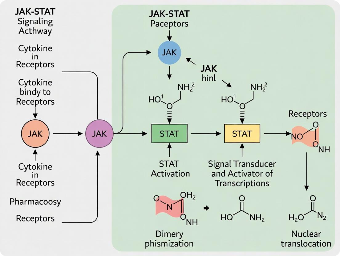
The Janus kinase-signal transducer and activator of transcription (JAK-STAT) pathway is a fundamental signaling cascade that transduces extracellular signals from cytokines, interferons, and growth factors into the nucleus, regulating gene expression. It is a principal mediator of critical physiological processes, including hematopoiesis, immune function, tissue repair, and inflammatory responses. Dysregulated activation of this pathway is a hallmark of numerous human diseases, including myeloproliferative neoplasms, autoimmune diseases (e.g., rheumatoid arthritis, psoriasis), and various cancers. Its role as a central hub makes it a prime target for therapeutic intervention, with several JAK inhibitors (jakinibs) now FDA-approved. This whitepaper provides an in-depth technical guide to the pathway's activation mechanics, aligned with a research thesis focused on elucidating the nuances of JAK-STAT signaling activation dynamics.
Core Pathway Activation Mechanism
The canonical JAK-STAT pathway activation is a rapid, membrane-to-nucleus signaling event.
- Cytokine Receptor Engagement: A ligand (e.g., IFN-γ, IL-6) binds to its cognate type I or II transmembrane receptor, inducing dimerization or conformational change of the receptor subunits.
- JAK Activation: Receptor-associated JAKs (JAK1, JAK2, JAK3, TYK2) are brought into proximity, leading to their trans-auto-phosphorylation on tyrosine residues and full kinase activation.
- Receptor Phosphorylation: Activated JAKs phosphorylate specific tyrosine residues on the intracellular tails of the receptor chains, creating docking sites for STAT proteins.
- STAT Recruitment and Phosphorylation: Cytosolic STAT monomers (STAT1-6) bind to the phospho-tyrosine sites via their Src homology 2 (SH2) domains. JAKs then phosphorylate a conserved tyrosine residue near the STAT C-terminus.
- STAT Dimerization and Nuclear Translocation: Phosphorylated STATs dissociate from the receptor, forming homo- or heterodimers via reciprocal phospho-tyrosine-SH2 domain interactions. These dimers are actively transported into the nucleus.
- Gene Transcription: Nuclear STAT dimers bind to specific promoter sequences (e.g., GAS elements for STAT1/3/5, ISRE for ISGF3) and recruit transcriptional co-activators, initiating target gene transcription.
Pathway Diagram: Canonical JAK-STAT Activation
Quantitative Data on JAK-STAT Components and Diseases
Table 1: JAK-STAT Family Members and Associated Pathologies
| Component | Family Members | Primary Associated Cytokines/Cues | Key Disease Associations |
|---|---|---|---|
| JAK Kinases | JAK1, JAK2, JAK3, TYK2 | IFNs, IL-2/4/6 family, EPO, TPO, G-CSF | RA, Psoriasis, MPNs, Allergies, Immunodeficiencies |
| STAT Proteins | STAT1, STAT2, STAT3, STAT4, STAT5A/B, STAT6 | IFNs (STAT1/2), IL-6 (STAT3), IL-12 (STAT4), IL-2/GH (STAT5), IL-4 (STAT6) | Cancers (STAT3/5), Autoimmunity, Immunodeficiencies |
| Negative Regulators | SOCS1-7, PIAS1-4, PTPs (SHP1/2, TC-PTP) | Feedback inhibition, STAT dephosphorylation | Loss contributes to constitutive activation in cancer. |
Table 2: Clinical Efficacy of Select JAK Inhibitors (Representative Data)
| Drug Name | Target Selectivity | Approved Indication(s) | Key Trial Efficacy Metric (Approx.) |
|---|---|---|---|
| Ruxolitinib | JAK1/JAK2 | Myelofibrosis, Polycythemia Vera | ~35-45% Spleen Volume Reduction (MF) |
| Tofacitinib | Pan-JAK (JAK3>JAK1>JAK2) | RA, Psoriatic Arthritis, UC | ~70% ACR20 Response in RA (vs ~30% placebo) |
| Upadacitinib | JAK1-selective | RA, Atopic Dermatitis, Crohn's | ~80% EASI75 in AD (vs ~16% placebo) |
| Baricitinib | JAK1/JAK2-selective | RA, Alopecia Areata, COVID-19 | ~70% SALT score ≤20 in AA (vs ~6% placebo) |
Detailed Experimental Protocols
Protocol 1: Assessing STAT Phosphorylation via Western Blot
- Objective: To detect ligand-induced tyrosine phosphorylation of STAT proteins.
- Materials: Cell line expressing target receptor, recombinant cytokine, lysis buffer (RIPA + phosphatase/protease inhibitors), anti-pSTAT (Y701 for STAT1, Y705 for STAT3), anti-total STAT antibody.
- Method:
- Stimulation: Serum-starve cells for 4-6 hrs. Stimulate with cytokine (e.g., 50ng/mL IFN-γ) for 15-30 mins. Include an unstimulated control.
- Lysis: Place cells on ice, wash with cold PBS, lyse in ice-cold buffer for 20 mins. Centrifuge at 14,000g for 15 mins at 4°C.
- Immunoblotting: Determine protein concentration. Load 20-40 µg of lysate per lane on an SDS-PAGE gel. Transfer to PVDF membrane.
- Detection: Block membrane, incubate with primary anti-pSTAT antibody overnight at 4°C. Wash, incubate with HRP-conjugated secondary antibody. Develop with ECL reagent.
- Reprobing: Strip membrane and reprobe with anti-total STAT antibody to confirm equal loading.
Protocol 2: JAK-STAT Pathway Reporter Gene Assay
- Objective: To functionally measure transcriptional output of the pathway.
- Materials: Luciferase reporter plasmid (e.g., pGAS-Luc for STAT1/3/5, pISRE-Luc for ISGF3), transfection reagent, Renilla control plasmid, dual-luciferase assay kit.
- Method:
- Transfection: Seed cells in 24-well plates. Co-transfect with the STAT-responsive firefly luciferase reporter plasmid and a constitutive Renilla luciferase control plasmid (e.g., pRL-TK) using appropriate transfection reagent.
- Stimulation: 24-48 hrs post-transfection, stimulate cells with cytokine for 6-12 hrs.
- Lysis and Measurement: Lyse cells per kit instructions. Measure firefly and Renilla luciferase activity sequentially in a luminometer.
- Analysis: Normalize firefly luminescence to Renilla luminescence for each well to control for transfection efficiency. Plot fold-change relative to unstimulated control.
Experimental Workflow Diagram: JAK-STAT Functional Assay Workflow
The Scientist's Toolkit: Key Research Reagent Solutions
Table 3: Essential Reagents for JAK-STAT Pathway Research
| Reagent Category | Specific Example(s) | Primary Function in Research |
|---|---|---|
| Recombinant Cytokines | Human/Mouse IFN-γ, IL-6, IL-4, EPO, Leptin | Ligand for specific receptor-JAK-STAT axis activation in stimulation experiments. |
| JAK Inhibitors (Tool Compounds) | Ruxolitinib (JAK1/2), Tofacitinib (pan-JAK), STATTIC (STAT3 inhibitor) | Pharmacological inhibition to probe pathway necessity, mechanism, and for control experiments. |
| Phospho-Specific Antibodies | Anti-pSTAT1 (Y701), Anti-pSTAT3 (Y705), Anti-pJAK2 (Y1007/1008) | Detection of pathway activation status via Western blot, flow cytometry, or immunofluorescence. |
| Reporter Plasmids | pGAS-Luciferase, pISRE-Luciferase | Measurement of transcriptional endpoint activity in functional cellular assays. |
| SOCS Overexpression/Knockdown Tools | SOCS1/SOCS3 expression vectors, siRNA/shRNA targeting SOCS | Investigation of negative feedback regulation mechanisms. |
| ChIP-Grade Antibodies | Anti-STAT1, Anti-STAT3 (for Chromatin Immunoprecipitation) | Identification of direct genomic binding sites and target genes. |
The Janus kinase (JAK)–signal transducer and activator of transcription (STAT) pathway is a principal signaling cascade that transmits information from extracellular polypeptide signals, primarily cytokines, interferons, and growth factors, directly to the nucleus, orchestrating gene expression programs governing immunity, cell proliferation, differentiation, and apoptosis. Framed within the broader thesis of JAK-STAT activation process research, this architectural overview deconstructs the core machinery: the transmembrane receptor complexes, the associated JAK kinases, and the STAT transcription factors. Understanding this architecture is foundational for deciphering pathway dysregulation in disease and for the rational design of targeted therapeutics.
Core Architectural Components
Transmembrane Cytokine Receptors
These receptors lack intrinsic enzymatic activity. Their architecture is defined by:
- Extracellular Domain: Binds specific ligands (cytokines). Common structures include cytokine receptor homology domains (CHRs) and fibronectin type III domains.
- Single-Pass Transmembrane Helix: Anchors the receptor.
- Intracellular Domain: Contains conserved box1/box2 motifs that serve as the docking platform for JAK kinases.
Cytokine receptors typically function as dimers. Ligand binding induces a conformational rearrangement (e.g., rotation, proximity) of the receptor subunits.
Janus Kinases (JAKs)
JAKs are non-receptor tyrosine kinases constitutively associated with the intracellular domain of cytokine receptors. The mammalian family has four members: JAK1, JAK2, JAK3, and TYK2. Their architecture features:
- FERM Domain: Mediates receptor association.
- SH2 Domain: Contributes to receptor and kinase regulation.
- Pseudokinase Domain: Regulatory; autoinhibitory but essential for proper activation.
- Tyrosine Kinase Domain: Catalytic unit; phosphorylates receptors and STATs.
Signal Transducers and Activators of Transcription (STATs)
STATs are latent cytoplasmic transcription factors. Seven members exist in mammals: STAT1, STAT2, STAT3, STAT4, STAT5a, STAT5b, and STAT6. Their domains include:
- N-terminal Domain: Facilitates tetramerization and cooperative DNA binding.
- Coiled-coil Domain: Involved in protein-protein interactions and nuclear import.
- DNA-binding Domain: Recognizes specific gamma-activated sequence (GAS) elements.
- Linker Domain: Stabilizes DNA binding.
- SH2 Domain: Critical for activation: mediates recruitment to phosphorylated receptor tails and STAT dimerization via reciprocal phosphotyrosine-SH2 interactions.
- Transactivation Domain (TAD): Contains a conserved tyrosine phosphorylation site (Y~701 in STAT1) and a serine phosphorylation site (S~727); recruits transcriptional co-activators.
The Activation Process: A Stepwise Mechanism
Step 1: Ligand-Induced Receptor Dimerization/Conformational Change. A cytokine binds to its cognate receptor, inducing proper alignment of two receptor subunits. This repositions the associated JAKs into a catalytically favorable proximity.
Step 2: JAK Transphosphorylation and Activation. The juxtaposed JAKs phosphorylate each other on tyrosine residues within their activation loops, relieving autoinhibition and achieving full catalytic activity.
Step 3: Receptor Tail Phosphorylation and STAT Recruitment. Activated JAKs phosphorylate specific tyrosine residues on the intracellular receptor tails, creating docking sites for STAT proteins via their SH2 domains.
Step 4: STAT Phosphorylation, Dimerization, and Nuclear Translocation. JAKs phosphorylate the conserved tyrosine residue in the STAT TAD. Phosphorylated STATs dissociate from the receptor and form reciprocal SH2-phosphotyrosine-mediated homo- or heterodimers.
Step 5: Nuclear Entry, DNA Binding, and Transcriptional Regulation. STAT dimers are actively transported into the nucleus via importins, bind to specific enhancer sequences in target gene promoters (e.g., GAS elements), and recruit co-activators (e.g., CBP/p300, histone acetyltransferases) to initiate transcription.
Title: JAK-STAT Pathway Activation Cascade
Table 1: Core JAK-STAT Family Members and Associated Ligands/Receptors
| Component | Family Members | Key Associated Ligands/Receptors | Chromosomal Location (Human) | Approx. Molecular Weight (kDa) |
|---|---|---|---|---|
| JAK Kinases | JAK1 | IFN-α/β/γ, IL-2, IL-6 family cytokines | 1p31.3 | 130-135 |
| JAK2 | EPO, TPO, GH, GM-CSF, IL-3 | 9p24.1 | 125-130 | |
| JAK3 | Common γ-chain cytokines (IL-2, IL-4, IL-7, IL-9, IL-15, IL-21) | 19p13.11 | 120-125 | |
| TYK2 | IFN-α/β, IL-12, IL-23 | 19p13.2 | 135-140 | |
| STAT Proteins | STAT1 | IFNs, EGF, PDGF | 2q32.2 | 84-91 |
| STAT2 | Type I IFNs (IFN-α/β) | 12q13.3 | 113 | |
| STAT3 | IL-6 family, EGF, Leptin | 17q21.2 | 79-86 | |
| STAT4 | IL-12, IL-23 | 2q32.2 | 85-89 | |
| STAT5a/5b | Prolactin, GH, EPO, IL-2 | 17q21.2 | 90-94 | |
| STAT6 | IL-4, IL-13 | 12q13.3 | 94 |
Table 2: Common Experimental Readouts for JAK-STAT Activity
| Assay Type | Target/Measurement | Common Quantitative Output | Typical Assay Platform |
|---|---|---|---|
| Phosphorylation | p-JAK (Tyr~1007/1008 for JAK2), p-STAT (Tyr~701 for STAT1) | Phosphorylation signal normalized to total protein (Fold-change over control) | Western Blot, ELISA, Flow Cytometry (Phospho-flow) |
| Nuclear Translocation | STAT-GFP fusion proteins | Nuclear-to-cytoplasmic fluorescence ratio | Live-Cell Imaging, Immunofluorescence |
| Transcriptional Activity | Luciferase reporter under GAS/ISRE promoter | Luciferase activity (RLU) normalized to control reporter | Dual-Luciferase Reporter Assay |
| Gene Expression | Downstream target genes (e.g., SOCS3, IRF1) | mRNA expression (e.g., ΔΔCt value vs. control) | RT-qPCR, RNA-Seq |
Experimental Protocols
Protocol 1: Assessing STAT Phosphorylation by Western Blot
Objective: To detect and quantify tyrosine phosphorylation of STAT proteins in cell lysates upon cytokine stimulation.
Detailed Methodology:
- Cell Stimulation: Seed cells in 6-well plates. Serum-starve (e.g., 2-24 hours) to reduce basal signaling. Stimulate with cytokine of interest (e.g., 10-100 ng/mL IFN-γ for STAT1) for a time course (e.g., 0, 5, 15, 30, 60 min).
- Lysis: Rapidly aspirate medium and lyse cells on ice with 200-300 µL RIPA buffer supplemented with phosphatase and protease inhibitors. Scrape and transfer to a microcentrifuge tube. Incubate on ice for 15-30 min, then centrifuge at 14,000 x g for 15 min at 4°C. Collect supernatant.
- Protein Quantification: Use BCA assay to determine lysate concentration. Adjust samples with Laemmli buffer to equal protein concentrations.
- SDS-PAGE and Transfer: Load 20-40 µg total protein per lane on a 8-10% polyacrylamide gel. Run at constant voltage. Transfer proteins to a PVDF or nitrocellulose membrane.
- Immunoblotting: Block membrane with 5% BSA in TBST for 1 hour. Incubate with primary antibody (e.g., anti-pSTAT1 Tyr701) diluted in blocking buffer overnight at 4°C. Wash with TBST, incubate with appropriate HRP-conjugated secondary antibody for 1 hour at RT. Develop using enhanced chemiluminescence (ECL) substrate and image.
- Reprobing: Strip membrane (optional) and re-probe with antibody against total STAT1 to confirm equal loading. Quantify band intensity using densitometry software.
Protocol 2: JAK-STAT Pathway Reporter Gene Assay
Objective: To functionally measure JAK-STAT pathway-induced transcriptional activity.
Detailed Methodology:
- Reporter Construct: Use a plasmid containing a firefly luciferase gene under the control of a promoter with multiple copies of a STAT-binding element (e.g., GAS or ISRE). A constitutive promoter-driven Renilla luciferase plasmid serves as transfection control.
- Cell Transfection: Seed cells in 24-well plates 24h prior. Co-transfect cells with the STAT-responsive firefly luciferase reporter and the Renilla control plasmid using a suitable transfection reagent (e.g., lipofection). Incubate for 24-48 hours.
- Stimulation: Stimulate cells with the appropriate cytokine or test inhibitor compound for a defined period (e.g., 6-24 hours).
- Luciferase Assay: Aspirate medium, wash with PBS, and lyse cells with Passive Lysis Buffer (Promega). Transfer lysate to a tube or assay plate. Measure firefly and Renilla luciferase activities sequentially using a dual-luciferase assay system and a luminometer.
- Data Analysis: Calculate the ratio of firefly luciferase activity (pathway reporter) to Renilla luciferase activity (transfection control) for each well. Express results as fold induction relative to unstimulated control.
Title: STAT Transcriptional Reporter Assay Workflow
The Scientist's Toolkit: Key Research Reagent Solutions
Table 3: Essential Reagents for JAK-STAT Pathway Research
| Reagent Category | Specific Example(s) | Function & Application |
|---|---|---|
| Recombinant Cytokines/Growth Factors | Human/Mouse IFN-γ, IL-6, EPO, GM-CSF | Ligand for specific receptor-JAK-STAT axis; used for pathway stimulation in experiments. |
| Selective JAK Inhibitors (Tool Compounds) | Ruxolitinib (JAK1/2), Tofacitinib (JAK1/3), AG490 (JAK2) | Pharmacologic probes to inhibit specific JAK activity; validate pathway dependency. |
| Phospho-Specific Antibodies | Anti-pSTAT1 (Tyr701), Anti-pSTAT3 (Tyr705), Anti-pJAK2 (Tyr1007/1008) | Detect activation-specific phosphorylation events via Western blot, flow cytometry, or IF. |
| STAT DNA-Binding ELISA Kits | TransAM STAT Family Kits (Active Motif) | Quantify active, DNA-binding STAT dimers in nuclear extracts in a 96-well format. |
| Luciferase Reporter Vectors | pGAS-Luc, pISRE-Luc (Addgene) | Measure STAT-mediated transcriptional activity in live cells. |
| SOCS Protein Expression Vectors | SOCS1, SOCS3 overexpression plasmids | Endogenous pathway negative regulators; used to study feedback inhibition. |
| JAK/STAT Deficient Cell Lines | JAK1-KO HEK293, STAT1-KO U3A cell lines | Isogenic controls to confirm protein-specific functions in genetic rescue/complementation assays. |
| Proteasome Inhibitors | MG-132, Bortezomib | Prevent STAT protein degradation; used to stabilize proteins for detection or study regulation. |
The JAK-STAT (Janus Kinase–Signal Transducer and Activator of Transcription) signaling pathway is a primary mechanism for transducing extracellular cytokine signals into intracellular transcriptional responses. This whitepaper focuses on the critical, initial triggering event: cytokine binding and subsequent receptor dimerization or oligomerization. This step is the allosteric linchpin that converts an extracellular ligand-receptor interaction into an intracellular tyrosine kinase activation event. Research into this precise molecular mechanism is foundational for developing targeted therapeutics for immune disorders, myeloproliferative neoplasms, and cancers where pathway dysregulation is prevalent.
Molecular Mechanism of the Initial Step
Cytokines of the helical bundle family (e.g., interleukins, interferons, colony-stimulating factors) initiate signaling by binding to specific single-pass transmembrane receptors. The prevailing model involves a sequential, cooperative process:
- Cytokine Architecture: Most cytokines are bivalent or multivalent, possessing at least two distinct receptor-binding epitopes (Site I and Site II/III).
- Initial Binding: The cytokine first engages with its primary, high-affinity receptor subunit via Site I.
- Receptor Dimerization/Oligomerization: This initial complex presents Site II/III, facilitating the recruitment of a second receptor subunit (which may be identical, forming a homodimer, or different, forming a heterodimer or multi-chain complex).
- Conformational Rearrangement: The bringing together of two receptor cytoplasmic domains induces a precise spatial reorientation of the pre-associated Janus Kinase (JAK) proteins, which are constitutively bound to the receptor's Box1/Box2 membrane-proximal regions. This proximity is essential for trans-activation.
The stoichiometry and specificity of this interaction are precise and vary by cytokine family, as summarized in Table 1.
Table 1: Quantitative Parameters for Select Cytokine-Receptor Complexes
| Cytokine (Example) | Receptor Composition | Binding Affinity (Kd) for Subunit 1 | Binding Affinity (Kd) for Subunit 2 | Final Complex Stoichiometry | Key JAKs Associated |
|---|---|---|---|---|---|
| Erythropoietin (EPO) | Homodimer (EPOR) | 0.5 - 1 nM | ~10 µM (weak, cytokine-mediated) | 1:2 (Cytokine:Receptor) | JAK2 |
| Interleukin-6 (IL-6) | α-chain (IL-6Rα) + gp130 (homodimer) | 10 - 100 pM (for IL-6Rα) | nM range (for gp130) | 1:1:2 (IL-6:IL-6Rα:gp130) | JAK1, JAK2, TYK2 |
| Interferon-γ (IFN-γ) | Heterotetramer (IFNGR1 + IFNGR2) | ~1 nM (for IFNGR1) | ~50 nM (for IFNGR2) | 1:2:2 (IFN-γ:IFNGR1:IFNGR2) | JAK1, JAK2 |
| Growth Hormone (GH) | Homodimer (GHR) | 0.1 - 1 nM | Weak, induced by first binding | 1:2 (Cytokine:Receptor) | JAK2 |
Key Experimental Protocols for Study
Surface Plasmon Resonance (SPR) for Binding Kinetics
Objective: Quantify the real-time kinetics (association/dissociation rates) and affinity of cytokine binding to immobilized receptor extracellular domains. Protocol:
- Immobilization: The extracellular domain (ECD) of one receptor subunit is covalently immobilized on a CMS sensor chip via amine coupling.
- Binding Analysis: Purified cytokine is flowed over the chip at various concentrations (e.g., 0.5 nM to 200 nM) in HBS-EP buffer (10 mM HEPES, 150 mM NaCl, 3 mM EDTA, 0.005% v/v Surfactant P20, pH 7.4).
- Data Processing: Sensorgrams are double-referenced. Binding curves are fitted to a 1:1 Langmuir binding model or a more complex two-state or bivalent analyte model using Biacore Evaluation Software to determine ka (association rate, M⁻¹s⁻¹), kd (dissociation rate, s⁻¹), and KD (equilibrium dissociation constant, kd/ka).
- Sequential Binding: To model dimerization, a second receptor ECD can be injected over the pre-formed cytokine-first receptor complex.
Co-Immunoprecipitation (Co-IP) & Western Blot for Complex Formation
Objective: Validate receptor dimerization in a cellular context upon cytokine stimulation. Protocol:
- Cell Stimulation: Serum-starve cells expressing tagged receptors (e.g., HA- and FLAG-tagged subunits) for 4-6 hours. Stimulate with cytokine (e.g., 10-100 ng/mL) for 0, 5, 15, and 30 minutes at 37°C.
- Lysis & Precipitation: Lyse cells in non-denaturing IP lysis buffer (e.g., 25 mM Tris, 150 mM NaCl, 1% NP-40, 1 mM EDTA, protease/phosphatase inhibitors). Clarify lysate. Incubate with anti-HA magnetic beads for 2 hours at 4°C.
- Wash & Elution: Wash beads 3-4 times with lysis buffer. Elute proteins with 2X Laemmli sample buffer containing β-mercaptoethanol.
- Detection: Resolve proteins by SDS-PAGE, transfer to PVDF membrane, and probe sequentially with anti-FLAG (to detect co-precipitated receptor) and anti-HA (to confirm IP efficiency) antibodies. Dimerization is indicated by the presence of the FLAG-tagged subunit in the HA IP only upon cytokine stimulation.
Bioluminescence Resonance Energy Transfer (BRET) for Live-Cell Proximity
Objective: Measure real-time, spatial proximity between receptor subunits in live cells. Protocol:
- Construct Design: Fuse receptor subunit A to a BRET donor (e.g., NanoLuc luciferase) and subunit B to a BRET acceptor (e.g., fluorescent protein HaloTag).
- Transfection & Plating: Co-transfect constructs into HEK293T cells and plate in a white 96-well plate.
- Reading & Stimulation: Add the NanoLuc substrate furimazine. Measure baseline luminescence (460 nm filter) and acceptor emission (e.g., 610 nm filter) using a microplate reader. Inject cytokine directly into wells and monitor the BRET ratio (Acceptor Emission / Donor Luminescence) over time.
- Analysis: An increase in BRET ratio indicates decreased distance between donor and acceptor (<10 nm), confirming cytokine-induced receptor subunit proximity.
Visualization of the Core Mechanism
Diagram 1: Cytokine-induced receptor dimerization and JAK proximity (72 chars)
The Scientist's Toolkit: Key Research Reagent Solutions
Table 2: Essential Reagents for Studying Cytokine Binding & Dimerization
| Reagent Category | Example Product/Kit | Function in Research |
|---|---|---|
| Recombinant Cytokines & ECDs | Human IL-6Rα Fc Chimera (R&D Systems), His-tagged EPO | Provide pure, bioactive ligands and soluble receptor domains for SPR, ELISA, and crystallization studies. |
| Tagged Expression Vectors | pCMV-HA Vector, pFLAG-CMV-2 | Enable transient or stable expression of receptor subunits with distinct epitope tags (HA, FLAG, Myc) for Co-IP experiments. |
| Co-IP & Detection Kits | Pierce Anti-HA Magnetic Beads, Anti-FLAG M2 Magnetic Beads | Magnetic bead-based systems for efficient immunoprecipitation of tagged proteins from cell lysates. |
| Live-Cell Proximity Assays | NanoBRET Protein:Protein Interaction System (Promega) | Integrated system including donor/acceptor vectors, substrates, and protocols for BRET-based dimerization assays in live cells. |
| Kinetic Analysis Software | Biacore Insight Evaluation Software, Scrubber-2 | Specialized software for fitting and analyzing kinetic data from SPR and other biosensor platforms. |
| Pathway Inhibitors (Controls) | Ruxolitinib (JAK1/2 inhibitor), Tocilizumab (IL-6Rα blocking antibody) | Used as negative controls to block downstream signaling or ligand binding, validating the specificity of the observed dimerization. |
Within the JAK-STAT signaling paradigm, the transition from cytokine-receptor engagement to downstream STAT protein phosphorylation is governed by a critical regulatory event: Janus kinase (JAK) transphosphorylation and kinase activation. This whitepaper, part of a broader thesis on the JAK-STAT activation process, dissects this molecular switch. Following receptor dimerization and JAK approximation, Step 2 involves the reciprocal phosphorylation of key tyrosine residues within the JAK activation loop, liberating the kinase domain from autoinhibition and creating docking sites for STAT proteins. This document provides a technical guide for researchers and drug development professionals, detailing the mechanisms, experimental interrogation, and quantitative dynamics of this process.
JAKs (JAK1, JAK2, JAK3, TYK2) are constitutively associated with the intracellular domains of cytokine receptors. In their basal state, the kinase domain is inhibited by the pseudokinase domain. Receptor dimerization induced by cytokine binding brings two JAK molecules into close proximity. This spatial rearrangement permits trans-phosphorylation, where one JAK phosphorylates its counterpart on a specific tyrosine residue (e.g., Y1038/Y1039 in JAK2) within the activation loop of the kinase domain. This event induces a conformational shift, destabilizing the autoinhibitory interaction and fully activating the kinase. The now-active JAKs subsequently phosphorylate tyrosine residues on the receptor cytoplasmic tails, creating docking platforms for SH2 domain-containing proteins like STATs.
Diagram 1: JAK Activation via Receptor Dimerization and Transphosphorylation
Quantitative Dynamics of Activation
The kinetics of JAK transphosphorylation are influenced by cytokine concentration, receptor density, and JAK isoform. The following table summarizes key quantitative parameters derived from recent studies.
Table 1: Quantitative Parameters of JAK2 Transphosphorylation
| Parameter | Value | Experimental System | Reference (Example) |
|---|---|---|---|
| Phosphorylation Rate Constant (k~act~) | 0.15 ± 0.03 min⁻¹ | HEK293 cells expressing EpoR & JAK2 | [1] |
| Half-time of Activation (t~1/2~) | ~4.6 minutes | Ba/F3 cells stimulated with Epo | [2] |
| Dissociation Constant (K~d~) for JAK2 Dimer | 0.8 µM | Purified JAK2 kinase domains (in vitro) | [3] |
| Phosphorylation Site (Human JAK2) | Y1007/Y1008 (Activation loop) | Mass spectrometry analysis | [4] |
| Inhibitor IC~50~ (ATP-competitive) | Ruxolitinib: 2.8 nM (JAK2) | Cell-free kinase assay | [5] |
Experimental Protocols for Analysis
Protocol: Immunoprecipitation and Western Blot for JAK Transphosphorylation
Objective: To detect and quantify transphosphorylation of specific JAK activation loop tyrosines. Materials: See "The Scientist's Toolkit" below. Method:
- Cell Stimulation: Serum-starve cytokine-responsive cells (e.g., HEL 92.1.7 for JAK2) for 4-6 hours. Stimulate with ligand (e.g., 10 U/mL EPO for erythropoietin) for a time course (0, 2, 5, 10, 30 min). Use a JAK inhibitor (e.g., 1 µM Ruxolitinib) as a negative control.
- Cell Lysis: Rapidly lyse cells in 500 µL ice-cold RIPA buffer supplemented with phosphatase and protease inhibitors. Centrifuge at 16,000 × g for 15 min at 4°C.
- Immunoprecipitation (IP): Pre-clear 500 µg of lysate with Protein A/G beads for 30 min. Incubate supernatant with 2 µg of anti-JAK antibody overnight at 4°C with gentle rotation. Add 40 µL bead slurry and incubate for 2 hours.
- Wash & Elution: Wash beads 3x with lysis buffer. Elute proteins by boiling in 2X Laemmli sample buffer for 5 min.
- Western Blot: Resolve proteins by SDS-PAGE (6-8% gel). Transfer to PVDF membrane. Block with 5% BSA in TBST. Probe with primary antibodies:
- Phospho-specific anti-pJAK (Y1007/1008 for JAK2) (1:1000, 4°C overnight).
- Total anti-JAK antibody (1:2000).
- Detection & Analysis: Use HRP-conjugated secondary antibodies (1:5000) and chemiluminescence. Quantify band intensity via densitometry; normalize pJAK signal to total JAK.
Protocol: In Vitro Kinase Assay with Recombinant JAKs
Objective: To measure direct transphosphorylation activity in a controlled system. Method:
- Reaction Setup: In a 50 µL reaction volume, combine:
- Kinase Buffer: 25 mM HEPES (pH 7.4), 10 mM MgCl~2~, 0.1 mM Na~3~VO~4~, 1 mM DTT.
- ATP: 10 µM ATP + 0.5 µCi [γ-³²P]ATP.
- Substrate: 200 ng recombinant inactive JAK kinase domain (or a peptide substrate like Poly(Glu,Tyr) 4:1).
- Enzyme: 100 ng of active recombinant JAK kinase domain.
- Incubate at 30°C for 30 minutes.
- Termination & Detection:
- For peptide substrate: Spot reaction mix onto phosphocellulose paper, wash extensively in 0.75% phosphoric acid, and measure incorporated radioactivity by scintillation counting.
- For protein substrate: Stop reaction with Laemmli buffer, run SDS-PAGE, dry gel, and visualize phosphorylation via autoradiography or phosphorimaging.
Diagram 2: Workflow for JAK Phosphorylation Analysis
The Scientist's Toolkit: Key Research Reagents
Table 2: Essential Reagents for JAK Transphosphorylation Studies
| Reagent | Function/Description | Example Product (Vendor) |
|---|---|---|
| Phospho-specific JAK Antibodies | Detect activated JAKs via pY sites (e.g., JAK1 pY1034/1035, JAK2 pY1007/1008). Critical for WB/IP. | Anti-phospho-JAK2 (Tyr1007/1008) (Cell Signaling, #3771) |
| Pan/JAK Isoform Antibodies | Immunoprecipitation or loading control for total JAK protein levels. | Anti-JAK2 Antibody (Invitrogen, MA5-32148) |
| Active Recombinant JAK Kinases | For in vitro kinase assays to study biochemistry and inhibitor screening. | Recombinant Human JAK2 kinase domain (active), (SignalChem, #J52-10G) |
| ATP-analog & Detection Kits | Enable measurement of kinase activity (luminescent/fluorescent). | ADP-Glo Kinase Assay (Promega, #V9101) |
| Selective JAK Inhibitors | Tool compounds for negative controls and mechanistic studies. | Ruxolitinib (JAK1/2i), Tofacitinib (JAK3i) (Selleckchem) |
| Cytokine Ligands | To stimulate specific JAK-dependent pathways in cellular models. | Recombinant Human Erythropoietin (EPO) (PeproTech, #100-64) |
| Phosphatase/Protease Inhibitors | Preserve phosphorylation state during cell lysis. | PhosSTOP & cOmplete (Roche) |
| JAK-deficient Cell Lines | Isogenic backgrounds for rescue experiments and validation. | γ2A (JAK1-deficient), ΔJAK2 HEK293 (generated via CRISPR) |
Pathophysiological & Therapeutic Implications
Dysregulated JAK transphosphorylation is a cornerstone of pathology. Gain-of-function mutations (e.g., JAK2 V617F) cause constitutive, cytokine-independent transphosphorylation, driving myeloproliferative neoplasms. Conversely, loss-of-function mutations impair immune signaling. Therapeutically, ATP-competitive inhibitors (e.g., Ruxolitinib) bind the active kinase domain, blocking transphosphorylation and substrate phosphorylation. Next-generation Type II inhibitors stabilize the inactive conformation, providing greater selectivity. Precise targeting of this step remains a central strategy in treating autoimmune diseases, cancers, and inflammatory disorders.
Diagram 3: Dysregulation and Inhibition of JAK Transphosphorylation
This whitepaper details the third critical phase in the JAK-STAT pathway activation cascade, a core focus of our broader thesis research. Following cytokine receptor engagement and JAK auto-/trans-phosphorylation (Step 1) and the creation of phospho-tyrosine docking sites on the receptor (Step 2), Step 3 involves the specific recruitment, phosphorylation, and subsequent dimerization of STAT (Signal Transducer and Activator of Transcription) proteins. This step transduces the extracellular signal into a direct nuclear command, making it a prime target for therapeutic intervention in autoimmune diseases, myeloproliferative neoplasms, and cancers.
The process is characterized by a sequence of highly specific protein-domain interactions:
- Recruitment: STAT monomers exist in the cytoplasm. Their Src homology 2 (SH2) domains recognize and bind to specific phospho-tyrosine (pY) motifs on the activated receptor-JAK complex.
- Phosphorylation: Once docked, a juxtaposed JAK kinase phosphorylates a conserved tyrosine residue on the STAT protein's C-terminal transactivation domain.
- Dimerization: Phosphorylation induces a critical conformational change. The STAT's own SH2 domain can now bind reciprocally to the pY of another STAT molecule, forming either homodimers or heterodimers.
- Release: The STAT dimer dissociates from the receptor complex, exposing its nuclear localization signal (NLS), and is now primed for nuclear translocation (Step 4 in the cascade).
Quantitative Data on STAT Isoforms
| STAT Isoform | Approx. Size (kDa) | Primary Phosphorylation Site (Tyrosine) | Common Dimer Forms | Key Activating Cytokines/Pathways |
|---|---|---|---|---|
| STAT1 | 91 | Y701 | Homodimer, STAT1-STAT2 | IFN-γ, IFN-α/β |
| STAT2 | 113 | Y690 | STAT1-STAT2 heterodimer | IFN-α/β |
| STAT3 | 88 / 79 (isoforms) | Y705 | Homodimer | IL-6 family, EGF, IL-10 |
| STAT4 | 85 | Y693 | Homodimer | IL-12 |
| STAT5a / 5b | ~90 | Y694 (5a) / Y699 (5b) | Homodimers, Heterodimers (5a/5b) | Prolactin, GH, IL-2, IL-3 |
| STAT6 | 94 | Y641 | Homodimer | IL-4, IL-13 |
Detailed Experimental Protocols
Protocol 1: Co-Immunoprecipitation (Co-IP) for STAT-Receptor/JAK Complex Analysis
- Objective: To validate STAT recruitment to the activated receptor complex.
- Methodology:
- Stimulate cells (e.g., HeLa, HepG2) with relevant cytokine (e.g., IFN-γ for STAT1, IL-6 for STAT3) for 5-15 minutes.
- Lyse cells in a non-denaturing IP lysis buffer (e.g., containing 1% NP-40, phosphatase, and protease inhibitors).
- Pre-clear lysate with Protein A/G beads for 30 min at 4°C.
- Incubate supernatant with antibody against the cytokine receptor or a tagged STAT protein overnight at 4°C.
- Add Protein A/G beads for 2 hours to capture immune complexes.
- Wash beads 3-4 times with lysis buffer.
- Elute proteins in 2X Laemmli buffer by boiling for 5 min.
- Analyze by Western Blot (WB) using antibodies against pJAK, pSTAT, total STAT, and the receptor.
Protocol 2: Phospho-STAT Detection by Flow Cytometry (Phosflow)
- Objective: To quantify STAT phosphorylation at the single-cell level across a population.
- Methodology:
- Stimulate suspension or dissociated adherent cells with ligand in a time-course (e.g., 0, 15, 30, 60 min).
- Immediately fix cells with pre-warmed 4% formaldehyde for 10 min at 37°C to preserve phosphorylation states.
- Permeabilize cells by adding ice-cold 100% methanol drop-wise while vortexing; incubate ≥30 min at -20°C.
- Wash cells and stain with fluorochrome-conjugated antibodies specific for pSTAT (e.g., pSTAT1-Y701, pSTAT3-Y705, pSTAT5-Y694) and relevant surface markers for 1 hour at RT.
- Analyze on a flow cytometer. Median fluorescence intensity (MFI) of the pSTAT channel indicates phosphorylation level.
Protocol 3: Electrophoretic Mobility Shift Assay (EMSA) for STAT Dimerization & DNA Binding
- Objective: To confirm functional STAT dimer formation by assessing its ability to bind consensus DNA sequences.
- Methodology:
- Prepare nuclear extracts from stimulated and control cells.
- Label a double-stranded DNA oligonucleotide containing the STAT consensus binding site (e.g., GAS element for STAT1/3/5; ISRE for ISGF3 [STAT1:STAT2:IRF9]) with [γ-32P]ATP.
- Incubate nuclear extract (5-20 µg protein) with labeled probe in binding buffer for 20-30 min at RT.
- Run the protein-DNA complexes on a non-denaturing polyacrylamide gel in 0.5X TBE buffer.
- Dry gel and expose to a phosphorimager screen. A "supershift" with an anti-STAT antibody confirms the dimer's identity.
Pathway & Workflow Visualizations
Diagram Title: STAT Activation Steps 1-3: Recruitment, Phosphorylation, Dimerization
Diagram Title: Co-IP Workflow for Analyzing STAT Recruitment
The Scientist's Toolkit: Key Research Reagent Solutions
| Reagent / Material | Function / Application | Key Considerations |
|---|---|---|
| Phospho-Specific STAT Antibodies (e.g., anti-pSTAT1 Y701, pSTAT3 Y705) | Detects activated STATs in WB, ICC/IHC, Flow. Critical for monitoring Step 3. | Verify species reactivity. Use with appropriate fixation (methanol for flow). |
| STAT SH2 Domain Inhibitors/Peptides | Competitively blocks STAT recruitment to pY sites. Used for mechanistic validation. | Cell-permeable variants are required for intracellular assays. |
| Recombinant Cytokines & Growth Factors (e.g., IFN-γ, IL-6, EGF) | Defined ligands to specifically activate pathways leading to STAT phosphorylation. | Use carrier protein (e.g., BSA) for low-concentration stocks. |
| JAK Inhibitors (e.g., Ruxolitinib, Tofacitinib) | Pharmacological tools to inhibit upstream kinase activity, preventing STAT phosphorylation. | Distinguish pan-JAK vs. isoform-selective inhibitors for experiment design. |
| STAT Reporter Cell Lines (e.g., with GAS/ISRE-luciferase construct) | Functional readout of STAT dimerization, nuclear translocation, and transcriptional activity. | Allows for high-throughput screening of modulators. |
| Protein A/G Magnetic Beads | For efficient Co-IP of STAT complexes. Reduce non-specific binding vs. agarose beads. | Choose based on antibody species and isotype for optimal binding. |
| Methanol & Cross-linking Fixatives (Formaldehyde, Paraformaldehyde) | Essential for preserving labile protein phosphorylation states prior to intracellular staining. | Methanol is standard for phospho-epitopes in flow cytometry. |
Within the comprehensive study of the JAK-STAT signaling pathway, Step 4 represents the culmination of the activation cascade, where the signal is converted into a sustained transcriptional response. Following receptor engagement, JAK-mediated phosphorylation, and STAT dimerization, the phosphorylated STAT dimers translocate to the nucleus. Here, they bind to specific regulatory sequences in DNA, recruiting transcriptional co-activators to modulate the expression of target genes, which dictate cellular outcomes such as proliferation, differentiation, and immune responses. This whitepaper details the molecular mechanisms, quantitative dynamics, experimental protocols, and essential tools for investigating this critical phase.
Molecular Mechanism and Quantitative Dynamics
Nuclear translocation is an energy-dependent process facilitated by the importin α/β system. The STAT dimer's nuclear localization signal (NLS), often exposed upon phosphorylation and dimerization, is recognized by importin-α. This complex is then transported through the nuclear pore via interaction with importin-β. Once in the nucleus, STAT dimers bind to palindromic sequences known as Gamma-Activated Sites (GAS) for STAT1, STAT3, STAT4, and STAT5, or interferon-stimulated response elements (ISRE) for STAT1 and STAT2 complexes.
The affinity and specificity of DNA binding, along with the duration of nuclear residence, are key regulatory points. Post-translational modifications (e.g., acetylation, methylation) and interactions with coregulators (e.g., CBP/p300, NCoA) fine-tune transcriptional activity. Signal termination is achieved via nuclear phosphatases (e.g., TC45), which dephosphorylate STATs, leading to their export to the cytoplasm via exportin (CRM1).
Table 1: Key Quantitative Parameters for STAT1 Nuclear Translocation and Transcription
| Parameter | Approximate Value / Range | Experimental Method | Reference Context |
|---|---|---|---|
| Time to max nuclear accumulation post-stimulation | 15-45 minutes | Live-cell imaging (FRAP/FLIP) | IFN-γ stimulation |
| Nuclear residency half-life (phosphorylated STAT1) | 30-90 minutes | Photobleaching assays | HeLa cells |
| Dissociation constant (Kd) for STAT1 dimer to GAS site | 1-10 nM | EMSA / Surface Plasmon Resonance | In vitro purified proteins |
| Transcriptional activation onset | 1-2 hours | RNA FISH / RT-qPCR | IFN-α/γ target genes |
| Peak mRNA levels of target genes (e.g., IRF1) | 4-8 hours | RT-qPCR time course | Primary fibroblasts |
Table 2: Core Transcriptional Co-regulators in JAK-STAT Signaling
| Co-regulator Protein | Function in STAT Transcription | Interacting STAT(s) |
|---|---|---|
| CBP / p300 | Histone acetyltransferase (HAT) activity; chromatin remodeling | STAT1, STAT2, STAT3, STAT5 |
| NCoA/SRC-1 | Recruits additional HAT activity; stabilizes transcription complex | STAT1, STAT3, STAT6 |
| Mediator Complex | Bridges transcription factors to RNA Polymerase II | All STATs |
| BRD4 | Binds acetylated histones/STATs; promotes transcriptional elongation | STAT3, STAT5 |
| HDACs (e.g., HDAC3) | Deacetylation; negative regulation of transcription | STAT1, STAT3 |
Experimental Protocols
Protocol 1: Subcellular Fractionation and Immunoblot for STAT Localization
Objective: To quantitatively assess STAT protein levels in cytoplasmic and nuclear compartments over time. Methodology:
- Stimulation & Harvest: Stimulate cells (e.g., with IFN-γ 10 ng/mL) for various times (0, 15, 30, 60, 120 min). Wash with ice-cold PBS.
- Hypotonic Lysis: Pellet cells. Resuspend in hypotonic buffer (10 mM HEPES pH 7.9, 1.5 mM MgCl2, 10 mM KCl, 0.5 mM DTT, protease/phosphatase inhibitors). Incubate on ice 15 min. Add 0.5% NP-40, vortex, centrifuge (10,000 g, 5 min). Supernatant = cytoplasmic fraction.
- Nuclear Extraction: Wash pellet. Resuspend in high-salt RIPA buffer (25 mM Tris pH 7.6, 150 mM NaCl, 1% NP-40, 1% sodium deoxycholate, 0.1% SDS) or commercial nuclear extraction kit. Sonicate briefly, incubate on ice 30 min, centrifuge (14,000 g, 15 min). Supernatant = nuclear fraction.
- Analysis: Perform immunoblot for STAT1 (pY701 and total), using markers like α-tubulin (cytoplasm) and Lamin B1 or Histone H3 (nucleus) for normalization.
Protocol 2: Chromatin Immunoprecipitation (ChIP) Assay for STAT-DNA Binding
Objective: To determine the in vivo binding of STAT proteins to specific promoter regions. Methodology:
- Cross-linking & Lysis: Stimulate cells. Fix with 1% formaldehyde for 10 min at RT. Quench with glycine. Lyse cells in SDS lysis buffer.
- Sonication: Sonicate chromatin to shear DNA to 200-1000 bp fragments. Centrifuge to remove debris.
- Immunoprecipitation: Pre-clear lysate with protein A/G beads. Incubate overnight at 4°C with antibody against target STAT (e.g., anti-STAT1 pY701) or control IgG. Capture immune complexes with beads.
- Washing & Elution: Wash beads sequentially with low-salt, high-salt, LiCl, and TE buffers. Elute complexes with elution buffer (1% SDS, 0.1M NaHCO3). Reverse cross-links by adding NaCl and heating at 65°C overnight.
- DNA Purification & Analysis: Treat with Proteinase K, purify DNA. Analyze target gene promoter occupancy by quantitative PCR (ChIP-qPCR) using primers specific for the GAS/ISRE element of interest (e.g., in the IRF1 promoter).
Visualization Diagrams
Diagram Title: STAT Nuclear Translocation and Transcription Initiation
Diagram Title: Chromatin Immunoprecipitation (ChIP) Workflow
The Scientist's Toolkit: Research Reagent Solutions
Table 3: Essential Reagents for Studying Nuclear Translocation and Transcription
| Reagent / Material | Function / Application | Example Product/Catalog |
|---|---|---|
| Phospho-specific STAT Antibodies | Detect activated, tyrosine-phosphorylated STATs in WB, IF, ChIP. Critical for tracking the active transcription factor. | Cell Signaling Tech #9167 (STAT1 pY701); #9145 (STAT3 pY705) |
| Nuclear-Cytoplasmic Fractionation Kit | Rapid, clean separation of cellular compartments for quantifying protein redistribution. | Thermo Fisher NE-PER Kit |
| Importin β1 (KPNA2) Inhibitor (Importazole) | Chemical inhibitor of the Importin β1-mediated nuclear import pathway. Used to functionally block STAT translocation. | Sigma-Aldrich SML1129 |
| CpG-free Luciferase Reporter Vector | To assay STAT-dependent promoter activity without confounding immune stimulation from vector-borne CpG motifs. | InvivoGen pCpGfree-basic |
| Live-Cell Imaging Dyes (HaloTag/ SNAP-tag Ligands) | For real-time visualization of STAT protein dynamics using tagged constructs (e.g., STAT1-HaloTag). | Promega HaloTag Janelia Fluor 646 |
| STAT-DNA Binding ELISA Kit | Quantitative, plate-based assay to measure STAT dimer binding to immobilized GAS consensus sequences. | Active Motif TransAM STAT Family Kits |
| BET Bromodomain Inhibitor (JQ1) | Inhibits BRD4, a key regulator of transcriptional elongation downstream of STATs. Useful for dissecting mechanism. | Cayman Chemical 11187 |
| RNase Inhibitors & cDNA Synthesis Kits | Essential for accurate quantification of nascent target gene mRNA transcripts via RT-qPCR. | Takara Bio PrimeScript RT reagent Kit |
The JAK-STAT signaling pathway is a principal mediator of cytokine and growth factor signaling, governing processes from immune response to hematopoiesis. Its precise regulation is critical to prevent pathological outcomes such as autoimmunity and cancer. This whitepaper details three core classes of negative regulators that fine-tune this pathway: Suppressors of Cytokine Signaling (SOCS) proteins, Protein Inhibitors of Activated STATs (PIAS), and the deubiquitinase USP7. Understanding their mechanisms is paramount for developing targeted therapeutics for inflammatory diseases, immune disorders, and cancers driven by dysregulated JAK-STAT signaling.
Core Regulatory Mechanisms
Suppressors of Cytokine Signaling (SOCS) Proteins
SOCS proteins (CIS and SOCS1-7) form a classic negative feedback loop. They are rapidly induced by STAT activation and inhibit signaling via two primary mechanisms: 1) acting as pseudo-substrates that block the JAK kinase active site (e.g., SOCS1), and 2) acting as adaptors for E3 ubiquitin ligase complexes, targeting associated proteins like JAKs and cytokine receptors for proteasomal degradation.
Protein Inhibitors of Activated STATs (PIAS)
The PIAS family (PIAS1, PIAS3, PIASx, PIASy) regulates signaling primarily at the level of the transcription factor. PIAS proteins inhibit STAT-mediated gene transcription by blocking DNA binding, promoting SUMOylation of STATs (and other pathway components), and recruiting transcriptional co-repressors.
Ubiquitin-Specific Peptidase 7 (USP7)
USP7 (HAUSP) is a deubiquitinating enzyme that stabilizes key proteins in the pathway. By removing ubiquitin chains, USP7 counteracts proteasomal targeting. Notably, it deubiquitinates and stabilizes SOCS3, creating a complex regulatory circuit, and also targets other pathway components like STAT3.
Table 1: Key Characteristics of JAK-STAT Regulatory Proteins
| Protein Family | Member Examples | Molecular Weight (kDa) | Primary Mechanism of Action | Effect on JAK-STAT | Associated Diseases if Dysregulated |
|---|---|---|---|---|---|
| SOCS | SOCS1, SOCS3 | ~25-30 | SH2 domain binding; E3 ligase recruitment | Inhibits JAK kinase activity; Targets receptors/JAKs for degradation | Inflammation, Cancer, Metabolic Disorders |
| PIAS | PIAS1, PIAS3 | ~60-80 | SUMO E3 ligase activity; Blocking DNA binding | Inhibits STAT transcriptional activity | Cancer, Immune Dysregulation |
| Deubiquitinase | USP7 | ~130 | Cysteine protease; Deubiquitination | Stabilizes SOCS3, STAT3; Modulates pathway output | Cancer, Neurological Disorders |
Table 2: Experimental Readouts for Regulatory Function Assessment
| Assay Type | Measured Parameter | Typical Control Value (Baseline) | Value with Regulator Overexpression | Value with Regulator Knockdown/KO |
|---|---|---|---|---|
| Phospho-STAT ELISA | p-STAT1/3/5 levels (AU) | 1.0 (Normalized) | 0.2 - 0.5 | 2.0 - 4.0 |
| Luciferase Reporter | STAT-driven luciferase activity (RLU) | 100,000 RLU | 10,000 - 30,000 RLU | 300,000 - 500,000 RLU |
| qPCR Target Gene | SOCS3 mRNA (Fold Change) | 1.0 | 10.0 - 50.0 (Feedback) | 0.1 - 0.3 |
| Protein Half-life (Cycloheximide) | SOCS3 t½ (minutes) | ~30-45 min | N/A | ~15-20 min (without USP7) |
Experimental Protocols
Protocol: Assessing SOCS3-Mediated JAK1 Degradation (Co-immunoprecipitation & Cycloheximide Chase)
Objective: To determine if SOCS3 expression promotes ubiquitin-mediated degradation of JAK1. Materials: HEK293T or relevant cell line, expression plasmids for JAK1, SOCS3, HA-Ubiquitin, anti-JAK1 antibody, cycloheximide, MG132. Procedure:
- Transfection: Co-transfect cells with JAK1, SOCS3, and HA-Ubiquitin plasmids. Include a control without SOCS3.
- Proteasome Inhibition (Optional): 6 hours before harvest, treat one set with MG132 (10 µM) to inhibit the proteasome.
- Pulse-Chase: 24h post-transfection, treat cells with cycloheximide (100 µg/mL) to inhibit new protein synthesis. Harvest cells at time points (0, 30, 60, 120 min).
- Immunoprecipitation: Lyse cells in RIPA buffer. Immunoprecipitate JAK1 using specific antibody conjugated to beads.
- Western Blot: Analyze immunoprecipitates and whole-cell lysates by WB for: JAK1 (to assess degradation), HA (to assess polyubiquitination), and SOCS3 (expression check). Analysis: Compare JAK1 half-life and ubiquitination levels in presence/absence of SOCS3 and MG132.
Protocol: Measuring PIAS1 Inhibition of STAT1 Transcriptional Activity (Luciferase Reporter Assay)
Objective: To quantify the inhibitory effect of PIAS1 on STAT1-driven transcription. Materials: Cell line responsive to IFN-γ (e.g., HeLa), GAS-Luc reporter plasmid, Renilla luciferase control plasmid, PIAS1 expression plasmid, recombinant IFN-γ. Procedure:
- Transfection: Seed cells in 24-well plates. Co-transfect with GAS-Luc reporter, Renilla plasmid, and increasing amounts of PIAS1 plasmid. Keep total DNA constant with empty vector.
- Stimulation: 24h post-transfection, stimulate cells with IFN-γ (10 ng/mL) for 6-8 hours.
- Lysis & Measurement: Lyse cells with passive lysis buffer. Measure firefly and Renilla luciferase activities using a dual-luciferase assay kit.
- Calculation: Normalize firefly luciferase activity to Renilla activity for each well. Express results as fold induction relative to unstimulated control. Analysis: Plot normalized luciferase activity against PIAS1 plasmid dose. IC50 can be calculated.
Protocol: Evaluating USP7 Stabilization of SOCS3 (Deubiquitination Assay)
Objective: To demonstrate USP7-mediated deubiquitination and stabilization of SOCS3. Materials: HEK293T cells, plasmids: Flag-SOCS3, Myc-Ubiquitin, HA-USP7 (wild-type and catalytically dead mutant C223S), anti-Flag antibody. Procedure:
- Transfection: Co-transfect cells with Flag-SOCS3, Myc-Ubiquitin, and either HA-USP7-WT, HA-USP7-C223S, or empty vector.
- Proteasome Inhibition: Treat cells with MG132 (10 µM) for 4-6 hours before harvest to accumulate ubiquitinated species.
- Immunoprecipitation: Harvest cells, lyse in denaturing buffer (e.g., with 1% SDS, diluted for IP). Immunoprecipitate Flag-SOCS3.
- Western Blot: Probe the immunoprecipitate with anti-Myc to detect SOCS3 ubiquitination, anti-Flag for total SOCS3, and anti-HA for USP7 expression. Analysis: Reduced Myc signal in the USP7-WT condition compared to control or C223S mutant indicates deubiquitination.
Signaling Pathway Diagrams
Diagram 1 Title: JAK-STAT Pathway Core with SOCS, PIAS, and USP7 Regulation
Diagram 2 Title: USP7-SOCS3 Regulatory Circuit Balancing JAK Inhibition
The Scientist's Toolkit: Research Reagent Solutions
Table 3: Essential Reagents for Studying JAK-STAT Regulation
| Reagent / Material | Primary Function & Utility | Example Product/Catalog # (Vendor Agnostic) |
|---|---|---|
| Phospho-STAT Specific Antibodies | Detecting pathway activation status via WB, IF, IP. Essential for measuring regulator effects. | Anti-pSTAT1 (Tyr701), Anti-pSTAT3 (Tyr705), Anti-pSTAT5 (Tyr694). |
| SOCS/PIAS/USP7 Expression Plasmids | Gain-of-function studies. Mutant constructs (kinase-dead, catalytic-dead) are critical controls. | WT and mutant (e.g., SOCS1 ΔSH2, PIAS1 ΔRING, USP7 C223S) mammalian expression vectors. |
| siRNA/shRNA Libraries | Loss-of-function studies to assess endogenous regulator role. | Validated siRNA pools targeting SOCS family, PIAS family, USP7. |
| Active Recombinant JAK Kinases | In vitro kinase assays to test direct SOCS inhibition. | Recombinant JAK1, JAK2, JAK3 (active). |
| GAS-Luciferase Reporter Plasmid | Quantifying STAT transcriptional output in live cells. | Plasmid containing a Gamma-Activated Sequence (GAS) upstream of firefly luc. |
| SUMOylation Assay Kit | Detecting PIAS-mediated STAT SUMOylation. Includes SUMO enzymes, detection antibodies. | Kit containing recombinant SAE1/SAE2, Ubc9, SUMO isoforms, Anti-SUMO antibodies. |
| USP7 Inhibitors (Small Molecule) | Pharmacological perturbation to study USP7 function in cells/animals. | P5091, FT671, HBX 19818. (Use with appropriate vehicle controls). |
| Proteasome Inhibitor (MG132) | Blocks degradation of ubiquitinated proteins, allowing accumulation for detection in ubiquitination assays. | MG132 (Z-Leu-Leu-Leu-al). |
| Cycloheximide | Inhibits protein translation; used in chase experiments to measure protein half-life. | Cycloheximide solution, cell culture grade. |
| Recombinant Cytokines (e.g., IFN-γ, IL-6) | Specific and controlled pathway activation for experiments. | High-purity, carrier-free recombinant human cytokines. |
Canonical vs. Non-Canonical Signaling in Autoimmunity and Cancer
This whitepaper, framed within a broader thesis on JAK-STAT signaling pathway activation process research, provides an in-depth technical comparison of canonical and non-canonical signaling pathways in the context of autoimmunity and cancer. Understanding the divergence and crosstalk between these signaling modes is crucial for developing targeted therapeutics that modulate immune responses and oncogenic progression.
Defining Canonical and Non-Canonical Pathways
Canonical signaling refers to the primary, well-characterized signaling cascade initiated by a ligand-receptor interaction, typically leading to a direct and linear transcriptional response. In the context of JAK-STAT, this involves cytokine binding to type I/II receptors, JAK-mediated receptor phosphorylation, STAT recruitment, phosphorylation, dimerization, and nuclear translocation to drive target gene expression.
Non-canonical signaling encompasses alternative, less linear pathways that diverge from the primary cascade. This includes: STAT functions independent of tyrosine phosphorylation (e.g., as transcriptional co-factors or in mitochondrial regulation), cross-talk with other major signaling pathways (e.g., NF-κB, MAPK, PI3K), and non-genomic STAT actions. These pathways are increasingly implicated in pathological persistence and therapeutic resistance.
Role in Autoimmunity
Dysregulated JAK-STAT signaling is a hallmark of autoimmune diseases. Canonical IFN-γ/STAT1 and IL-6/STAT3 pathways drive T helper 1 (Th1) and T helper 17 (Th17) differentiation, respectively, promoting inflammation. Non-canonical signaling, such as STAT5's role in stabilizing regulatory T-cells (Tregs) via metabolic regulation or unphosphorylated STAT3 (U-STAT3) amplifying inflammatory gene expression, contributes to loss of tolerance and chronicity.
Table 1: Key JAK-STAT Pathways in Selected Autoimmune Diseases
| Disease | Dominant Cytokine(s) | Key STAT(s) | Canonical Role | Non-Canonical Involvement |
|---|---|---|---|---|
| Rheumatoid Arthritis | IL-6, GM-CSF, IFNs | STAT3, STAT1, STAT5 | Th17 differentiation, synovial fibroblast activation, osteoclastogenesis. | U-STAT3 sustains IL-6 production; STAT3-mitochondrial crosstalk promotes cell survival. |
| Systemic Lupus Erythematosus | Type I IFNs (IFN-α/β), IL-12 | STAT1, STAT4, STAT3 | "Interferon signature" gene upregulation, B-cell hyperactivity, autoantibody production. | STAT1 cooperates with IRF9 in unphosphorylated complexes; STAT3 modulates metabolic fitness of autoreactive B-cells. |
| Multiple Sclerosis | IL-12, IL-23, IFN-γ | STAT4, STAT3, STAT1 | Th1/Th17 cell differentiation, blood-brain barrier disruption. | STAT5b phosphorylation in Tregs is impaired, reducing suppressive capacity. |
| Psoriasis | IL-23, IL-17, IFN-α | STAT3, STAT1 | Keratinocyte hyperproliferation, IL-17 production. | STAT3 interacts with NF-κB subunits to amplify pro-inflammatory gene expression. |
Role in Cancer
In oncology, persistent canonical JAK-STAT signaling (e.g., via constitutively active mutants or autocrine loops) drives proliferation, survival, and immune evasion. Non-canonical pathways provide alternative mechanisms for tumor progression and resistance. For instance, STAT3 can transcriptionally upregulate PD-L1 or interact with HIF1α to adapt to hypoxia, while STAT5 can regulate DNA repair mechanisms.
Table 2: JAK-STAT Signaling Alterations in Cancer Types
| Cancer Type | Common Alterations | Primary STAT | Canonical Oncogenic Role | Non-Canonical Oncogenic Role |
|---|---|---|---|---|
| Myeloproliferative Neoplasms (MPNs) | JAK2 V617F, CALR mutations | STAT5, STAT3 | Constitutive erythropoiesis/megakaryopoiesis, cytokine-independent growth. | STAT5 modulates Bcl-xL localization to mitochondria; STAT3 promotes epigenetic reprogramming. |
| Breast Cancer (ER-) | IL-6/JAK/STAT3 autocrine loop, STAT3 amplifications. | STAT3 | Stem cell maintenance, angiogenesis, inhibition of apoptosis. | STAT3 interacts with PKM2 to regulate Warburg effect; nuclear STAT3 acts as a chromatin remodeler. |
| Head & Neck SCC | EGFR/JAK/STAT3 axis, STAT3 mutations. | STAT3 | Cell cycle progression, invasion. | U-STAT3 drives malignant transformation independent of phosphorylation; crosstalk with Wnt/β-catenin. |
| T-cell Leukemia/Lymphoma | STAT3/5B gain-of-function mutations, IL-2/JAK/STAT5. | STAT5, STAT3 | Clonal expansion of malignant T-cells. | STAT5 regulates expression of endogenous retroelements, impacting genomic instability. |
Experimental Methodologies for Pathway Delineation
Protocol 1: Differentiating Canonical vs. Non-Canonical STAT Activation
Objective: To determine if a phenotypic outcome is driven by tyrosine-phosphorylated STAT dimers (canonical) or by alternative mechanisms. Key Reagents: See "Scientist's Toolkit" below. Procedure:
- Stimulation & Inhibition: Treat cells (primary immune cells or cancer lines) with cytokine of interest (e.g., IL-6, IFN-γ) for varying times (5min-24h). Include controls with JAK inhibitors (e.g., Ruxolitinib, Tofacitinib) or STAT tyrosine phosphorylation inhibitors.
- Cell Fractionation: At designated time points, perform subcellular fractionation to isolate cytoplasmic, nuclear, and mitochondrial fractions.
- Immunoblotting: Analyze fractions by Western blot.
- Canonical Readout: Probe for pY-STAT (e.g., pY705-STAT3, pY701-STAT1) in cytoplasmic/nuclear fractions. Rapid, transient nuclear localization post-stimulation indicates canonical signaling.
- Non-Canonical Readout: Probe for total STAT in all fractions. Persistent nuclear or novel mitochondrial localization of total STAT in the absence of pY-STAT, especially after inhibitor treatment, suggests non-canonical trafficking.
- Co-Immunoprecipitation (Co-IP): Immunoprecipitate total STAT from nuclear/mitochondrial fractions of inhibitor-treated cells. Blot for known non-canonical interactors (e.g., NF-κB p65, IRF9, PKM2).
- Functional Assay: Perform siRNA knockdown of the STAT and repeat stimulation. Use qPCR to measure target genes known to be regulated by both canonical (e.g., SOCS3 for STAT3) and non-canonical (e.g., NFKBIA via STAT3-p65 interaction) mechanisms.
Protocol 2: Assessing Pathway Crosstalk in a Disease Model
Objective: To map interaction between JAK-STAT and another pathway (e.g., NF-κB) in an autoimmune or cancer context. Procedure:
- Dual-Luciferase Reporter Assay: Co-transfect cells with a STAT-responsive reporter (e.g., 4xM67 pLuc TKS3) and an NF-κB-responsive reporter (e.g., pNF-κB-Luc), plus a Renilla control.
- Stimulation: Stimulate with TNF-α (primarily NF-κB) and/or a STAT-activating cytokine (e.g., IL-6). Include JAK/STAT or NF-κB (e.g., IKK inhibitor) pathway-specific inhibitors.
- Measurement: Measure firefly and Renilla luciferase activity. Co-activation of both reporters by a single stimulus indicates potential crosstalk.
- Chromatin Immunoprecipitation (ChIP): Perform ChIP for STAT3 and p65 on shared target gene promoters (e.g., CCL2) under co-stimulation conditions. Sequential ChIP (Re-ChIP) can confirm simultaneous co-occupancy.
- Validation in Primary Cells: Isolate CD4+ T-cells (autoimmunity) or patient-derived organoids (cancer). Treat with stimuli/inhibitors and perform RNA-Seq. Pathway enrichment analysis (GSEA) for STAT and NF-κB target genes will reveal overlapping transcriptional programs.
Signaling Pathway Visualizations
The Scientist's Toolkit: Key Research Reagent Solutions
Table 3: Essential Reagents for Studying Canonical vs. Non-Canonical Signaling
| Reagent Category | Specific Example(s) | Function & Application |
|---|---|---|
| Phospho-Specific Antibodies | Anti-pY701-STAT1, Anti-pY705-STAT3, Anti-pY694-STAT5 | Detect activated (tyrosine-phosphorylated) STATs via WB, IF, or flow cytometry. Critical for measuring canonical signaling. |
| Total STAT Antibodies | Anti-STAT1/3/5 (pan-specific) | Detect STAT protein regardless of phosphorylation state. Essential for quantifying expression, localization shifts, and IP in non-canonical studies. |
| JAK Inhibitors (Tool Compounds) | Ruxolitinib (JAK1/2), Tofacitinib (JAK1/3), AZD1480 (JAK2) | Pharmacologically inhibit canonical pathway activation. Used to isolate phosphorylation-independent (non-canonical) functions. |
| STAT Inhibitors | Stattic (SH2 domain inhibitor), S3I-201 | Inhibit STAT dimerization/function. Useful for distinguishing STAT-dependent vs. -independent effects downstream of receptors. |
| Pathway-Specific Reporter Constructs | p4xM67-TK-Luc (STAT3/5), pISRE-Luc (STAT1/2), pNF-κB-Luc | Luciferase-based reporters to quantify transcriptional activity of specific pathways in live cells, ideal for crosstalk experiments. |
| Recombinant Cytokines/Growth Factors | Human/mouse IL-6, IFN-γ, IL-2, TNF-α, Oncostatin M | Defined ligands to specifically activate JAK-STAT and related pathways with precision. |
| Subcellular Fractionation Kits | Mitochondria Isolation Kits, Nuclear/Cytoplasmic Fractionation Kits | Enable clean separation of organelles to assess non-canonical STAT localization (e.g., mitochondria, nucleus without phosphorylation). |
| ChIP-Validated Antibodies & Kits | Validated STAT ChIP-grade antibodies, Micrococcal Nuclease-based ChIP kits | Allow for mapping of STAT binding to chromatin, including in contexts where it may act as a co-factor without direct DNA binding. |
From Theory to Bench: Essential Techniques to Monitor and Manipulate JAK-STAT Activation
Within the intricate study of the JAK-STAT signaling pathway, the detection of phosphorylation events is paramount. This pathway, critical for cytokine-mediated regulation of immune response, cell proliferation, and differentiation, is initiated by ligand-receptor binding, leading to Janus kinase (JAK) auto-phosphorylation and subsequent phosphorylation of STAT proteins. Monitoring these phosphorylation steps is essential for understanding pathway dynamics in both physiological and pathological contexts, such as autoimmune diseases and cancer, and for developing targeted therapeutics like JAK inhibitors. This guide provides an in-depth technical comparison of three cornerstone methodologies: Western blot, Phos-tag gel electrophoresis, and phospho-flow cytometry.
Comparative Analysis of Detection Methods
The choice of method depends on the experimental needs for throughput, sensitivity, resolution, and quantitative capability. The following table summarizes the key characteristics of each technique.
Table 1: Comparative Analysis of Phosphorylation Detection Methods
| Parameter | Western Blot | Phos-tag Gels | Phospho-flow Cytometry |
|---|---|---|---|
| Throughput | Low to moderate (1-10s of samples) | Low to moderate (1-10s of samples) | High (1000s of samples) |
| Sensitivity | Moderate (requires sufficient protein load) | Moderate-High | Very High (single-cell detection) |
| Spatial Resolution | Yes (determines protein size) | Yes (shifts based on phosphorylation state) | No (cell-level) |
| Multiplexing Capability | Limited (typically 2-3 phospho-targets per blot) | Limited (per gel) | High (10+ phospho-proteins simultaneously) |
| Quantitative Nature | Semi-quantitative | Semi-quantitative | Fully Quantitative (median fluorescence intensity) |
| Single-Cell Resolution | No (population average) | No (population average) | Yes |
| Key Application | Validation, size-based separation | Resolving phospho-isoforms without antibodies | Profiling heterogeneous cell populations |
Detailed Methodologies
Western Blot for Phospho-Protein Detection
This is the gold standard for validating phosphorylation events, relying on phospho-specific antibodies.
- Sample Preparation: Lyse cells (e.g., cytokine-stimulated T cells) in RIPA buffer supplemented with phosphatase inhibitors (e.g., sodium orthovanadate, β-glycerophosphate) and protease inhibitors. Determine protein concentration via BCA assay.
- Gel Electrophoresis: Load 20-50 µg of total protein per lane on an SDS-PAGE gel (8-12% acrylamide, depending on target protein size). Run at constant voltage until the dye front migrates off the gel.
- Membrane Transfer: Transfer proteins from gel to PVDF membrane using wet or semi-dry transfer apparatus.
- Immunoblotting:
- Block membrane with 5% BSA in TBST for 1 hour.
- Incubate with primary phospho-specific antibody (e.g., anti-pSTAT1 (Tyr701), anti-pSTAT3 (Tyr705)) diluted in blocking buffer, overnight at 4°C.
- Wash and incubate with HRP-conjugated secondary antibody for 1 hour at room temperature.
- Develop using enhanced chemiluminescence (ECL) substrate and image.
- Stripping and Reprobing: To confirm equal loading, membranes are often stripped and reprobed for total protein (e.g., total STAT1) or a housekeeping protein (e.g., β-Actin).
Phos-tag Gel Electrophoresis
This technique utilizes Phos-tag acrylamide, a compound that binds phosphate groups, to retard the migration of phosphorylated proteins in a phosphate concentration-dependent manner, allowing separation of phospho-isoforms.
- Gel Casting: Prepare a standard separating gel solution (e.g., 7.5% acrylamide). Critical Step: Add 25-100 µM Phos-tag acrylamide stock and an equimolar amount of MnCl₂ to the solution before polymerization. The Mn²⁺ coordinates the interaction between Phos-tag and the phosphate group.
- Sample Preparation: Prepare lysates as for Western blot, but omit EDTA and other chelators that interfere with Mn²⁺.
- Electrophoresis and Transfer: Run gels at constant current. Note that the migration is slower than conventional SDS-PAGE. Transfer proteins to membrane as usual.
- Immunoblotting: Probe with an antibody that recognizes the target protein irrespective of its phosphorylation state (i.e., a pan-antibody). Multiple shifted bands will appear, representing the protein with 0, 1, 2, etc., phosphate groups.
Phospho-flow Cytometry (Phospho-flow)
This method combines intracellular staining for phospho-epitopes with flow cytometry, enabling high-throughput, single-cell analysis of signaling networks.
- Cell Stimulation & Fixation: Stimulate cells (e.g., whole blood or PBMCs) with cytokine (e.g., IFN-γ, IL-6). Rapidly fix cells by adding an equal volume of pre-warmed 4% paraformaldehyde directly to the culture medium. Incubate for 10-15 minutes at 37°C. This step freezes phosphorylation states.
- Permeabilization: Pellet cells, resuspend in ice-cold 100% methanol, and incubate at -20°C for at least 30 minutes. Methanol permeabilizes membranes and exposes intracellular epitopes.
- Staining:
- Wash cells thoroughly in staining buffer (PBS + 2% FBS).
- Aliquot cells into a 96-well plate.
- Incubate with surface marker antibodies (e.g., CD3, CD4) for 20-30 minutes on ice.
- Wash, then incubate with phospho-specific antibodies (e.g., anti-pSTAT1-Alexa Fluor 647, anti-pSTAT5-PE) for 30-60 minutes at room temperature.
- Wash and resuspend in buffer for acquisition.
- Data Acquisition & Analysis: Acquire data on a flow cytometer capable of detecting 8+ colors. Use fluorescence minus one (FMO) controls to set gates for phospho-protein positivity. Analyze median fluorescence intensity (MFI) of phospho-staining within defined cell subsets (e.g., CD4+ T cells).
Visualizing the JAK-STAT Pathway and Techniques
Title: JAK-STAT Signal Transduction Pathway Steps
Title: Core Workflows for Three Phosphorylation Detection Methods
The Scientist's Toolkit: Essential Research Reagents
Table 2: Key Reagents for Phosphorylation Analysis in JAK-STAT Research
| Reagent Category | Specific Example | Function in Experiment |
|---|---|---|
| Phosphatase Inhibitors | Sodium orthovanadate, β-glycerophosphate | Critical in lysis buffers to prevent dephosphorylation of proteins after cell disruption. |
| Phospho-Specific Antibodies | Anti-pSTAT3 (Tyr705), Anti-pJAK2 (Tyr1007/1008) | Primary antibodies that selectively bind the phosphorylated epitope of the target protein for detection by WB or flow. |
| Phos-tag Acrylamide | Phos-tag Acrylamide AAL-107 | Gel additive that binds phosphorylated residues, causing mobility shifts during electrophoresis. |
| Cross-Linking Fixatives | Paraformaldehyde (PFA) | Rapidly cross-links proteins, "freezing" intracellular phosphorylation states for phospho-flow. |
| Methanol | 100% Methanol (ice-cold) | Permeabilizes fixed cells for intracellular antibody access in phospho-flow protocols. |
| Fluorochrome-Conjugated Antibodies | Anti-pSTAT5-PE, Anti-CD4-FITC | Enable multiplexed detection of phospho-proteins and cell surface markers by flow cytometry. |
| Cytokine Stimuli | Recombinant Human IFN-γ, IL-6 | Ligands used to specifically activate the JAK-STAT pathway in experimental models. |
| JAK/STAT Inhibitors (Controls) | Ruxolitinib (JAK1/2 inhibitor), Stattic (STAT3 inhibitor) | Pharmacological tools to inhibit pathway activation, serving as negative controls. |
The JAK-STAT signaling pathway is a principal mechanism for transducing extracellular cytokine and growth factor signals into transcriptional responses within the nucleus. A critical, rate-limiting step in this pathway is the phosphorylation-dependent dimerization and subsequent nuclear translocation of Signal Transducers and Activators of Transcription (STAT) proteins. This whitepaper provides an in-depth technical guide for assessing these two pivotal events—dimerization and localization—utilizing three cornerstone methodologies: Co-immunoprecipitation (Co-IP), Förster Resonance Energy Transfer (FRET), and Immunofluorescence (IF). Accurate assessment of these processes is fundamental for research into immune function, cellular development, and oncogenesis, where dysregulated STAT activation is a common feature.
Core Methodologies: Protocols and Applications
Co-immunoprecipitation (Co-IP) for Detecting STAT Dimerization
Co-IP is a biochemical method used to identify stable protein-protein interactions, such as STAT dimer formation post-phosphorylation.
Detailed Protocol:
- Cell Lysis: Culture and stimulate cells (e.g., with IFN-γ for STAT1). Lyse cells in a non-denaturing ice-cold lysis buffer (e.g., RIPA with 1% NP-40, supplemented with phosphatase and protease inhibitors).
- Pre-clearing: Incubate lysate with control IgG and Protein A/G beads for 30-60 minutes at 4°C. Centrifuge to remove non-specifically binding proteins.
- Immunoprecipitation: Incubate pre-cleared supernatant with an antibody specific for the STAT protein of interest (e.g., anti-STAT1) conjugated to beads, or with the antibody followed by bead addition. Rotate overnight at 4°C.
- Washing: Pellet beads and wash 3-5 times with ice-cold lysis buffer to remove unbound proteins.
- Elution and Analysis: Elute bound proteins using 2X Laemmli sample buffer by boiling for 5-10 minutes. Analyze by SDS-PAGE and Western blotting. Probe the membrane first for the co-precipitating partner (e.g., p-STAT1 or STAT1) and then re-probe for the immunoprecipitated protein to confirm pull-down efficiency.
Data Interpretation: A positive interaction is indicated by the presence of the partner STAT protein in the IP sample, but not in the IgG control IP.
Förster Resonance Energy Transfer (FRET) for Live-Cell Dimerization Kinetics
FRET measures nanometer-scale proximity between two fluorescently tagged proteins, ideal for quantifying dynamic dimerization in live cells.
Detailed Protocol (Microscopy-based Acceptor Photobleaching FRET):
- Construct Preparation: Create fusion constructs of the STAT protein with FRET donor (e.g., CFP, mTurquoise2) and acceptor (e.g., YFP, mVenus).
- Cell Transfection: Co-transfect cells with both STAT-Donor and STAT-Acceptor constructs.
- Image Acquisition: Stimulate cells and image using a confocal microscope. Acquire pre-bleach donor and acceptor channel images.
- Acceptor Photobleaching: Select a region of interest (ROI) and bleach the acceptor fluorophore using high-intensity laser light at the acceptor's excitation wavelength.
- Post-bleach Acquisition: Re-image the donor channel.
- Calculation: Calculate FRET efficiency E for each pixel/cell using the formula: E = (Ipost – Ipre) / I_post, where I is donor intensity. Increased donor fluorescence post-bleach indicates FRET.
Data Interpretation: A higher FRET efficiency signifies closer proximity (<10 nm) and probable dimerization.
Immunofluorescence (IF) for STAT Subcellular Localization
IF visualizes and quantifies the translocation of STAT proteins from the cytoplasm to the nucleus upon activation.
Detailed Protocol:
- Cell Culture and Stimulation: Seed cells on glass coverslips. Stimulate with appropriate ligand and include unstimulated controls.
- Fixation and Permeabilization: Fix cells with 4% paraformaldehyde for 15 min at RT. Permeabilize with 0.1-0.5% Triton X-100 for 10 min.
- Blocking and Staining: Block with 5% BSA or serum for 1 hour. Incubate with primary antibody (e.g., anti-STAT3, anti-p-STAT3) overnight at 4°C.
- Secondary Detection: Incubate with fluorophore-conjugated secondary antibody (e.g., Alexa Fluor 488, 568) and nuclear counterstain (DAPI or Hoechst) for 1 hour at RT.
- Mounting and Imaging: Mount coverslips and image using a fluorescence or confocal microscope.
- Quantification: Use image analysis software (e.g., ImageJ, CellProfiler) to define nuclear and cytoplasmic regions based on DAPI. Measure mean fluorescence intensity in each compartment. Calculate Nuclear/Cytoplasmic (N/C) ratio.
Data Interpretation: An increase in the N/C ratio upon stimulation indicates STAT nuclear translocation.
Table 1: Comparison of Core Methodologies for Assessing STAT Dimerization & Localization
| Method | Primary Readout | Spatiotemporal Resolution | Throughput | Key Quantitative Output |
|---|---|---|---|---|
| Co-IP | Physical protein association | End-point, population-level | Low-Moderate | Presence/Absence on Western blot; band intensity (semi-quantitative). |
| FRET | Protein proximity (<10 nm) | Real-time, single-cell | Moderate | FRET Efficiency (E), typically 5-35% for dimers. |
| Immunofluorescence | Subcellular distribution | Fixed time-point, single-cell | Moderate-High | Nuclear/Cytoplasmic (N/C) Fluorescence Intensity Ratio. |
Table 2: Example Quantitative FRET & IF Data from Simulated Experiments
| Condition | FRET Efficiency (%) Mean ± SD | IF N/C Ratio Mean ± SD | Implied Biological State |
|---|---|---|---|
| Unstimulated | 8.2 ± 2.1 | 0.7 ± 0.2 | Monomeric, cytoplasmic STAT. |
| Cytokine Stimulated (15 min) | 25.7 ± 4.3 | 3.2 ± 0.8 | Dimerized, nuclear-translocated STAT. |
| JAK Inhibitor + Cytokine | 9.8 ± 1.9 | 0.9 ± 0.3 | Inhibition prevents phosphorylation, dimerization, and translocation. |
The Scientist's Toolkit: Research Reagent Solutions
Table 3: Essential Reagents for STAT Dimerization and Localization Studies
| Reagent/Category | Example Product/Description | Function in Experiment |
|---|---|---|
| Phospho-specific STAT Antibodies | Anti-p-STAT1 (Tyr701), Anti-p-STAT3 (Tyr705) | Critical for detecting activated, dimerization-competent STATs in Co-IP and IF. |
| Validated Co-IP Antibodies | High-affinity, monoclonal anti-STAT antibodies | Ensure efficient and specific immunoprecipitation of target STAT with minimal background. |
| FRET-Optimized Fluorescent Proteins | mTurquoise2 (donor), mVenus (acceptor) | Bright, photostable FP pairs with optimized spectral overlap for sensitive FRET measurements. |
| Cell Stimulation Ligands | Recombinant IFN-γ, IL-6, EGF | Defined cytokines/growth factors to activate specific JAK-STAT pathways. |
| JAK/STAT Pathway Inhibitors | Ruxolitinib (JAK1/2 inhibitor), Stattic (STAT3 inhibitor) | Essential negative controls to confirm specificity of observed dimerization/translocation. |
| Microscopy Mounting Media | Antifade mounting media with DAPI | Preserves fluorescence, reduces photobleaching, and provides nuclear counterstain for IF. |
| Live-Cell Imaging Media | Phenol-red free media with HEPES buffer | Maintains cell health during live FRET imaging without interfering with fluorescence signals. |
Diagrams of Signaling Pathways and Workflows
Title: JAK-STAT Pathway Activation Leading to STAT Dimerization & Translocation
Title: Experimental Strategy Decision Tree for STAT Analysis
Title: Core Workflows for FRET and Immunofluorescence Experiments
The study of Janus kinase-signal transducer and activator of transcription (JAK-STAT) signaling is fundamental to understanding cellular responses to cytokines and growth factors. A critical endpoint in this pathway is the transcriptional activation of specific target genes. Within a broader thesis on JAK-STAT signaling pathway activation, quantifying this transcriptional output is paramount. STAT-specific luciferase reporter assays, such as those employing a pSTAT1-luc construct, provide a sensitive, quantitative, and high-throughput method to measure STAT-dependent transcription, offering insights into pathway activity, kinetics, and modulation by pharmacological agents.
Principle of the STAT-Specific Luciferase Reporter Assay
The assay employs a plasmid vector containing a firefly luciferase gene under the control of a minimal promoter and tandem repeats of a specific STAT-binding element (e.g., GAS for STAT1). Upon pathway activation, phosphorylated STAT dimers translocate to the nucleus, bind this element, and drive luciferase expression. The resultant luminescent signal is proportional to STAT transcriptional activity. A co-transfected Renilla luciferase plasmid under a constitutive promoter normalizes for transfection efficiency.
Diagram 1: JAK-STAT pathway leading to luciferase reporter activation.
Detailed Experimental Protocol
Materials and Cell Preparation
- Cell Line: HEK293, HeLa, or relevant cytokine-responsive lines (e.g., HepG2).
- Reporter Plasmid: pSTAT1-TA-luc (e.g., from commercial sources like Addgene or Promega), containing multiple GAS elements upstream of a minimal promoter and firefly luc gene.
- Control Plasmid: Renilla luciferase plasmid (e.g., pRL-TK or pRL-SV40).
- Transfection Reagent: Polyethylenimine (PEI), lipofectamine, or similar.
- Stimuli: Cytokine (e.g., IFN-γ at 10-100 ng/ml for STAT1), small molecule inhibitor/agonist.
- Luciferase Assay Kit: Dual-Luciferase Reporter Assay System (Promega) or equivalent.
- Equipment: Luminometer, cell culture incubator, sterile tissue cultureware.
Step-by-Step Method
- Day 1: Seeding Cells. Plate cells in 24-well or 96-well plates at 50-80% confluency in complete growth medium without antibiotics.
- Day 2: Transfection. For each well, prepare DNA mix: 0.5 µg pSTAT1-luc plasmid + 0.05 µg Renilla control plasmid in serum-free medium. Add transfection reagent per manufacturer's protocol. Incubate 15-30 min, then add dropwise to cells. Incubate 6-24h.
- Day 3: Stimulation. Replace medium with fresh medium containing the desired cytokine, inhibitor, or vehicle control. Incubate for an optimized period (typically 6-24 hours).
- Day 3/4: Lysate Preparation. Aspirate medium. Wash cells gently with PBS. Add 1X Passive Lysis Buffer (from kit). Rock plates for 15 min at RT.
- Luminescence Measurement: Program luminometer for a 2-second pre-measurement delay and a 10-second measurement period.
- Transfer 20 µL of lysate to a luminometer tube or plate.
- Inject 50 µL of Luciferase Assay Reagent II (LAR II), measure Firefly luminescence.
- Inject 50 µL of Stop & Glo Reagent, measure Renilla luminescence.
- Data Analysis: Calculate the ratio of Firefly luminescence to Renilla luminescence for each well. Express data as fold activation relative to unstimulated control wells.
Key Controls and Optimization
- Negative Control: Reporter plasmid with mutated STAT-binding sites.
- Positive Control: Known potent cytokine (e.g., IFN-γ for pSTAT1-luc).
- Specificity Control: Co-treatment with a JAK-STAT pathway inhibitor (e.g., Ruxolitinib).
- Normalization: The Renilla luciferase control corrects for cell viability and transfection variance.
Diagram 2: Workflow for STAT-specific luciferase reporter assay.
Quantitative Data Presentation
Table 1: Representative Data from a STAT1 Reporter Assay with Pharmacological Inhibition
| Experimental Condition | IFN-γ (50 ng/ml) | Mean Firefly Luminescence (RLU) | Mean Renilla Luminescence (RLU) | Normalized Ratio (Firefly/Renilla) | Fold Activation vs. Unstimulated |
|---|---|---|---|---|---|
| Unstimulated Control | - | 1.5 x 10³ | 2.0 x 10⁴ | 0.075 | 1.0 |
| IFN-γ Stimulated | + | 1.2 x 10⁵ | 2.1 x 10⁴ | 5.714 | 76.2 |
| IFN-γ + Ruxolitinib (1 µM) | + | 5.0 x 10³ | 1.9 x 10⁴ | 0.263 | 3.5 |
| Mutated Reporter + IFN-γ | + | 2.1 x 10³ | 2.2 x 10⁴ | 0.095 | 1.3 |
RLU: Relative Light Units. Data is illustrative.
Table 2: Comparison of STAT-Specific Reporter Constructs
| STAT Isoform | Typical Inducing Cytokine | Consensus Binding Element (in Reporter) | Common Reporter Plasmid Name | Dynamic Range (Typical Fold Induction) |
|---|---|---|---|---|
| STAT1 | IFN-γ, IFN-α/β | GAS (TTCCNGGAA) | pSTAT1-TA-luc, pGAS-luc | 10 - 100 |
| STAT3 | IL-6, OSM | GAS (TTCCNGGAA) | pSTAT3-TA-luc, pAPRE-luc | 5 - 50 |
| STAT5 | Prolactin, GH | GAS (TTCNNGAA) | pSTAT5-luc | 10 - 80 |
| STAT6 | IL-4, IL-13 | GAS (TTCNNGAA) | pSTAT6-luc | 20 - 60 |
The Scientist's Toolkit: Key Research Reagent Solutions
| Item/Category | Example Product/Description | Function in Assay |
|---|---|---|
| STAT-Specific Reporter Plasmids | pSTAT1-TA-luc (Cignal Reporter); pGAS-luc (Promega, Addgene). | Core sensor element; drives luciferase expression upon specific STAT binding. |
| Constitutive Control Reporter | pRL-TK (Renilla luc under HSV-TK promoter); pRL-SV40. | Internal control for normalization of transfection efficiency and cell viability. |
| Dual-Luciferase Assay System | Dual-Luciferase Reporter Assay System (Promega, Cat.# E1910). | Provides optimized buffers for sequential measurement of Firefly and Renilla luciferase from a single sample. |
| Cytokine Stimuli | Recombinant Human IFN-γ (PeproTech); IL-6 (R&D Systems). | Activates the upstream JAK-STAT pathway leading to specific STAT phosphorylation/dimerization. |
| JAK-STAT Inhibitors | Ruxolitinib (JAK1/2i); STATTIC (STAT3 inhibitor); Fludarabine (STAT1 inhibitor). | Tool compounds to demonstrate pathway specificity and for mechanistic studies. |
| Transfection Reagent | Lipofectamine 3000 (Thermo Fisher); Polyethylenimine (PEI) Max (Polysciences). | Introduces reporter and control plasmids into mammalian cells. |
| Positive Control Plasmid | pGL4.75[hRluc/CMV] (Promega). | A strong Renilla construct for optimizing transfection conditions independently of the STAT reporter. |
| Lysis Buffer | 1X Passive Lysis Buffer (included in kit) or homemade PLB. | Gently lyses cells to release luciferase enzymes while maintaining activity. |
The JAK-STAT signaling pathway is a critical mediator of cytokine signaling, governing processes such as immune response, hematopoiesis, and cell growth. Dysregulation of this pathway is implicated in numerous diseases, including myeloproliferative neoplasms, autoimmune disorders, and cancers. A core thesis in modern pathway research is to systematically deconvolute the complex regulatory networks governing JAK-STAT activation, feedback inhibition, and crosstalk with other signaling cascades. Functional genomics approaches, specifically pooled CRISPR screens and arrayed siRNA knockdown, have become indispensable for the unbiased, genome-scale identification of novel pathway modulators—including positive regulators, negative feedback nodes, and synthetic lethal partners. This technical guide details the application of these methods within JAK-STAT pathway research, providing current protocols, data interpretation frameworks, and essential reagents.
Core Functional Genomics Technologies
Pooled CRISPR-Cas9 Screens
This approach enables the systematic knockout of thousands of genes in a pooled population of cells to identify genes affecting a phenotype of interest, such as STAT phosphorylation or reporter gene expression.
Detailed Protocol: CRISPR Knockout Screen for JAK-STAT Modulators
- Library Design & Cloning: Utilize a genome-wide lentiviral sgRNA library (e.g., Brunello or Brie). For focused studies, custom libraries targeting kinomes, phosphatases, or known signaling components are used.
- Virus Production & Titering: Produce lentivirus in HEK293T cells using a 3-plasmid system (psPAX2, pMD2.G, and sgRNA library plasmid). Determine functional titer via puromycin selection on target cells.
- Cell Infection & Selection: Infect target cells (e.g., Ba/F3-EPOR, HEL, or cytokine-dependent cell lines) at a low MOI (~0.3) to ensure single sgRNA integration. Culture under puromycin selection for 5-7 days to generate the mutant pool.
- Phenotypic Selection:
- For Positive Regulators: Stimulate the pool with a sub-saturating dose of cytokine (e.g., EPO, IL-3, IFN-γ). Cells lacking a positive regulator will show reduced STAT phosphorylation/survival. Isolate the bottom 10-20% of cells via FACS using an anti-pSTAT antibody after 15-30 minutes of stimulation.
- For Negative Regulators: Stimulate the pool and isolate the top 10-20% of pSTAT-high cells. Knockout of a negative regulator (e.g., SOCS1, SOCS3, PTPN1) will result in hyper-phosphorylation.
- Alternative: Use a STAT-responsive GFP reporter cell line. Sort GFP-high and GFP-low populations after cytokine induction.
- Genomic DNA Extraction & NGS: Extract gDNA from pre-selection and post-sorted populations. Perform PCR amplification of the integrated sgRNA cassette with indexed primers for multiplexing. Sequence on an Illumina platform.
- Bioinformatic Analysis: Align reads to the library reference. Use algorithms like MAGeCK or CERES to calculate sgRNA depletion/enrichment and identify statistically significant hit genes (FDR < 0.1).
Arrayed siRNA Knockdown
This method allows for targeted gene silencing in a well-by-well format, suitable for high-content imaging and multi-parameter signaling assays.
Detailed Protocol: Arrayed siRNA Screen for JAK-STAT Crosstalk
- Plate Design: Use 96- or 384-well plates pre-arrayed with siRNAs targeting genes of interest (e.g., a kinase inhibitor library). Include non-targeting siRNA (negative control) and siRNA against JAK1/JAK2 (positive control for pathway inhibition).
- Reverse Transfection: Plate cells in a transfection reagent/siRNA complex. For adherent lines (e.g., HeLa, HEK293), use lipid-based reagents. For suspension lines, use electroporation or specialized reagents.
- Incubation & Stimulation: Incubate for 72-96 hours to allow for maximal protein knockdown. Serum-starve cells if necessary, then stimulate with the relevant cytokine for a defined period (e.g., 30 min for peak pSTAT).
- Assay Readout:
- Immunofluorescence: Fix, permeabilize, and stain for pSTAT (Y701 for STAT1, Y705 for STAT3) and a nuclear marker (DAPI). Image on a high-content imager.
- Dual-Luciferase Reporter Assay: Co-transfect with a STAT-responsive firefly luciferase reporter and a constitutive Renilla luciferase control. Measure luminescence after stimulation.
- Data Analysis: Normalize raw values to plate controls. Calculate Z-scores or strictly standardized mean difference (SSMD) to rank hits. Confirm hits with independent siRNAs or pharmacological inhibitors.
Data Presentation: Comparative Analysis of Functional Genomics Approaches
Table 1: Quantitative Comparison of CRISPR vs. siRNA Screening for JAK-STAT Research
| Parameter | Pooled CRISPR-KO Screen | Arrayed siRNA-KD Screen |
|---|---|---|
| Genetic Perturbation | Permanent knockout (frameshift indel) | Transient knockdown (mRNA degradation) |
| Screening Scale | Genome-wide (~20k genes) | Focused libraries (e.g., kinome, druggable genome) |
| Typical Duration | 3-4 weeks (incl. sorting & NGS) | 1-2 weeks |
| Primary Readout | DNA sequencing of sgRNA abundance | Fluorescence, luminescence, absorbance |
| Key Advantage | Identifies essential genes; no off-target transcriptional effects | Faster; amenable to multi-parameter phenotypic analysis |
| Key Limitation | False positives from copy-number effects; complex deconvolution | Transient effect; potential for siRNA off-target effects |
| Optimal JAK-STAT Application | Discovery of novel essential positive regulators & synthetic lethal interactions | Profiling crosstalk & dose-dependent modulation of signaling dynamics |
| Typical Hit Validation Rate | 60-80% (after orthogonal validation) | 40-70% (depends on library design) |
Table 2: Example Hit Genes from JAK-STAT Functional Genomics Screens
| Gene Identified | Screen Type | Proposed Role in JAK-STAT Pathway | Phenotype Upon Perturbation | Potential Therapeutic Relevance |
|---|---|---|---|---|
| USP9X | CRISPR-KO (Positive Regulator) | Deubiquitinase stabilizing JAK2 | Reduced pSTAT5; cytokine-independent growth arrest | Target in JAK2-V617F+ MPNs |
| PTPN2 | CRISPR-KO (Negative Regulator) | Phosphatase dephosphorylating JAK/STAT | Hyper-phosphorylation of STAT1/3; increased inflammatory gene expression | Immuno-oncology target to enhance IFN-γ signaling |
| TBK1 | siRNA-KD (Crosstalk Node) | Kinase phosphorylating STAT1 on S708 | Altered STAT1 dimerization dynamics & specific gene subset expression | Target in autoimmune disease & cancer |
| BCL2 | CRISPR-KO (Synthetic Lethal) | Anti-apoptotic protein | Cell death specifically in JAK2-V617F mutant cells upon knockout | Rationale for BCL2 inhibitor (Venetoclax) combination therapy |
The Scientist's Toolkit: Research Reagent Solutions
Table 3: Essential Reagents for JAK-STAT Functional Genomics
| Item | Function & Explanation | Example Product/Catalog |
|---|---|---|
| Genome-wide sgRNA Library | A pooled plasmid library expressing sgRNAs targeting all human or mouse protein-coding genes. Essential for unbiased discovery. | Addgene: Brunello Human Library (73179) |
| Lentiviral Packaging Mix | Plasmids (psPAX2, pMD2.G) for producing 3rd generation, replication-incompetent lentivirus to deliver sgRNAs. | Addgene: psPAX2 (12260), pMD2.G (12259) |
| Validated siRNA Library | Pre-arrayed, sequence-verified siRNAs in multi-well plates for targeted, high-confidence knockdown screens. | Horizon Discovery: ON-TARGETplus Human Kinase siRNA Library |
| JAK/STAT Phospho-Specific Antibodies | Antibodies for detecting activated pathway components via flow cytometry or immunofluorescence (e.g., pSTAT1, pSTAT3, pSTAT5). | CST: Phospho-STAT1 (Tyr701) (58D6) |
| STAT Reporter Cell Line | Engineered cell line with a STAT-responsive element driving a fluorescent (GFP) or luminescent (Luciferase) reporter gene. | BPS Bioscience: STAT3 Responsive Luciferase Reporter Cell Line (60626) |
| CRISPR Screen Analysis Software | Bioinformatics pipeline for identifying enriched/depleted sgRNAs from NGS data. | MAGeCK (https://sourceforge.net/p/mageck) |
| NGS Library Prep Kit | Kit for amplifying integrated sgRNA sequences from genomic DNA and attaching indexes for Illumina sequencing. | Illumina: Nextera XT DNA Library Prep Kit |
Visualizations
JAK-STAT Pathway and Functional Genomics Interrogation Points
Workflow: Pooled CRISPR Screen for JAK-STAT Modulators
Logic of Modulator Identification via Screening
The JAK-STAT signaling pathway is a primary mechanism for transducing extracellular cytokine and growth factor signals into transcriptional responses within the nucleus. A critical, rate-limiting step in pathway activation is the nucleocytoplasmic shuttling of STAT (Signal Transducer and Activator of Transcription) proteins. In the canonical model, cytokine-induced receptor engagement activates JAK kinases, leading to STAT phosphorylation, dimerization, and subsequent nuclear import. Once in the nucleus, STATs regulate target gene expression before being exported back to the cytoplasm, completing the cycle. Live-cell imaging of this dynamic shuttling process provides unparalleled, quantitative insights into the spatial and temporal regulation of signaling, offering a powerful tool for probing pathway kinetics, mechanisms of drug action, and aberrant signaling in disease.
Core Methodologies for Live-Cell STAT Imaging
Fluorescent Protein Tagging of STAT
The foundational requirement is the expression of a STAT protein fused to a fluorescent protein (FP) such as GFP, mCherry, or the brighter tagGFP2. For optimal results, the FP is typically attached to the N- or C-terminus of STAT via a flexible linker to minimize functional interference. Stable cell line generation (e.g., in HEK293, HeLa, or cytokine-responsive cell lines like HepG2) is preferred over transient transfection to ensure uniform, physiological expression levels and minimize experimental variability.
Microscopy Platforms and Configuration
Imaging is performed on an inverted, laser-scanning confocal or spinning-disk confocal microscope equipped with an environmental chamber (maintaining 37°C, 5% CO₂, and humidity). A high-numerical-aperture (≥1.4 NA) 60x or 63x oil-immersion objective is essential for capturing fine subcellular detail. Key configurations include:
- Laser Lines: 488 nm for GFP/tagGFP2, 561 nm for mCherry.
- Emission Filters: Appropriate bandpass filters (e.g., 500-550 nm for GFP, 570-620 nm for mCherry).
- Acquisition Settings: Low laser power and high detector gain to minimize photobleaching and phototoxicity during time-lapse imaging. A typical experiment captures images every 30-60 seconds for 60-120 minutes post-stimulation.
Stimulation and Perturbation
Cells are serum-starved prior to imaging to establish a baseline. During imaging, a defined cytokine (e.g., IFN-γ, IL-6 at 10-100 ng/mL) is added. For inhibitor studies, cells may be pre-treated with small molecules targeting:
- JAK Kinases: e.g., Ruxolitinib (1-10 µM).
- Nuclear Export: e.g., Leptomycin B (10-20 nM), an inhibitor of Exportin-1 (CRM1).
Quantitative Image Analysis
Quantification of nucleocytoplasmic shuttling involves measuring fluorescence intensity in manually or automatically segmented nuclear and cytoplasmic regions over time. Key calculated parameters include:
Table 1: Key Quantitative Metrics for STAT Shuttling Analysis
| Metric | Formula/Purpose | Interpretation |
|---|---|---|
| Nuclear-to-Cytoplasmic Ratio (N/C Ratio) | Mean Nuclear Intensity / Mean Cytoplasmic Intensity |
>1 indicates nuclear accumulation. Tracks import/export kinetics. |
| Time to Peak Nuclear Accumulation | Time from stimulus to maximum N/C ratio. | Measures speed of signal transduction. |
| Nuclear Accumulation Rate | Slope of the initial linear increase in N/C ratio. | Reflects efficiency of phosphorylation and import. |
| Nuclear Retention Half-Time | Time for N/C ratio to decay to half its peak value after stimulus removal. | Measures the rate of nuclear export and complex dissociation. |
| Fraction of Nuclear STAT | Nuclear Intensity / (Nuclear + Cytoplasmic Intensity) |
Alternative metric for nuclear partitioning. |
Detailed Experimental Protocol: Visualizing IFN-γ-Induced STAT1 Shuttling
Aim: To capture and quantify the real-time nuclear import and export of STAT1 in response to interferon-gamma (IFN-γ).
Materials:
- HeLa or HEK293 cell line stably expressing STAT1-tagGFP2.
- Lab-Tek II chambered coverglass.
- Live-cell imaging medium (FluoroBrite DMEM, 10% FBS, 4mM L-Glutamine).
- Recombinant human IFN-γ (stock at 100 µg/mL in PBS/BSA).
- Confocal microscope with environmental control.
Procedure:
- Cell Seeding: Plate STAT1-tagGFP2 cells in a chambered coverglass at 70% confluency 24 hours before imaging.
- Serum Starvation: Replace medium with serum-free imaging medium 4-6 hours prior to experiment to synchronize cells in a basal state.
- Microscope Setup: Pre-warm the environmental chamber to 37°C and 5% CO₂ for at least 1 hour. Position the sample.
- Baseline Acquisition: Define 5-10 fields of view. Acquire a z-stack (3-5 slices, 1 µm step) every 60 seconds for 10-15 minutes to establish a pre-stimulation baseline.
- Stimulation: Pause acquisition. Add IFN-γ directly to the chamber for a final concentration of 50 ng/mL. Gently mix. Resume acquisition immediately.
- Time-Lapse Imaging: Continue acquiring images every 60 seconds for 90-120 minutes post-stimulation.
- Optional Export Phase: To monitor export, perform a medium exchange to remove cytokine and add an inhibitor of new phosphorylation (e.g., a JAK inhibitor) and continue imaging.
- Image Analysis: Use software (e.g., ImageJ/Fiji, Volocity, or Imaris) to segment nuclei (based on DIC or Hoechst co-stain) and cytoplasm. Plot the N/C ratio over time for each cell. Calculate metrics from Table 1. Present data as mean ± SEM from ≥30 cells per condition.
Visualizing the Pathway and Workflow
Diagram 1: JAK-STAT Activation & STAT Shuttling Cycle
Diagram 2: Live-Cell Imaging Experimental Workflow
The Scientist's Toolkit: Research Reagent Solutions
Table 2: Essential Materials for Live-Cell STAT Imaging Experiments
| Item | Example Product/Catalog # | Function & Critical Notes |
|---|---|---|
| Fluorescent STAT Construct | STAT1-GFP plasmid (Addgene #8689), STAT3-tagGFP2 lentiviral vector. | Provides the visualizable probe. Tag position and linker design are crucial for preserving native function and localization. |
| Cell Culture Vessel for Imaging | Ibidi µ-Slide, Lab-Tek II Chambered Coverglass. | Optically clear, sterile, and compatible with high-NA objectives. |
| Live-Cell Imaging Medium | FluoroBrite DMEM (Thermo Fisher), Leibovitz's L-15 Medium. | Low autofluorescence, maintains pH without CO₂ (L-15), or is optimized for use with CO₂. |
| Cytokine Stimulant | Recombinant Human IFN-γ (PeproTech #300-02), IL-6 (PeproTech #200-06). | High-purity, carrier-protein stabilized aliquots to ensure consistent, specific pathway activation. |
| Kinase/Pathway Inhibitors | Ruxolitinib (Selleckchem S1378), Leptomycin B (Cayman Chemical 10004976). | Pharmacological tools to dissect mechanism. Leptomycin B is a potent, specific CRM1 inhibitor for blocking export. |
| Nuclear Counterstain (Optional) | Hoechst 33342 (Thermo Fisher H3570), SiR-DNA (Spirochrome SC007). | Vital dye for automated nuclear segmentation. Use at lowest effective concentration to minimize phototoxicity. |
| Transfection/Lentiviral Reagents | Lipofectamine 3000, FuGENE HD, or lentiviral packaging systems. | For generating stable cell lines. Lentiviral systems often provide more consistent, long-term expression. |
| Analysis Software | ImageJ/Fiji (Open Source), MetaMorph, Imaris, Volocity. | For segmentation, intensity measurement, and kinetic plotting. Fiji plugins like "Time Series Analyzer" are highly useful. |
This whitepaper provides an in-depth technical guide to BioID and APEX proximity labeling techniques, contextualized within a thesis focused on elucidating the dynamic protein-protein interactions governing JAK-STAT signaling pathway activation. Understanding these spatiotemporally regulated interactomes is critical for identifying novel therapeutic targets in oncology and autoimmune diseases.
Technical Principles & Core Methodologies
BioID (Proximity-Dependent Biotin Identification)
BioID utilizes a promiscuous mutant of the Escherichia coli biotin ligase (BirA), fused to a protein of interest (bait). In the presence of excess biotin, BirA biotinylates proximal endogenous proteins (prey) within a 10-20 nm radius. Biotinylated proteins are subsequently purified using streptavidin beads and identified via mass spectrometry.
APEX (Ascorbate Peroxidase)
APEX2, an engineered soybean ascorbate peroxidase, catalyzes the biotinylation of proximal proteins using biotin-phenol and H₂O₂. This reaction is extremely rapid (≤1 min), enabling the capture of transient interactions with high temporal resolution within specific subcellular compartments.
Comparative Analysis
The quantitative characteristics of both techniques are summarized below.
Table 1: Quantitative Comparison of BioID and APEX
| Feature | BioID | APEX2 |
|---|---|---|
| Labeling Radius | ~10-20 nm | <20 nm |
| Optimal Labeling Time | 15-24 hours | 1 minute |
| Enzyme Size | ~35 kDa | ~28 kDa |
| Catalytic Requirement | ATP | H₂O₂ |
| Endogenous Biotin Interference | High (requires stringent controls) | Low |
| Temporal Resolution | Low (hours) | Very High (seconds/minutes) |
| Typical # of High-Confidence Prey IDs | 100-400 | 200-500 |
Experimental Protocols for JAK-STAT Interactome Mapping
Protocol 1: BioID Experiment for JAK1 Proximity Interactome
- Construct Generation: Clone human JAK1 cDNA into a mammalian expression vector (e.g., pcDNA3.1) C-terminally fused to BirA*-FLAG via a flexible linker (e.g., GGSGG).
- Cell Transfection & Biotinylation: Transfect HEK293T or STAT-dependent cell line (e.g., HepG2) using polyethylenimine (PEI). 24h post-transfection, add 50 µM biotin to culture medium. Incubate for 18-24 hours.
- Cell Lysis & Streptavidin Capture: Harvest cells in RIPA lysis buffer (50 mM Tris-HCl pH 7.5, 150 mM NaCl, 1% NP-40, 0.5% sodium deoxycholate, 0.1% SDS) supplemented with protease inhibitors. Clarify lysate by centrifugation. Incubate supernatant with pre-washed streptavidin-coated magnetic beads for 3h at 4°C.
- Stringent Washes: Wash beads sequentially with: i) RIPA buffer, ii) 1 M KCl, iii) 0.1 M Na₂CO₃, iv) 2 M urea in 10 mM Tris-HCl pH 8.0, and v) RIPA buffer again.
- On-Bead Digestion & MS Sample Prep: Reduce with 5 mM DTT, alkylate with 10 mM iodoacetamide, and digest with sequencing-grade trypsin overnight. Desalt peptides using C18 StageTips.
Protocol 2: APEX2 Labeling of Activated STAT3 Nuclear Interactome
- Stable Cell Line Generation: Lentivirally transduce cells with STAT3 N-terminally fused to APEX2 and a nuclear localization signal (NLS). Select with puromycin (2 µg/mL) for 7 days.
- Biotin-Phenol Loading & Stimulation: Incubate cells with 500 µM biotin-phenol in culture medium for 30 min. Stimulate with cytokine (e.g., 50 ng/mL IL-6) for 15 min to induce STAT3 nuclear translocation.
- Rapid Peroxide Labeling & Quenching: Add 1 mM H₂O₂ for exactly 1 minute. Immediately quench by aspirating medium and washing cells with ice-cold quencher solution (10 mM sodium ascorbate, 10 mM sodium azide, 5 mM Trolox in PBS).
- Lysis & Purification: Lyse cells in RIPA buffer with 0.1% SDS. Sonicate briefly. Perform streptavidin pull-down with stringent high-salt washes (as in BioID protocol).
- LC-MS/MS & Data Analysis: Analyze peptides by LC-MS/MS (e.g., Orbitrap Fusion). Process raw files with MaxQuant, searching against the human UniProt database. Apply SAINTexpress for statistical significance (BFDR < 0.05).
Visualizing Pathways and Workflows
Diagram 1: Core JAK-STAT Signaling Pathway
Diagram 2: BioID Experimental Workflow
Diagram 3: APEX2 Experimental Workflow
The Scientist's Toolkit: Research Reagent Solutions
Table 2: Essential Reagents for Proximity Labeling Experiments
| Reagent | Function in Experiment | Example Product/Source |
|---|---|---|
| BirA* Expression Vector | Encodes the promiscuous biotin ligase for BioID fusion. | pcDNA3.1-BirA*-FLAG (Addgene #74223) |
| APEX2 Expression Vector | Encodes the engineered ascorbate peroxidase for APEX fusion. | pcDNA3-APEX2-NES (Addgene #72480) |
| Biotin (for BioID) | Substrate for BirA*; becomes activated to biotin-5'-AMP. | Sigma-Aldrich B4639 |
| Biotin-Phenol (for APEX) | Membrane-permeable substrate for APEX2-mediated biotinylation. | Iris Biotech BIO-325 |
| High-Capacity Streptavidin Beads | Capture biotinylated proteins with high specificity and capacity. | Pierce Streptavidin Magnetic Beads |
| Competitive Elution Buffer | Elutes biotinylated proteins using excess free biotin (gentler than boiling in SDS). | 2 mM biotin in PBS with 0.02% SDS |
| SAINTexpress Software | Statistical framework for scoring specific proximal interactors from MS data. | CRAN/ GitHub (Choi et al., Nat Methods 2011) |
| Control Cell Lines | Expresses the labeling enzyme (BirA* or APEX2) alone, without bait. Essential for background subtraction. | Generated in-house via lentiviral transduction |
The Janus Kinase (JAK) and Signal Transducer and Activator of Transcription (STAT) signaling pathway is a critical intracellular communication route for cytokines, interferons, and growth factors. Aberrant activation of this pathway is implicated in a wide range of pathologies, including autoimmune diseases (e.g., rheumatoid arthritis, psoriasis), myeloproliferative neoplasms, and certain cancers. Therefore, targeting this pathway with small-molecule inhibitors represents a major therapeutic strategy. High-throughput screening (HTS) serves as the foundational engine for discovering novel, potent, and selective inhibitors of JAK kinases and the STAT protein-protein interactions. This guide details the technical application of HTS within the broader context of elucidating and intervening in the JAK-STAT activation cascade.
The JAK-STAT Signaling Pathway: A Primer for Screening Context
Understanding the pathway mechanics is essential for designing relevant HTS assays. The canonical pathway is initiated by extracellular cytokine binding to its cognate receptor, inducing dimerization and trans-phosphorylation of receptor-associated JAKs. The activated JAKs then phosphorylate specific tyrosine residues on the receptor cytoplasmic tails, creating docking sites for latent cytosolic STAT proteins. Upon recruitment, STATs are phosphorylated by JAKs, leading to their dimerization, nuclear translocation, and DNA binding to regulate gene transcription.
Diagram 1: Canonical JAK-STAT signaling pathway activation.
HTS Assay Strategies for JAK and STAT Inhibitors
HTS campaigns can target different nodes in the pathway. The primary focus has been on JAK kinase enzymatic activity, while disrupting STAT dimerization or DNA binding presents a more challenging but promising frontier.
JAK Kinase Inhibition Assays
These are the most established HTS formats, measuring the compound's ability to inhibit JAK's phosphate transfer from ATP to a peptide or protein substrate.
Key Assay Types:
- Biochemical Kinase Assays: Use purified JAK kinase domain (e.g., JAK1, JAK2, JAK3, TYK2). Detection methods include luminescence (e.g., ADP-Glo), fluorescence polarization (FP), time-resolved fluorescence resonance energy transfer (TR-FRET), or radiometric ([³³P]ATP) formats.
- Cellular Phospho-STAT Assays: Measure inhibition of STAT phosphorylation (e.g., pSTAT1, pSTAT3, pSTAT5) in cytokine-stimulated cell lines using ELISA, electrochemiluminescence (Meso Scale Discovery), or high-content imaging. This confirms cell permeability and on-target activity.
STAT Inhibitor Assays
- STAT Dimerization Assays: Utilize fluorescence complementation (BiFC) or protein-fragment complementation assays (PCA).
- STAT-DNA Binding Assays: Employ electrophoretic mobility shift assays (EMSAs) in a high-throughput format or reporter gene assays with a STAT-responsive luciferase construct.
Table 1: Common HTS Assay Platforms for JAK-STAT Inhibitor Discovery
| Assay Target | Assay Type | Readout Method | Throughput | Advantages | Disadvantages |
|---|---|---|---|---|---|
| JAK Kinase Activity | Biochemical (Purified Enzyme) | ADP-Glo / Luminescence | Ultra-High (100K+/day) | Low cost, minimal interference, direct target engagement | No cellular context, may miss allosteric inhibitors |
| JAK Kinase Activity | Biochemical (Purified Enzyme) | TR-FRET (Phospho-peptide Ab) | High (50K+/day) | Homogeneous, excellent S/N ratio | Requires specific antibody |
| Pathway Activation (pSTAT) | Cellular (Whole Cells) | ELISA / ECL (Meso Scale Discovery) | Medium-High (10-50K/day) | Cellular context, measures upstream inhibition | More variable, compound interference possible |
| Pathway Activation (Reporter) | Cellular (Whole Cells) | Luciferase Reporter Gene | High (50K+/day) | Functional readout, good S/N | Reporter artifacts, false positives from cytotoxicity |
| STAT Dimerization | Cellular (Whole Cells) | Bimolecular Fluorescence Complementation (BiFC) | Medium (1-10K/day) | Direct measurement of protein-protein interaction | Slow fluorophore maturation, irreversible signal |
| STAT-DNA Binding | Biochemical/Cellular | Fluorescence Polarization (FP-DNA Probe) | High (50K+/day) | Direct DNA-binding inhibition | Requires purified STAT or cell lysates |
Detailed Experimental Protocols
Protocol 4.1: Biochemical HTS for JAK2 Inhibition using ADP-Glo
Objective: Identify ATP-competitive inhibitors of JAK2 kinase activity in a 384-well format.
Materials:
- Recombinant human JAK2 kinase domain (e.g., Carna Biosciences, SignalChem).
- ATP (variable concentration for Km determination).
- Poly(Glu,Tyr) 4:1 peptide substrate (e.g., Sigma).
- ADP-Glo Kinase Assay Kit (Promega).
- Test compounds (in DMSO, 10 mM stock).
- 384-well low-volume white plates.
Procedure:
- Compound Dispensing: Pin-transfer 50 nL of test compound (or DMSO control) to assay plates. Final DMSO concentration should be ≤1%.
- Enzyme/Substrate Addition: Prepare 2X reaction mix containing JAK2 (final 1-5 nM) and peptide substrate (final 0.1-0.2 µg/µL) in kinase buffer (e.g., 40 mM Tris pH 7.4, 20 mM MgCl₂, 0.1 mg/mL BSA, 1 mM DTT). Dispense 5 µL/well.
- Reaction Initiation: Add 5 µL/well of 2X ATP solution (final ATP concentration at apparent Km, typically ~10 µM). Start reaction.
- Incubation: Incubate plate at 25°C for 60 minutes.
- ADP Detection: Add 10 µL/well of ADP-Glo Reagent to stop kinase reaction and deplete remaining ATP. Incubate 40 min at 25°C.
- Kinase Detection Reagent: Add 20 µL/well of Kinase Detection Reagent to convert ADP to ATP and generate luminescence. Incubate 30-60 min.
- Readout: Measure luminescence on a plate reader (e.g., PerkinElmer EnVision).
- Data Analysis: Calculate % inhibition relative to DMSO (positive control) and staurosporine or known JAK2i (negative control). Fit dose-response curves to determine IC₅₀ values.
Protocol 4.2: Cellular pSTAT3 HTS Assay using Electrochemiluminescence
Objective: Identify cell-active inhibitors of IL-6-induced STAT3 phosphorylation in a 96-well format.
Materials:
- HEK293 or HepG2 cells.
- Human recombinant IL-6.
- MSD MULTI-SPOT Phospho(Total)-STAT3 (Tyr705) Plate (Meso Scale Discovery).
- Dilution Buffer, Lysis Buffer, Read Buffer T (MSD).
- SULFO-TAG labeled anti-STAT3 detection antibody (MSD).
- Test compounds.
Procedure:
- Cell Seeding: Seed cells in 96-well tissue culture plates at 20,000 cells/well in growth medium. Incubate overnight.
- Compound Treatment: Pre-treat cells with test compounds (diluted in medium, final DMSO ≤0.5%) for 60 minutes.
- Pathway Stimulation: Stimulate cells with IL-6 (final 10-50 ng/mL) for 15-30 minutes.
- Cell Lysis: Aspirate medium, add 50 µL/well of MSD Lysis Buffer with protease/phosphatase inhibitors. Shake for 5-10 minutes.
- Assay Plate Incubation: Transfer 25 µL of lysate to the MSD Phospho-STAT3 plate. Seal and incubate with shaking for 2 hours at RT.
- Detection: Add 25 µL/well of SULFO-TAG anti-STAT3 detection antibody. Incubate with shaking for 2 hours at RT.
- Wash and Read: Wash plate 3x with MSD Wash Buffer. Add 150 µL/well of Read Buffer T. Read immediately on an MSD MESO SECTOR Imager.
- Data Analysis: Normalize phospho-STAT3 signals to total STAT3 signal per well. Calculate % inhibition relative to IL-6 stimulated DMSO controls.
Diagram 2: Typical HTS workflow for JAK-STAT inhibitor discovery.
The Scientist's Toolkit: Essential Research Reagent Solutions
Table 2: Key Reagents and Materials for JAK-STAT HTS
| Category | Item / Product Example | Function / Explanation |
|---|---|---|
| Enzymes & Proteins | Recombinant JAK1, JAK2, JAK3, TYK2 kinase domains (Carna, SignalChem) | Purified catalytic domains for biochemical screening assays. Essential for primary target engagement studies. |
| Cell Lines | Engineered Reporter Lines (HEK293-STAT-Luc, U3A-STAT-GFP), Disease-Relevant Lines (HEL, SET-2) | Cellular systems for pathway activation, reporter gene assays, and phenotypic screening in relevant genetic backgrounds. |
| Detection Kits | ADP-Glo Kinase Assay (Promega), HTRF KinEASE STK Kit (Cisbio), MSD Phospho/Total STAT Kits | Homogeneous, robust assay platforms for quantifying kinase activity or phosphorylation states with high signal-to-noise. |
| Key Cytokines | Recombinant Human IFN-γ, IL-6, IL-2, GM-CSF, EPO (R&D Systems, PeproTech) | Specific ligands to activate distinct JAK-STAT pathway branches (e.g., IFN-γ for JAK1/2-STAT1, IL-6 for JAK1/2-STAT3). |
| Positive Controls | Tofacitinib (JAK1/3), Ruxolitinib (JAK1/2), Stattic (STAT3 dimerization inhibitor) | Well-characterized tool compounds for assay validation, as positive controls for inhibition, and for benchmarking new hits. |
| Screening Plates | 1536-well or 384-well low-volume, white/black assay plates (Corning, Greiner) | Microplates optimized for miniaturized, automated liquid handling and specific optical readouts (luminescence/fluorescence). |
| Automation | Liquid Handlers (Beckman Coulter Biomek), Plate Dispensers (Multidrop), Plate Readers (PerkinElmer EnVision, MSD) | Instruments essential for consistent reagent addition, compound handling, and high-throughput signal detection. |
The JAK-STAT signaling pathway is a principal transduction mechanism for a wide array of cytokines, growth factors, and hormones, governing critical processes like immunity, cell proliferation, and apoptosis. Upon ligand binding, receptor-associated Janus kinases (JAKs) phosphorylate each other and the receptor cytoplasmic tails, creating docking sites for Signal Transducer and Activator of Transcription (STAT) proteins. STATs are subsequently phosphorylated on conserved tyrosine residues, leading to dimerization, nuclear translocation, and modulation of target gene expression. This rapid, direct signaling makes the phosphorylation status of STAT proteins an exceptionally sensitive and proximal indicator of pathway activation.
Within the thesis framework of elucidating JAK-STAT activation dynamics, the quantitation of phospho-STAT (pSTAT) levels emerges as a cornerstone for translational research. It provides a direct molecular readout of target engagement and pathway modulation by therapeutic agents, bridging preclinical discovery and clinical application. This whitepaper details the development and implementation of pSTATs as pharmacodynamic (PD) biomarkers in clinical trials.
Core Principles of pSTATs as PD Biomarkers
A robust PD biomarker must demonstrate: Proximity to the drug target, Specificity for the pathway modulated, Dynamic Range (change upon intervention), and Technical Reproducibility. pSTATs fulfill these criteria:
- Direct Readout: Phosphorylation is the immediate consequence of JAK activation.
- Mechanistic Specificity: Different cytokines and drugs induce distinct pSTAT signatures (e.g., IL-6 → pSTAT3; IFN-α → pSTAT1/2).
- Temporal Relevance: Levels can fluctuate rapidly (minutes to hours), enabling precise kinetic studies.
- Quantifiable: Amenable to various high-fidelity assay platforms.
Key Methodologies for pSTAT Analysis
Phospho-Specific Flow Cytometry (Intracellular Staining)
This protocol enables single-cell, multiplexed pSTAT analysis in heterogeneous populations (e.g., peripheral blood mononuclear cells - PBMCs).
Detailed Protocol:
- Stimulation: Incubate fresh whole blood or PBMCs with the target therapeutic agent or a pathway-specific stimulant (e.g., IL-2, GM-CSF) in vitro, or collect ex vivo samples from dosed subjects. Include vehicle controls.
- Fixation: Terminate signaling at precise timepoints (e.g., 15-30 min) by adding 1X Phosflow Lyse/Fix Buffer (pre-warmed to 37°C). Mix and incubate for 10 min at 37°C.
- Permeabilization: Centrifuge, discard supernatant. Resuspend cell pellet in ice-cold Phosflow Perm Buffer III. Incubate on ice for 30 min, then wash with FACS buffer.
- Staining: Aliquot cells into tubes. Add Fc block (10-15 min). Stain with a cocktail of surface antibodies (CD3, CD4, CD8, CD14, CD19, etc.) for 20-30 min at RT in the dark. Wash.
- Intracellular Staining: Stain with fluorochrome-conjugated phospho-specific antibodies (e.g., anti-pSTAT1-Alexa Fluor 488, anti-pSTAT5-PE) for 30-60 min at RT in the dark.
- Acquisition & Analysis: Wash, resuspend, and acquire on a flow cytometer. Analyze median fluorescence intensity (MFI) of pSTAT signals within defined cell subsets using Boolean gating.
Multiplex Immunoassay (Luminex/Meso Scale Discovery)
Ideal for high-throughput, quantitative analysis of specific pSTATs in cell lysates.
Detailed Protocol:
- Cell Lysis: After stimulation, pellet cells and lyse in a validated lysis buffer (containing phosphatase/protease inhibitors) for 20 min on ice. Clarify by centrifugation.
- Assay Setup: For a multiplex assay (e.g., MILLIPLEX MAP), add standards, controls, and samples to a pre-coated magnetic bead plate. Incubate overnight at 4°C with shaking.
- Detection: Wash beads and add biotinylated detection antibody cocktail (2 hr, RT). Wash, then add streptavidin-phycoerythrin (1 hr, RT).
- Reading: Wash, resuspend in reading buffer, and analyze on a Luminex instrument. Data are reported as fluorescence intensity or concentration (pg/mL) interpolated from a standard curve.
Western Blotting
Provides confirmation of protein size and modification, though lower throughput.
Detailed Protocol:
- Sample Prep: Lyse cells in RIPA buffer. Determine protein concentration, prepare samples with Laemmli buffer containing DTT.
- Electrophoresis & Transfer: Load 20-40 µg protein per lane on an SDS-PAGE gel (8-12%). Run and transfer to PVDF membrane.
- Immunoblotting: Block membrane, then incubate with primary antibodies (anti-pSTAT and anti-total STAT) overnight at 4°C. Wash, incubate with HRP-conjugated secondary antibodies.
- Detection: Develop using ECL reagent and image. Quantify band density via densitometry. pSTAT levels are normalized to total STAT protein.
Table 1: Representative pSTAT Dynamic Range in Clinical Studies
| Therapeutic Class | Target | Analyzed pSTAT | Tissue/Sample | Mean Fold-Change from Baseline (Range) | Assay Platform | Key Trial Phase |
|---|---|---|---|---|---|---|
| JAK1 Inhibitor | JAK1 | pSTAT1, pSTAT3 | PBMCs (ex vivo) | -70% to -90% (IL-6 stimulation) | Phospho-flow | Phase II (RA) |
| TKI (Ruxolitinib) | JAK1/JAK2 | pSTAT5 | Whole Blood (ex vivo) | -85% (Epo stimulation) | Phospho-flow | Approved (MF) |
| STAT3 Decoy | STAT3 Dimerization | pSTAT3 | Tumor Biopsy | -50% to -65% | IHC / WB | Phase I/II (HNC) |
| IL-6R Antibody | IL-6R | pSTAT3 | Serum (inflammatory markers) | -60% (vs. placebo) | Multiplex ELISA | Phase III (COVID-19) |
Table 2: Comparative Analysis of Key pSTAT Assay Platforms
| Parameter | Phospho-Flow Cytometry | Multiplex Immunoassay | Western Blot | Quantitative IHC |
|---|---|---|---|---|
| Sample Type | Whole blood, PBMCs | Cell Lysates, Tissue Homogenates | Cell/Tissue Lysates | FFPE Tissue Sections |
| Throughput | High | Very High | Low | Medium |
| Single-Cell Resolution | Yes (Multiplexed) | No | No | Yes (Spatial) |
| Quantification | MFI | Concentration (pg/mL) | Band Densitometry | H-Score, Digital Pathology |
| Key Advantage | Cellular Heterogeneity | Multiplexing, Sensitivity | Size Verification, Specificity | Spatial Context |
| Primary Use | Immune Cell PD, Dose-Finding | Soluble/Bulk Analysis, Screening | Mechanism Confirmation | Tumor Microenvironment |
Essential Diagrams
Diagram 1: JAK-STAT Pathway Activation & pSTAT Formation
Diagram 2: pSTAT PD Biomarker Integration in Clinical Workflow
The Scientist's Toolkit: Research Reagent Solutions
Table 3: Essential Reagents for pSTAT Analysis
| Item | Function & Critical Specification | Example Vendor/Product |
|---|---|---|
| Phospho-Specific Flow Antibodies | Detect pSTATs (Y701 for STAT1, Y694 for STAT5, etc.) in fixed/permeabilized cells. Must be validated for intracellular staining. | BD Phosflow, Cell Signaling Technology |
| Lysing/Fixation & Permeabilization Buffers | Preserve transient phosphorylation (fix) and allow intracellular antibody access (perm). Compatibility is key. | BD Phosflow Lyse/Fix Buffer 555, Perm Buffer III |
| Validated Lysis Buffer (with inhibitors) | Extract phosphoproteins while preventing dephosphorylation/degradation during lysis. | CST Cell Lysis Buffer, RIPA + PhosSTOP/Complete Protease Inhibitor |
| Multiplex pSTAT Immunoassay Kits | Simultaneously quantify multiple pSTATs/analytes from a single lysate sample. | MILLIPLEX MAP Magnetic Bead Panel, Meso Scale Discovery (MSD) U-PLEX |
| Recombinant Cytokines/Stimulants | Used for in vitro pathway stimulation to demonstrate on-target inhibition or assay dynamic range. | PeproTech, R&D Systems |
| Phosphatase Inhibitors | Critical additives to lysis/buffer solutions to preserve pSTAT signals post-collection. | Sodium orthovanadate, PhosSTOP (Roche) |
| Viability Dye (for Flow) | Distinguish live cells from dead cells during analysis, as dead cells can exhibit non-specific pSTAT staining. | Fixable Viability Dye eFluor 780 |
| Total STAT Antibodies | Used for normalization in Western Blots or to calculate a pSTAT/total STAT ratio, controlling for protein load. | Cell Signaling Technology, Santa Cruz Biotechnology |
Resolving Experimental Hurdles: A Troubleshooting Guide for JAK-STAT Pathway Analysis
Within the broader thesis on JAK-STAT signaling pathway activation, a critical and frequent technical challenge is the reliable detection of phosphorylated STAT proteins (phospho-STAT). This step is fundamental for assessing pathway activation in response to cytokines, growth factors, or drug treatments. A low signal-to-noise ratio (SNR) plagues many experiments, leading to inconclusive data, poor reproducibility, and misinterpretation of biological states. This whitepaper delves into the root causes of low SNR in phospho-STAT detection and provides a detailed, actionable guide for optimization, ensuring robust data for research and drug development.
The JAK-STAT Signaling Pathway Context
The JAK-STAT pathway is a principal mechanism for translating extracellular signals into transcriptional programs. Its core activation process involves cytokine binding to receptors, JAK kinase activation, STAT protein phosphorylation, dimerization, nuclear translocation, and target gene regulation. Accurate detection of phospho-STAT is the definitive readout for the activation state of this pathway at a specific time point.
Diagram Title: Core JAK-STAT Pathway Activation Process
Root Causes of Low Signal-to-Noise in Phospho-STAT Detection
Low SNR manifests as weak specific phospho-STAT signal obscured by high background staining or non-specific bands. Primary causes include:
- Inadequate Signal Generation: Poor antibody affinity, suboptimal epitope accessibility, or insufficient target protein abundance.
- Excessive Background Noise: Non-specific antibody binding, incomplete blocking, endogenous phosphatase/kinase activity post-lysis, or cross-reactivity.
- Sample Preparation Artifacts: Inconsistent cell lysis, improper handling leading to rapid dephosphorylation, or protein degradation.
- Detection System Limitations: Inefficient reporter enzymes or fluorophores, improper film exposure times (ECL), or saturated detectors.
Optimization Strategies and Detailed Protocols
Pre-Lysis: Stabilizing the Phospho-Proteome
Critical Step: Arrest phosphorylation/dephosphorylation dynamics instantly. Protocol:
- Rapid Aspiration of culture medium.
- Immediate Addition of pre-warmed (37°C) lysis buffer directly to cells in situ. DO NOT wash with PBS first, as this can trigger signaling changes.
- Alternatively, for suspension cells, add a 10X concentrate of phosphatase/protease inhibitors directly to the culture, mix, and incubate for 5 minutes before pelleting and lysis.
Lysis Buffer Optimization
A rigorous lysis buffer is non-negotiable. The table below compares common components.
Table 1: Critical Lysis Buffer Components for Phospho-STAT Analysis
| Component | Function & Rationale | Recommended Concentration/Type |
|---|---|---|
| Detergent | Solubilizes membrane proteins and complexes. | 1% NP-40, 0.5% Sodium Deoxycholate, or 1% Triton X-100. RIPA buffer is common. |
| Phosphatase Inhibitors | Blocks serine/threonine & tyrosine phosphatases. Critical. | 2-10 mM NaF, 1 mM Na3VO4, 10 mM β-glycerophosphate. Use commercial cocktails. |
| Protease Inhibitors | Prevents STAT degradation. | 1 mM PMSF or commercial EDTA-free cocktails. |
| Salt | Modulates ionic strength for protein solubilization. | 150 mM NaCl. |
| Buffering Agent | Maintains pH stability. | 20-50 mM Tris-HCl or HEPES, pH 7.4-7.6. |
| Chelating Agents | Inhibits metalloproteases; can affect some kinases. | 1-5 mM EDTA or EGTA (use with caution). |
| Nuclease | Reduces sample viscosity from DNA. | Benzonase (25 U/mL) highly recommended. |
Antibody Selection and Validation
This is the most critical variable. Use the following criteria:
Table 2: Antibody Selection Criteria for Phospho-STAT Detection
| Criterion | Recommendation | Verification Experiment |
|---|---|---|
| Specificity | Monoclonal preferred. Must recognize only the phosphorylated epitope. | Stimulate cells with cognate cytokine (e.g., IFN-γ for STAT1) vs. unstimulated control. Band should appear only in stimulated lane. Pre-incubate antibody with phospho-peptide to block signal. |
| Sensitivity | High affinity to detect low-abundance pSTAT. | Perform a time course or dose-response; antibody should show graded signal. |
| Application Validation | Antibody datasheet must list your application (WB, IHC, ICC, Flow). | Follow the recommended protocol as a starting point. |
| Host Species | Choose based on secondary antibody compatibility. | Rabbit or mouse are most common. |
| Phospho-site Specific | Confirm it targets the correct residue (e.g., STAT3 Tyr705). | Check literature and product datasheet. |
Electrophoresis and Transfer Optimization
- Gel Percentage: 8-10% SDS-PAGE gels effectively separate STAT proteins (80-95 kDa).
- Transfer: Use wet transfer at 4°C for 60-90 minutes at constant voltage (100V) for complete transfer of STAT proteins. PVDF membrane is preferred for phospho-proteins due to high affinity and durability.
Blocking and Immunodetection
- Blocking Buffer: 5% BSA in TBST is superior to non-fat milk for phospho-tyrosine detection, as milk contains casein phosphoproteins that increase background.
- Antibody Incubation: Primary phospho-antibody incubation: use at manufacturer's suggested dilution in 5% BSA/TBST, incubate overnight at 4°C with gentle agitation. Secondary antibody: use high-quality HRP or fluorescent conjugates for 1 hour at RT.
Normalization and Data Analysis
Always normalize phospho-STAT signal to total STAT protein to control for loading and total protein expression changes. Workflow Diagram:
Diagram Title: Phospho-STAT Western Blot Optimization Workflow
The Scientist's Toolkit: Essential Research Reagents
Table 3: Key Reagent Solutions for Phospho-STAT Research
| Item | Function & Role in Optimization |
|---|---|
| Phosphatase Inhibitor Cocktails (e.g., PhosSTOP) | Prevents dephosphorylation during and after lysis. Non-negotiable for preserving signal. |
| Protease Inhibitor Cocktails (EDTA-free) | Prevents degradation of STAT proteins, ensuring accurate total protein normalization. |
| High-Affinity, Phospho-Specific Antibodies | Primary drivers of signal specificity. Validate for your specific STAT isoform and residue. |
| BSA (Fraction V), Fatty-Acid Free | Optimal blocking agent for phospho-tyrosine detection, minimizing background vs. milk. |
| Recombinant Cytokines/Growth Factors | Positive controls for pathway activation (e.g., IL-6 for STAT3, IFN-α for STAT1/2). |
| PVDF Transfer Membrane | Robust membrane with high protein binding capacity, ideal for sequential probing. |
| HRP or Fluorescent Secondary Antibodies | High-quality conjugates for sensitive detection. Choose based on imaging system. |
| Enhanced Chemiluminescent (ECL) Substrate | For HRP detection. Use ultra-sensitive formulations for low-abundance targets. |
| Signal Normalization Antibodies (Total STAT) | Antibodies against non-phosphorylated STAT for loading control. Must be from different host species than phospho-Ab. |
Within the study of the JAK-STAT signaling pathway, a cornerstone of cytokine signaling, oncology, and immunology research, the accurate detection of pathway activation is paramount. This activation is primarily measured through the phosphorylation states of JAK kinases and STAT transcription factors. The research community relies heavily on phospho-specific antibodies for techniques like western blotting, immunofluorescence, and flow cytometry. However, significant challenges persist regarding antibody specificity, leading to irreproducible data and erroneous conclusions. This guide addresses these specificity issues, framing solutions within the broader thesis of rigorous JAK-STAT pathway activation research, and provides a validated toolkit for researchers and drug development professionals.
Antibodies against phospho-STATs and JAKs are prone to several key issues:
- Cross-Reactivity: Antibodies may recognize similar phospho-epitopes on other proteins (e.g., p-STAT3 (Tyr705) antibody detecting p-STAT1 (Tyr701)) or non-phosphorylated forms of the target.
- Lot-to-Lot Variability: Performance can differ significantly between different production lots of the same antibody clone.
- Context-Dependent Performance: An antibody validated for western blot may not work for immunohistochemistry, and vice versa.
- Off-Target Binding: Non-specific binding to unrelated proteins, often due to improper blocking or antibody concentration.
The consequences include false-positive activation signals, failure to detect true activation, and ultimately, flawed biological interpretations that can derail downstream research and drug development efforts.
Validation Best Practices: A Multi-Pronged Approach
Relying solely on manufacturer data is insufficient. A rigorous, in-house validation strategy is required.
Core Validation Experiments
A. Genetic Knockdown/Knockout (Gold Standard)
- Protocol: Use siRNA, shRNA, or CRISPR-Cas9 to deplete the target protein (e.g., STAT3) in your cell line.
- Application: Treat control and knockout cells with the relevant cytokine (e.g., IL-6 for STAT3). Perform western blotting.
- Expected Result: The phospho-specific signal should be abolished in the knockout cells, while total protein loading controls confirm knockdown. Any remaining signal is non-specific.
B. Pharmacological Inhibition
- Protocol: Pre-treat cells with a specific, well-characterized kinase inhibitor (e.g., JAK inhibitor Ruxolitinib for JAK-STAT pathways) prior to cytokine stimulation.
- Application: Analyze lysates via western blot.
- Expected Result: The phospho-signal should be dramatically reduced or eliminated, confirming specificity for the pathway-induced phosphorylation event.
C. Peptide Competition Assay
- Protocol: Pre-incubate the phospho-specific antibody with a 10-fold molar excess of the phospho-peptide used to generate the antibody (or a corresponding non-phospho peptide as a control) for 1-2 hours at 4°C before applying to the blot.
- Application: Western blot.
- Expected Result: Signal should be blocked only by the phospho-peptide, not the non-phospho control.
D. Target Overexpression
- Protocol: Transiently transfect cells with a plasmid expressing a constitutively active form of the target (e.g., STAT3-Y705F mutant) or the wild-type protein.
- Application: Compare phosphorylation signals in transfected vs. untransfected cells via western blot or immunofluorescence.
- Expected Result: A strong increase in specific signal in expressing cells.
Method-Specific Optimization
Western Blotting:
- Use Tris-Glycine or Bis-Tris gels appropriate for the target protein size.
- Blocking: Use 5% BSA in TBST for phospho-antibodies to avoid interference from phosphoproteins in milk.
- Antibody Dilution: Perform a dilution series to find the optimal signal-to-noise ratio.
- Controls: Always include lysate from unstimulated cells (negative control) and lysate from specifically stimulated cells (positive control). Include a total protein antibody to confirm equal loading and knockdown efficiency.
Immunofluorescence/Immunohistochemistry:
- Include isotype controls and secondary antibody-only controls.
- Use genetic or pharmacological inhibition in situ to confirm signal specificity.
- Correlate fluorescence intensity with western blot data from similarly treated samples.
Table 1: Common Specificity Issues and Validation Strategies for Key Targets
| Target | Common Specificity Issue | Recommended Validation Strategy | Key Control Experiment |
|---|---|---|---|
| p-STAT1 (Tyr701) | Cross-reactivity with p-STAT3 (Tyr705). | 1. STAT1 knockout cells.2. IFN-γ stimulation, not IL-6. | IFN-γ stimulated HeLa vs. unstimulated. |
| p-STAT3 (Tyr705) | Cross-reactivity with p-STAT1 (Tyr701); background in some cell types. | 1. STAT3 knockout (e.g., A4 cells).2. IL-6 + sIL-6R stimulation.3. Inhibitor (Stattic, Ruxolitinib). | IL-6-stimulated HepG2 vs. Ruxolitinib pre-treated. |
| p-STAT5 (Tyr694) | Detects both STAT5A and STAT5B. | 1. Individual STAT5A/5B knockdown.2. Prolactin or IL-3 stimulation. | IL-3 stimulated TF-1 cells. |
| p-JAK1 (Tyr1034/1035) | High background; low signal-to-noise. | 1. JAK1 knockout cells.2. Use fresh lysates with extended phosphatase inhibition. | IFN-α stimulated cells with/without JAK1 inhibitor. |
| p-JAK2 (Tyr1007/1008) | Most reliable, but can have non-specific bands. | 1. JAK2 knockout (γ2A cells).2. EPO stimulation in hematopoietic cells. | EPO-stimulated UT-7 cells. |
Table 2: Example Validation Results for an Anti-p-STAT3 (Y705) Antibody
| Validation Method | Experimental Condition | Band Intensity (Relative Units) | Specificity Conclusion |
|---|---|---|---|
| Genetic KO | WT HeLa + IL-6 | 1.00 | Validated Signal |
| STAT3 KO HeLa + IL-6 | 0.05 | ||
| Pharmacological Inhibition | HepG2 + IL-6 | 1.00 | Validated Signal |
| HepG2 + Ruxolitinib + IL-6 | 0.15 | ||
| Peptide Competition | Standard Blot | 1.00 | Specific Blocking |
| + Phospho-peptide | 0.10 | ||
| + Non-phospho-peptide | 0.95 |
Essential JAK-STAT Pathway & Experimental Workflow
JAK-STAT Canonical Signaling Pathway
Antibody Specificity Validation Decision Workflow
The Scientist's Toolkit: Key Research Reagent Solutions
Table 3: Essential Reagents for JAK-STAT Phospho-Specificity Research
| Reagent Category | Specific Example(s) | Function in Validation |
|---|---|---|
| Validated Cell Lines | STAT1/3/5 KO lines (e.g., A4-STAT3 KO), JAK1/2 KO lines (e.g., γ2A). | Genetic negative controls to confirm antibody specificity. |
| Specific Agonists | Human IL-6 + sIL-6R, IFN-γ, EPO, GM-CSF, Oncostatin M. | To specifically and robustly activate target JAK-STAT nodes for positive controls. |
| Targeted Inhibitors | Ruxolitinib (JAK1/2), Tofacitinib (JAK1/3), Stattic (STAT3), AG490 (JAK2). | Pharmacological tools to suppress phosphorylation and confirm signal identity. |
| Phosphatase Inhibitors | Sodium orthovanadate, Sodium fluoride, PhosSTOP tablets. | Preserve labile phospho-epitopes during lysis and sample preparation. |
| Blocking Reagents | Bovine Serum Albumin (BSA), Fraction V. | Preferred blocking agent for phospho-antibodies to reduce background. |
| Competing Peptides | Phospho- and non-phospho peptides matching the antibody epitope. | Direct competition assay to test epitope specificity. |
| Loading Controls | Antibodies against total STAT/JAK, β-Actin, GAPDH, Vinculin. | Normalize for protein loading and knockdown efficiency. |
| Positive Control Lysates | Commercial or in-house lysates from strongly stimulated cells. | Benchmark for antibody performance and lot-to-lot comparison. |
Accurate delineation of JAK-STAT pathway activation is non-negotiable for high-quality research. The challenge of phospho-antibody specificity is significant but surmountable through a systematic, multi-faceted validation approach. By integrating genetic, pharmacological, and biochemical controls as standard practice, researchers can generate reliable, interpretable data. This rigor strengthens the broader thesis of JAK-STAT signaling research, ensuring that foundational insights into cellular communication, disease mechanisms, and therapeutic targeting are built upon a solid experimental foundation.
1. Introduction: Context in JAK-STAT Signaling Research
The JAK-STAT signaling pathway is a critical mediator of cellular responses to cytokines, interferons, and growth factors, governing processes like immune regulation, hematopoiesis, and inflammation. Research into its activation dynamics is foundational for understanding disease mechanisms and developing targeted therapies. A core challenge in this research is the precise experimental control of pathway stimulation. This guide provides an in-depth technical framework for optimizing three pivotal parameters: cytokine concentration, stimulation time course, and serum starvation pre-treatment. These optimizations are essential for generating reproducible, physiologically relevant, and interpretable data on STAT phosphorylation, dimerization, nuclear translocation, and gene expression.
2. The Scientist's Toolkit: Key Research Reagent Solutions
| Reagent/Category | Function/Application in JAK-STAT Studies |
|---|---|
| Recombinant Cytokines (e.g., IL-6, IFN-γ, IL-2) | High-purity, carrier-free proteins are essential for specific receptor engagement without unintended signaling artifacts. |
| Phospho-Specific Antibodies (pSTAT1, pSTAT3, pSTAT5) | Enable detection of activated STATs via Western Blot, Flow Cytometry, or immunofluorescence. Critical for time-course assays. |
| STAT Inhibitors (e.g., Ruxolitinib, Tofacitinib) | JAK kinase inhibitors used as negative controls to confirm the specificity of observed phosphorylation events. |
| Serum-Free Cell Culture Media | Formulations (e.g., DMEM/F-12 without serum) used during starvation and stimulation to eliminate confounding growth factors. |
| Protease & Phosphatase Inhibitor Cocktails | Added to lysis buffers to preserve the post-translational modification state of proteins during sample preparation. |
| Nuclear/Cytoplasmic Fractionation Kits | Allow for separate analysis of STAT localization, confirming nuclear translocation post-activation. |
| Dual-Luciferase Reporter Assay Systems | Quantify STAT transcriptional activity using reporter constructs containing STAT-responsive promoter elements. |
3. Quantitative Data Summary: Optimization Parameters
Table 1: Typical Cytokine Concentration Ranges for JAK-STAT Stimulation in Common Cell Lines
| Cytokine | Target STAT | Common Cell Line | Effective Concentration Range | Typical Peak pSTAT Time |
|---|---|---|---|---|
| Human IFN-γ | STAT1 | HeLa, THP-1 | 10 - 100 ng/mL | 15 - 30 min |
| Human IL-6 | STAT3 | HepG2, M1 Cells | 10 - 50 ng/mL | 15 - 30 min |
| Human IL-2 | STAT5 | NK-92, T Cells | 20 - 100 IU/mL | 5 - 15 min |
| Human EPO | STAT5 | UT-7/EPO | 5 - 20 U/mL | 10 - 20 min |
Table 2: Serum Starvation Protocol Variables and Recommendations
| Parameter | Standard Protocol | Rationale & Alternative Considerations |
|---|---|---|
| Duration | 4 - 16 hours (Overnight) | Depletes serum-induced basal signaling. Shorter (2-4h) for sensitive cells; avoid >24h to prevent stress responses. |
| Serum Level | 0% - 0.5% FBS | Complete (0%) starvation maximizes sensitivity but may reduce cell viability for some primary cells. |
| Media Change | Recommended pre-stimulation | Removes residual secreted factors and metabolic waste, ensuring a consistent baseline. |
| Validation | Measure basal pSTAT levels | A successful starvation protocol should yield minimal detectable pSTAT via Western blot. |
4. Detailed Experimental Protocols
Protocol 4.1: Optimized Serum Starvation and Cytokine Stimulation for Western Blot Analysis
A. Serum Starvation
- Cell Preparation: Seed cells in complete growth medium to reach 70-80% confluence at the time of starvation.
- Wash: Aspirate medium and gently wash cells with 1X PBS (pre-warmed to 37°C) to remove serum components.
- Starvation Media: Replace with pre-warmed, serum-free media or media containing 0.5% FBS. Incubate cells in a standard culture incubator (37°C, 5% CO₂) for the predetermined duration (e.g., 6h).
- Inspection: Visually inspect cells for signs of excessive stress (e.g., rounding, detachment).
B. Cytokine Stimulation & Time-Course Harvest
- Stimulant Preparation: Dilute cytokine stock to a 2X final concentration in the same serum-free media used for starvation. Keep on ice.
- Stimulation: Rapidly remove starvation media and add the 2X cytokine solution. Gently swirl the plate to ensure even distribution. For the "0-minute" control, add an equivalent volume of cytokine-free media.
- Time Points: Incubate for the desired durations (e.g., 0, 5, 15, 30, 60, 120 min). Workflow must be precise.
- Termination: At each time point, rapidly aspirate media and immediately lyse cells on the plate with 1X SDS-PAGE Laemmli lysis buffer supplemented with 1mM Na₃VO₄ and protease inhibitors. Scrape and transfer lysates to a microcentrifuge tube.
- Sample Processing: Heat denature lysates at 95°C for 5-10 minutes, then store at -80°C or proceed to Western blot.
Protocol 4.2: Phospho-STAT Flow Cytometry for Single-Cell Analysis
- Stimulation: Perform serum starvation and cytokine stimulation in suspension or adherent cells as in Protocol 4.1.
- Fixation: At each time point, harvest cells (using gentle dissociation for adherent cells) and immediately fix by adding an equal volume of pre-warmed 4% formaldehyde/PBS. Incubate for 10-15 min at 37°C.
- Permeabilization: Pellet cells, wash with PBS, then resuspend in ice-cold, 100% methanol. Vortex and incubate at -20°C for at least 30 minutes.
- Staining: Pellet cells, wash twice with FACS buffer (PBS + 2% FBS). Resuspend in FACS buffer containing a titrated amount of fluorochrome-conjugated anti-pSTAT antibody.
- Acquisition: Incubate for 1h at RT in the dark, wash, and resuspend in FACS buffer. Analyze on a flow cytometer within 24h.
5. Visualizing Key Concepts and Workflows
Diagram 1: JAK-STAT Activation & Experimental Workflow
Diagram 2: Typical pSTAT Signal Time Course Dynamics
Within the broader research thesis on the JAK-STAT signaling pathway activation process, a critical and often confounding factor is the induction of negative feedback regulators, primarily the Suppressor of Cytokine Signaling (SOCS) family proteins. This whitepaper provides an in-depth technical guide to managing pathway feedback in prolonged stimulation experiments, which are essential for understanding the dynamics of signal transduction, desensitization, and cellular adaptation. Accurate modeling of therapeutic interventions in immunology and oncology requires explicit accounting for this intrinsic feedback loop.
The SOCS Feedback Mechanism in JAK-STAT Signaling
The JAK-STAT pathway is activated by cytokine binding to its cognate receptor, inducing JAK kinase trans-phosphorylation and activation. STAT proteins are then recruited, phosphorylated, dimerize, and translocate to the nucleus to drive target gene expression. Among these target genes are the SOCS genes. SOCS proteins function via a classic negative feedback loop: they bind to phosphorylated JAKs or receptor chains via their SH2 domain, inhibiting kinase activity, and often target associated proteins for proteasomal degradation via their SOCS-box domain. This mechanism rapidly attenuates signaling, making prolonged stimulation experiments fundamentally different from acute stimulation.
Key Quantitative Data on SOCS Kinetics and Impact
The following table summarizes critical quantitative data on SOCS protein induction and their functional impact on pathway attenuation, as established in recent literature.
Table 1: SOCS Protein Induction Kinetics and Functional Impact
| SOCS Protein | Primary Inducing Cytokine(s) | mRNA Induction Peak (Post-Stimulation) | Protein Induction Peak (Post-Stimulation) | Key Mechanism of Action | Measured Impact on pSTAT Half-Life |
|---|---|---|---|---|---|
| SOCS1 | IFN-γ, IL-2, IL-12 | 30-60 min | 1-2 hours | Binds JAK catalytic cleft; Targets JAKs for degradation | Reduces pSTAT1 duration by ~70% |
| SOCS3 | IL-6, LIF, Leptin | 30-45 min | 1-3 hours | Binds gp130 receptor site; Inhibits JAK proximity | Reduces pSTAT3 duration by ~60-80% |
| CIS (SOCS2) | EPO, GH, IL-2, IL-3 | 60-90 min | 2-4 hours | Competes with STAT5 for receptor binding sites | Reduces pSTAT5 amplitude by ~50% |
| SOCS2 | GH | 2-4 hours | 4-8 hours | Regulates GH receptor stability; Complex dual role | Context-dependent enhancement or inhibition |
Experimental Protocols for Accounting for SOCS Induction
Protocol: Establishing a SOCS Induction Time-Course
Objective: To correlate the timeline of STAT phosphorylation decay with SOCS protein expression during prolonged stimulation. Materials: Relevant cell line (e.g., HepG2 for IL-6, T-cells for IL-2), recombinant cytokine, cell culture reagents, lysis buffer, antibodies for pSTAT, total STAT, target SOCS protein (e.g., SOCS3), and GAPDH/actin. Procedure:
- Serum-starve cells for 4-6 hours.
- Stimulate with a saturating cytokine concentration (e.g., 50 ng/mL IL-6).
- Harvest cell lysates at time points: 0, 15, 30, 60, 120, 240, 480 minutes post-stimulation.
- Perform Western blot analysis probing sequentially for pSTAT, total STAT, and the relevant SOCS protein.
- Quantify band intensity via densitometry. Normalize pSTAT to total STAT and SOCS to loading control.
- Plot kinetics: pSTAT levels (normalized) and SOCS protein levels vs. time.
Protocol: Genetic Inhibition of SOCS Function
Objective: To demonstrate the direct role of SOCS induction in signal attenuation. Methods: Utilize siRNA knockdown or CRISPR-Cas9 knockout. siRNA Knockdown Procedure:
- Design/Source validated siRNA pools targeting the SOCS gene of interest (e.g., SOCS3) and a non-targeting control (NTC).
- Transfect cells using an appropriate transfection reagent (e.g., Lipofectamine RNAiMAX) per manufacturer protocol.
- Incubate for 48-72 hours to allow for protein depletion.
- Serum-starve and then stimulate with cytokine for the prolonged time course (e.g., 0-480 min).
- Analyze lysates via Western blot for pSTAT and confirm SOCS knockdown efficiency. Expected Result: SOCS-depleted cells will sustain pSTAT signaling significantly longer than control cells.
Protocol: Pharmacological Modulation with Proteasome Inhibitor
Objective: To test the SOCS-box-mediated degradation arm of the feedback mechanism. Materials: MG132 or bortezomib proteasome inhibitor, DMSO vehicle control. Procedure:
- Pre-treat serum-starved cells with 10 µM MG132 or vehicle for 1 hour.
- Add cytokine stimulus (e.g., IL-6) to the culture medium containing the inhibitor/vehicle.
- Harvest lysates at acute (30 min) and prolonged (180 min) time points.
- Perform Western blot for pSTAT, SOCS protein, and a known proteasome target (e.g., IκBα) as a positive control for inhibitor activity. Interpretation: If SOCS action involves proteasomal degradation, proteasome inhibition may lead to SOCS protein accumulation and potentially enhanced or altered signal attenuation.
Visualizing the Pathway and Experimental Logic
Diagram 1: JAK-STAT Pathway with SOCS Negative Feedback Loop
Diagram 2: Experimental Workflow for SOCS Feedback Analysis
The Scientist's Toolkit: Key Research Reagent Solutions
Table 2: Essential Reagents for SOCS Feedback Experiments
| Reagent Category | Specific Example(s) | Function in Experiment | Key Considerations |
|---|---|---|---|
| Cytokines/Activators | Recombinant human IL-6, IFN-γ, IL-2 | Provides specific JAK-STAT pathway stimulus for prolonged experiments. | Use carrier-free, high-purity grade. Determine saturating concentration via dose-response. |
| SOCS Detection Antibodies | Anti-SOCS1, Anti-SOCS3 (WB, ICC validated) | Detects induced SOCS protein levels. Critical for correlating with pSTAT decay. | Many SOCS antibodies have poor specificity. Use KO/KD lysates for validation. |
| Phospho-STAT Antibodies | Phospho-STAT1 (Tyr701), STAT3 (Tyr705), STAT5 (Tyr694) | Measures pathway activation output. Primary readout for attenuation. | Must be paired with total STAT antibody for normalization. |
| Genetic Modulation Tools | siRNA pools (SOCS1/3), CRISPR sgRNAs, SOCS-expression plasmids | Genetically inhibits or enforces SOCS expression to establish causality. | Include non-targeting controls. Monitor off-target effects. |
| Pharmacologic Inhibitors | Proteasome inhibitor (MG132), JAK inhibitor (Ruxolitinib) | Probes degradation mechanism or provides pathway control. | Use appropriate vehicle controls. Optimize dose for efficacy vs. toxicity. |
| Cell Lines | HepG2 (IL-6/STAT3), TF-1 (EPO/STAT5), Primary T cells (IL-2/STAT5) | Model systems with well-characterized SOCS induction profiles. | Primary cells may show more physiologic feedback than immortalized lines. |
| Assay Kits | Luminex/multiplex phospho-STAT assays, qPCR primers for SOCS genes | Enables high-throughput or parallel quantification of signals. | Useful for screening but may lack the dynamic range of Western blot for kinetics. |
Within the critical research on the JAK-STAT signaling pathway activation process, the selection of an appropriate cellular model is a fundamental determinant of experimental validity and translational relevance. The JAK-STAT pathway, a principal mechanism for cytokine and growth factor signaling, exhibits profound cell line-specific variability. Immortalized lines like HEK293T offer reproducibility and ease of manipulation, while primary immune cells (e.g., T cells, macrophages) provide physiological fidelity but introduce donor variability. This guide provides a technical framework for model selection, grounded in current data and methodologies, to ensure findings accurately reflect the biology of interest.
Core Characteristics and Quantitative Comparison
The following tables summarize the defining attributes, JAK-STAT pathway components, and functional outputs of HEK293T cells versus primary human immune cells.
Table 1: General Model Characteristics
| Characteristic | HEK293T (Human Embryonic Kidney) | Primary Human Immune Cells (e.g., PBMCs, T cells) |
|---|---|---|
| Origin & Nature | Immortalized, transformed cell line. | Isolated directly from donor blood/tissue. |
| Genetic Stability | Clonal, stable but aneuploid. | Genetically diverse, subject to senescence. |
| Proliferation | Rapid, unlimited; easy to culture. | Limited ex vivo lifespan; requires stimulation. |
| Cost & Accessibility | Low cost, readily available from repositories. | Higher cost, requires ethical approval & fresh isolation. |
| Donor Variability | None (single genetic background). | High (genetic, epigenetic, and health status differences). |
| Key Applications in JAK-STAT | Pathway reconstitution, protein overexpression, siRNA screening, mechanistic studies. | Physiological signaling, biomarker discovery, immunomodulatory drug testing. |
Table 2: JAK-STAT Pathway Expression and Response Profile
| Parameter | HEK293T | Primary CD4+ T Cells |
|---|---|---|
| JAK Family Expression | Moderate JAK1, low JAK2/TYK2, very low JAK3. | High JAK1/JAK3 (T cells), high JAK2 (myeloid cells). |
| STAT Family Expression | Endogenous STAT1, STAT3, STAT5; levels variable. | Comprehensive, cell-subset specific (e.g., STAT4 in Th1). |
| Cytokine Receptor Repertoire | Limited endogenous receptors; often transfected. | Full native repertoire (e.g., IL-2R, IL-4R, IFN-γR). |
| SOCS Protein Feedback | Often impaired or absent. | Intact, rapid feedback inhibition. |
| Typical Activation Kinetics (e.g., STAT5 Phosphorylation) | Sustained upon transfection of receptor/JAK. | Transient, peaks at 15-30 min post-cytokine stimulation. |
| Baseline p-STAT Levels | Low. | Can be elevated due to ex vivo handling. |
Experimental Protocols for JAK-STAT Analysis
Protocol: Transient Reconstitution in HEK293T Cells
This protocol is ideal for structure-function studies of specific JAK-STAT pathway components.
- Cell Seeding: Seed HEK293T cells in 6-well plates at 60-70% confluence in DMEM + 10% FBS.
- Transfection: 24h later, transfert with plasmids encoding a cytokine receptor (e.g., EpoR), a JAK kinase (e.g., JAK2), and a STAT protein (e.g., STAT5-GFP fusion) using a calcium phosphate or PEI method. Include empty vector controls.
- Starvation & Stimulation: 36-48h post-transfection, serum-starve cells for 4-6h in low-serum (0.5% FBS) media. Stimulate with appropriate ligand (e.g., EPO for EpoR/JAK2) for 0, 15, 30, 60 min.
- Lysis & Analysis: Lyse cells in RIPA buffer with phosphatase/protease inhibitors. Analyze by SDS-PAGE and immunoblotting for p-STAT (e.g., pY694-STAT5), total STAT, and p-JAK.
Protocol: JAK-STAT Activation in Primary Human T Cells
This protocol assesses physiological pathway activation.
- Isolation: Isolate peripheral blood mononuclear cells (PBMCs) from donor blood via density gradient centrifugation (Ficoll-Paque).
- T Cell Enrichment: Isolate untouched CD4+ or CD8+ T cells using negative selection magnetic bead kits.
- Resting: Rest cells overnight in RPMI 1640 + 10% human AB serum + 1% Pen/Strep.
- Stimulation: Stimulate cells with recombinant human IL-2 (20 ng/mL) or IL-6 (50 ng/mL) for 0, 5, 15, 30, 45 min. Include a JAK inhibitor control (e.g., 100 nM Ruxolitinib added 1h prior).
- Fixation & Staining (for Phospho-Flow Cytometry): Immediately fix cells with 1.6% paraformaldehyde for 10 min at 37°C. Permeabilize with ice-cold 100% methanol for 30 min on ice. Stain with antibodies against CD3, CD4, and p-STAT5 (pY694) or p-STAT3 (pY705). Analyze by flow cytometry, gating on live, single CD3+CD4+ cells.
Visualizing Key Concepts
Decision Flow for Selecting JAK-STAT Model Systems
Core JAK-STAT Pathway Activation & Feedback
Workflow Comparison: HEK293T vs Primary T Cell Assays
The Scientist's Toolkit: Research Reagent Solutions
Table 3: Essential Reagents for JAK-STAT Pathway Studies
| Reagent / Material | Function & Purpose | Example(s) / Notes |
|---|---|---|
| Validated Phospho-Specific Antibodies | Detection of activated (phosphorylated) JAK and STAT proteins by WB or flow cytometry. | Anti-p-STAT1 (Y701), anti-p-STAT3 (Y705), anti-p-STAT5 (Y694), anti-p-JAK2 (Y1007/1008). Critical for assessing pathway activation dynamics. |
| Recombinant Cytokines & Growth Factors | Ligands to specifically activate cytokine receptors and initiate JAK-STAT signaling. | Human IL-2, IL-6, IFN-γ, EPO, GM-CSF. Use carrier-free, high-purity grades for cell stimulation. |
| JAK Inhibitors (Tool Compounds) | Pharmacological inhibition to confirm JAK-dependence of observed signaling. | Ruxolitinib (JAK1/2 inhibitor), Tofacitinib (JAK1/3 inhibitor). Use in dose-response experiments. |
| Cell Isolation Kits (Primary Cells) | Isolation of specific immune cell subsets with minimal activation. | Negative selection magnetic kits for human CD4+ T cells, CD8+ T cells, or monocytes. Preserves native receptor expression. |
| Transfection Reagents (for HEK293T) | Introduction of plasmids encoding JAK-STAT pathway components. | Polyethylenimine (PEI), calcium phosphate, or commercial lipids (e.g., Lipofectamine 3000). HEK293T are highly transfectable. |
| Phosphatase & Protease Inhibitor Cocktails | Preserve phosphorylation states and protein integrity during cell lysis. | Essential add-on to lysis buffers (e.g., RIPA) to prevent dephosphorylation/degradation of signaling proteins. |
| Flow Cytometry Antibody Panels | Multiplexed analysis of cell type and signaling state in heterogeneous primary samples. | Combine lineage markers (CD3, CD4, CD8, CD14) with phospho-STAT antibodies for phospho-flow cytometry. |
The Janus kinase-signal transducer and activator of transcription (JAK-STAT) signaling pathway is a principal mechanism for transducing extracellular cytokine and growth factor signals into transcriptional responses within the nucleus, governing processes like immunity, proliferation, and apoptosis. A critical component of researching this pathway's activation dynamics involves the accurate capture of protein phosphorylation states and total protein levels of key signaling molecules, particularly the STAT family of transcription factors. Upon pathway stimulation, JAKs phosphorylate STATs, which then dimerize and translocate to the nucleus. However, during cell lysis for subsequent western blotting, immunoprecipitation, or phospho-protein array analysis, endogenous proteases and phosphatases are released, rapidly degrading STAT proteins and stripping them of their essential phosphate groups. This leads to significant experimental artifacts: loss of signal, high background, and irreproducible data. Therefore, the optimization of cell lysis buffers with tailored cocktails of protease and phosphatase inhibitors is not merely a preparatory step but a foundational requirement for valid research into the kinetics and magnitude of JAK-STAT pathway activation.
Core Challenges in STAT Protein Stabilization During Lysis
The degradation and dephosphorylation of STAT proteins post-lysis occur within minutes. The primary adversaries are:
- Serine/Threonine and Cysteine Proteases: Cleave STAT proteins, leading to smeared or multiple lower-molecular-weight bands on western blots.
- Tyrosine Phosphatases: Directly remove the phosphate from phosphorylated STAT (pSTAT), obliterating the key readout of pathway activation.
- Acid/Alkaline Phosphatases: Exhibit broad activity against phospho-proteins.
- Metalloproteases: Require specific inhibition.
The efficacy of inhibition is influenced by buffer composition (e.g., RIPA vs. NP-40 based), lysis duration, temperature, and cell/tissue type.
Quantitative Comparison of Inhibitor Efficacy
The following table summarizes key inhibitors, their targets, and recommended working concentrations based on recent literature and product datasheets.
Table 1: Essential Protease and Phosphatase Inhibitors for STAT Protein Preservation
| Inhibitor Class | Specific Agent | Target Enzyme(s) | Mechanism | Recommended Working Concentration | Critical Notes for STAT Work |
|---|---|---|---|---|---|
| Serine Protease Inhibitor | PMSF (or safer alternative: AEBSF) | Serine proteases (e.g., chymotrypsin, trypsin) | Irreversible sulfonylation of active site serine. | 0.1 - 1 mM | PMSF is unstable in aqueous solution; add immediately before use. AEBSF is more stable and less toxic. |
| Cysteine Protease Inhibitor | E-64 | Cysteine proteases (e.g., cathepsins B, L) | Irreversible, specific epoxide inhibitor. | 1 - 10 µM | Essential for lysates from immune cells and tissues high in lysosomal proteases. |
| Aspartic Protease Inhibitor | Pepstatin A | Aspartic proteases (e.g., cathepsin D, pepsin) | Competitive inhibitor. | 1 - 10 µM | Requires dissolution in DMSO or methanol. |
| Metalloprotease Inhibitor | EDTA or EGTA | Metalloproteases (e.g., MMPs, calpains) | Chelates divalent cations (Zn²⁺, Ca²⁺, Mg²⁺). | 1 - 10 mM | EDTA is broader; EGTA is more Ca²⁺-specific. Can affect some protein interactions. |
| Broad-Spectrum Protease Inhibitor | Commercially prepared cocktails (e.g., Roche cOmplete, EDTA-free) | Mix of inhibitors targeting serine, cysteine, aspartic, and metalloproteases. | Combined mechanisms. | Per manufacturer (e.g., 1 tablet/10-50 ml) | Convenient and consistent. "EDTA-free" is critical for phospho-protein studies requiring metal ions. |
| Tyrosine Phosphatase Inhibitor | Sodium Orthovanadate (Na3VO4) | Tyrosine-specific phosphatases (PTPs) | Reversible competitive inhibitor, mimics phosphate. | 0.1 - 1 mM | Must be activated (heated to pH 10, cycled between pH 10 and 4) to form the inhibitory metavanadate polymers. |
| Ser/Thr Phosphatase Inhibitor | Sodium Fluoride (NaF) | Serine/Threonine phosphatases (PP1, PP2A) | General inhibitor. | 5 - 50 mM | Often used in combination with β-glycerophosphate. |
| Ser/Thr & Alkaline Phosphatase Inhibitor | β-Glycerophosphate | Ser/Thr phosphatases, Alkaline phosphatases | Competitive substrate analog. | 10 - 50 mM | Reduces background dephosphorylation. |
| Broad-Spectrum Phosphatase Inhibitor | Commercially prepared cocktails (e.g., PhosSTOP) | Comprehensive mix targeting tyrosine, serine/threonine, acid, and alkaline phosphatases. | Combined mechanisms. | Per manufacturer (e.g., 1 tablet/10 ml) | Highly recommended for reliable preservation of pSTAT signals. |
Detailed Experimental Protocol for Optimized Cell Lysis
Objective: To harvest whole cell protein lysates from cytokine-stimulated cells with preserved STAT protein integrity and phosphorylation status.
Reagents & Buffer Formulation:
- Base Lysis Buffer: 50 mM Tris-HCl (pH 7.4), 150 mM NaCl, 1% NP-40 (or Triton X-100). Alternatives: RIPA buffer for stringent lysis, but may disrupt some complexes.
- Inhibitor-Enhanced Lysis Buffer (prepare fresh, keep on ice):
- To 10 mL of cold Base Lysis Buffer, add:
- 1 tablet of EDTA-free protease inhibitor cocktail.
- 1 tablet of broad-spectrum phosphatase inhibitor cocktail.
- (Alternatively, from stock solutions: 1 mM AEBSF, 10 µM E-64, 1 µM Pepstatin A, 5 mM EDTA, 1 mM activated Sodium Orthovanadate, 10 mM NaF, 25 mM β-Glycerophosphate).
- To 10 mL of cold Base Lysis Buffer, add:
Procedure:
- Stimulation & Washing: Stimulate cells (e.g., with IFN-γ, IL-6) for the desired time (e.g., 15-30 min for peak pSTAT). Immediately place culture dish on ice. Aspirate medium and wash cells gently twice with ice-cold 1X Phosphate-Buffered Saline (PBS).
- Rapid Lysis: Aspirate PBS completely. Add cold Inhibitor-Enhanced Lysis Buffer directly to the cells (e.g., 100 µL per 1x10⁶ cells in a 6-well plate). Tilt plate to ensure complete coverage.
- Scraping & Collection: Using a pre-cooled cell scraper, rapidly detach cells and transfer the viscous lysate to a pre-chilled 1.5 mL microcentrifuge tube.
- Incubation and Clarification: Incubate tubes on a rotator at 4°C for 20-30 minutes. Avoid longer periods to minimize residual enzymatic activity.
- Centrifugation: Centrifuge at 16,000 x g for 15 minutes at 4°C to pellet insoluble debris (nuclei, cytoskeleton).
- Protein Quantification & Storage: Immediately transfer the clarified supernatant to a fresh, pre-chilled tube. Perform protein quantification (Bradford or BCA assay). For analysis, add 4X Laemmli sample buffer, boil for 5 minutes at 95-100°C, and store at -20°C or -80°C. For non-denaturing applications (e.g., co-IP), snap-freeze aliquots in liquid nitrogen and store at -80°C.
The Scientist's Toolkit: Essential Research Reagent Solutions
Table 2: Key Reagents for JAK-STAT Lysis Buffer Optimization
| Reagent / Kit Name | Vendor Examples | Primary Function in STAT Research |
|---|---|---|
| EDTA-free Protease Inhibitor Cocktail Tablets | Roche cOmplete, MilliporeSigma | Broad-spectrum protection against proteolytic degradation of STAT proteins and upstream receptors/JAKs. EDTA-free version preserves metal-dependent interactions. |
| Phosphatase Inhibitor Cocktail Tablets | Roche PhosSTOP, ThermoFisher Halt | Essential for preserving the labile phosphorylated tyrosine on STATs, the key marker of pathway activation. |
| Active Sodium Orthovanadate Solution | Cell Signaling Technology, New England Biolabs | Specific, potent inhibition of protein tyrosine phosphatases (PTPs) that directly dephosphorylate pSTAT. |
| NP-40 Alternative Detergent | ThermoFisher (IGEPAL CA-630) | Mild, non-ionic detergent for efficient membrane protein (e.g., cytokine receptor) solubilization while maintaining protein-protein interactions. |
| Phospho-STAT Specific Antibodies | Cell Signaling Technology, Abcam, CST | For detection of activated STATs (e.g., anti-pSTAT1 (Tyr701), anti-pSTAT3 (Tyr705)). Specificity and sensitivity are paramount. |
| Total STAT Antibodies | Cell Signaling Technology, Santa Cruz Biotechnology | For normalization of phospho-STAT signals and assessing total protein levels. |
| Precast Protein Gels | Bio-Rad, ThermoFisher | For high-resolution separation of STAT proteins (typically 8-10% gels) and their potential degradation fragments. |
| Enhanced Chemiluminescence (ECL) Substrate | Bio-Rad Clarity, ThermoFisher SuperSignal | High-sensitivity detection for low-abundance phospho-proteins and total STATs. |
Visualizing Pathways and Workflows
Diagram 1: JAK-STAT Activation & Lysis Threat.
Diagram 2: STAT-Preserving Cell Lysis Workflow.
Within the broader research into the JAK-STAT signaling pathway activation process, a critical analytical challenge is distinguishing direct, canonical activation from signal amplification or modulation through crosstalk with the MAPK and PI3K pathways. This guide details the experimental frameworks and data interpretation strategies required to make this distinction, which is paramount for understanding disease mechanisms and developing targeted therapeutics.
The following tables consolidate key quantitative readouts used to infer pathway activity and crosstalk.
Table 1: Phosphorylation Events as Primary Direct Activation Markers
| Pathway | Direct Phosphorylation Target (Residue) | Indicates Direct Activation When: | Common Detection Method |
|---|---|---|---|
| JAK-STAT | STAT3 (Tyr705) | Rapid (5-30 min), cytokine-stimulated increase. Not blocked by MEK/PI3Ki. | Western Blot, Phospho-flow |
| MAPK/ERK | ERK1/2 (Thr202/Tyr204) | Rapid (2-15 min) increase following growth factor stimulus. | ELISA, Multiplex Immunoassay |
| PI3K-AKT | AKT (Ser473) | Rapid (5-20 min) increase following growth factor/insulin stimulus. | Electrochemiluminescence |
| Crosstalk Indicator | STAT3 (Ser727) | Phosphorylated in response to PMA or growth factors; often ERK-dependent. | Phospho-specific Flow Cytometry |
Table 2: Pharmacological Inhibition Profiles for Pathway Dissection
| Inhibitor | Primary Target | Concentration Range (Typical) | Expected Outcome for Interpreting Crosstalk |
|---|---|---|---|
| Ruxolitinib | JAK1/2 | 0.1 - 1 μM | Blocks direct JAK-STAT activation. Persistent STAT3 Tyr705 phosphorylation suggests alternative upstream. |
| U0126 / Trametinib | MEK1/2 | 10 μM (U0126) / 10-100 nM (Trametinib) | Blocks ERK-mediated STAT3 Ser727 phosphorylation. Helps isolate JAK-specific signals. |
| LY294002 / GDC-0941 | PI3K (Pan) / PI3Kα/δ | 10-50 μM (LY) / 0.1-1 μM (GDC) | Attenuates AKT activity and its potential feedback loops onto JAK-STAT. |
| AG490 | JAK2 | 50 - 100 μM | Older tool inhibitor; used to confirm JAK2-specific contributions. |
Experimental Protocols for Decoupling Crosstalk
Protocol 2.1: Time-Course with Sequential Inhibition
- Objective: Decouple rapid direct phosphorylation from slower secondary, crosstalk-mediated events.
- Method:
- Serum-starve cells (e.g., HEK293, HepG2, or primary lymphocytes) for 4-6 hours.
- Pre-treat cells with DMSO (control), MEK inhibitor (U0126, 10 μM), or PI3K inhibitor (LY294002, 20 μM) for 60 minutes.
- Stimulate with cytokine (e.g., IL-6, 50 ng/mL) or growth factor (e.g., EGF, 100 ng/mL).
- Lyse cells at t = 0, 5, 15, 30, 60, and 120 minutes post-stimulation.
- Analyze lysates via Western blot probing for: p-STAT3 (Y705), p-STAT3 (S727), p-ERK1/2, p-AKT (S473), and total loading controls.
- Interpretation: Direct JAK-STAT activation shows rapid pY705 STAT3 induction insensitive to U0126/LY294002. ERK/PI3K crosstalk is indicated by a delayed or inhibited pS727 STAT3 signal in inhibitor-treated samples.
Protocol 2.2: siRNA Knockdown Validation
- Objective: Genetically confirm the contribution of a specific pathway node to an observed phosphorylation event.
- Method:
- Transiently transfect cells with siRNA targeting JAK2, STAT3, MEK1, or a non-targeting control (scramble) using a lipid-based transfection reagent (e.g., Lipofectamine RNAiMAX).
- Incubate for 48-72 hours to achieve maximal protein knockdown.
- Stimulate cells with the ligand of interest (e.g., IFN-γ, 20 ng/mL).
- Lyse cells and analyze by multiplex bead-based immunoassay (e.g., Luminex) to simultaneously quantify p-STAT1, p-STAT3, p-ERK, and p-AKT.
- Interpretation: If STAT3 phosphorylation is abolished by JAK2 siRNA but only reduced by MEK1 siRNA, it confirms direct activation with modulatory crosstalk.
Pathway and Workflow Visualizations
Title: JAK-STAT Activation Sources: Direct vs. Crosstalk
Title: Experimental Workflow for Decoupling Pathway Crosstalk
The Scientist's Toolkit: Key Research Reagent Solutions
Table 3: Essential Reagents for Crosstalk Experiments
| Reagent / Material | Function & Application in This Context | Example Product / Cat. # (Representative) |
|---|---|---|
| Phospho-Specific Antibodies | Detecting pathway-specific activation states (pY-STAT, pS-STAT, pERK, pAKT). Critical for Western blot, IF, and flow cytometry. | CST #9145 (p-STAT3 Y705), CST #9134 (p-STAT3 S727) |
| Selective Pathway Inhibitors | Pharmacologically dissecting pathway contributions. Used in pre-treatment protocols. | Selleckchem S1378 (Ruxolitinib), Selleckchem S2673 (U0126) |
| siRNA/shRNA Libraries | Genetic knockdown of specific pathway components (JAKs, STATs, MEK, PI3K) to validate signaling hierarchies. | Dharmacon ON-TARGETplus siRNA pools |
| Multiplex Bead-Based Assay Kits | Simultaneously quantifying multiple phospho-proteins from a single small-volume lysate. Enables correlative analysis. | Milliplex MAP Signaling Panels (MilliporeSigma) |
| Recombinant Cytokines/Growth Factors | Precise and consistent stimulation of target pathways (JAK-STAT vs. RTK-MAPK). | PeproTech or R&D Systems recombinant human proteins |
| Cell Lines with Reporter Constructs | Stable lines with STAT-response element (SRE) driving luciferase/GFP. Allows functional readout of transcriptional output. | BAF3/STAT-GFP reporter cells, HEK293-SRE-Luc |
| Proteome Profiler Arrays | Screening for broad phosphorylation changes across multiple pathways to identify novel crosstalk nodes. | R&D Systems Phospho-Kinase Array Kit |
| Proteasome Inhibitors (MG132) | Used to prevent protein degradation during long stimulation time-courses, ensuring accurate phospho-protein detection. | Selleckchem S2619 |
Research into the Janus Kinase-Signal Transducer and Activator of Transcription (JAK-STAT) signaling pathway is fundamental to understanding cytokine-mediated responses, immune regulation, and oncogenesis. The complexity and dynamic nature of this pathway—involving ligand-receptor engagement, JAK activation, STAT phosphorylation, dimerization, nuclear translocation, and target gene transcription—demands rigorous experimental standardization. Inconsistent cell lines, variable reagent lots, uncalibrated assays, and a lack of reference materials lead to irreproducible data, hindering biomarker validation and drug development. This whitepaper provides a technical guide for implementing internal controls and reference standards to ensure reproducibility in JAK-STAT pathway research.
Core Challenges in JAK-STAT Pathway Experimentation
Quantifying pathway activation involves multiple readouts: phospho-protein levels via Western blot or flow cytometry, nuclear localization via imaging, and gene expression via qPCR or RNA-seq. Key sources of variability include:
- Cellular Context: Cell type, passage number, culture conditions, and serum batch affect baseline STAT activity.
- Stimulation Conditions: Cytokine (e.g., IFN-γ, IL-6) concentration, duration, and temperature.
- Sample Processing: Lysis buffer composition, protease/phosphatase inhibitor efficacy, and time from stimulation to fixation/lysis.
- Reagent Variability: Antibody lot-to-lot differences in affinity and specificity for phospho-epitopes.
A Framework for Standardization
Internal Controls for Data Normalization
Internal controls account for technical variability within each experiment. They must be validated to remain invariant under the experimental conditions.
Table 1: Essential Internal Controls for JAK-STAT Experiments
| Assay Type | Control Target | Purpose | Validation Requirement |
|---|---|---|---|
| Western Blot | Total Protein (e.g., STAT1, JAK1) | Normalizes for protein loading & phospho-signal. | Verify expression is unchanged by stimulation. |
| Western Blot | Housekeeping Protein (e.g., GAPDH, β-Actin) | Normalizes for total protein load. | Confirm stability across all conditions. |
| qPCR | Reference Genes (e.g., GAPDH, HPRT, 18S rRNA) | Normalizes for cDNA input & RT efficiency. | Use geometric mean of ≥2 validated stable genes. |
| Flow Cytometry | Fluorescent Beads / Unstimulated Cells | Standardizes instrument PMT voltages & gating. | Run daily for cytometer setup & tracking. |
| Phospho-Flow | Isotype Control / Fluorescence Minus One (FMO) | Sets gates for positive phospho-signal. | Required for each antibody panel. |
Reference Standards for Cross-Experiment & Cross-Lab Calibration
Reference standards are well-characterized materials used to calibrate measurements and enable comparison across time and labs.
Table 2: Hierarchy of Reference Standards
| Standard Type | Description | Example in JAK-STAT Research | Implementation |
|---|---|---|---|
| Primary Reference Standard | Internationally defined, highest metrological quality. | WHO International Standard for IFN-γ (bioactivity). | Calibrate cytokine stocks used for stimulation. |
| Certified Reference Material (CRM) | Characterized by metrological procedure, supplied with certificate. | CRM for phosphorylated peptide (e.g., pSTAT1-Y701). | Validate phospho-specific antibody binding affinity. |
| Reference Cell Line | Genetically and phenotypically defined cell population. | Engineered cell line with inducible, calibrated STAT activity (e.g., STAT1-GFP fusion). | Include in every experiment as a positive control and signal calibrator. |
| Process Control | Sample to monitor entire experimental workflow. | Fixed, permeabilized cell pellet with known pSTAT levels. | Process alongside test samples from lysis/staining through analysis. |
Detailed Experimental Protocols
Protocol: Generating a pSTAT Reference Cell Pellet for Flow Cytometry
This protocol creates a stable, daily-use control for phospho-flow cytometry.
- Cell Culture: Grow a suitable cell line (e.g., human peripheral blood mononuclear cells (PBMCs) or U937 cells) to 80% confluency.
- Stimulation: Split cells into two aliquots. Stimulate one aliquot with a saturating dose of cytokine (e.g., 50 ng/mL IFN-γ for 15 minutes at 37°C). Leave the second aliquot unstimulated.
- Fixation: Immediately add an equal volume of pre-warmed (37°C) 4% paraformaldehyde (PFA) to each aliquot. Incubate for 10 minutes at 37°C.
- Permeabilization: Pellet cells (500 x g, 5 min), wash once with PBS. Resuspend pellet in 1 mL of ice-cold 100% methanol. Vortex gently and store at -20°C for ≥30 minutes (stable for years).
- Quality Control: On day of use, pellet an aliquot of both stimulated and unstimulated cells, wash twice in staining buffer, and stain with anti-pSTAT1 (Y701) antibody. Analyze on flow cytometer. The stimulated aliquot should show a clear, high median fluorescence intensity (MFI) shift compared to unstimulated.
- Use: Include small aliquots of both fixed/stained and fixed/unstained pellets in every phospho-flow experiment for protocol and instrument calibration.
Protocol: Quantitative Western Blot with a Calibrated Lysate Curve
This protocol minimizes blot-to-blot variance for phospho-protein quantification.
- Prepare Calibration Curve: Generate a lysate from cytokine-saturated cells (high phospho-signal). Perform a serial dilution (e.g., 1:1, 1:2, 1:4, 1:8) in lysis buffer from unstimulated cells (low phospho-signal). This creates a standard curve of known relative phospho-protein abundance.
- Electrophoresis & Transfer: Load calibration curve and experimental samples on the same gel. Perform standard SDS-PAGE and transfer to PVDF membrane.
- Immunoblotting: Probe membrane sequentially:
- Primary Antibody 1: Anti-phospho-STAT (e.g., pSTAT3-Y705). Incubate, wash, incubate with HRP-conjugated secondary antibody, develop, and image.
- Strip Membrane: Use a mild stripping buffer (e.g., 15 min in Restore PLUS Western Blot Stripping Buffer).
- Primary Antibody 2: Anti-total STAT3. Repeat detection.
- Analysis: For each lane of the calibration curve, calculate the ratio of pSTAT band intensity / total STAT band intensity. Plot ratio vs. dilution factor. Use this curve to interpolate the relative phosphorylation level of unknown experimental samples from their measured pSTAT/tSTAT ratio.
Visualizing the Pathway and Workflow
Diagram 1: Core JAK-STAT Signaling Pathway Activation
Diagram 2: Standardized Experimental Workflow with QC Gate
The Scientist's Toolkit: Research Reagent Solutions
Table 3: Essential Research Reagents for Standardized JAK-STAT Studies
| Reagent Category | Specific Example | Function & Importance for Standardization |
|---|---|---|
| Cytokines (Primary Standards) | Recombinant Human IFN-γ, NIBSC code: 82/587 (WHO IS) | Provides internationally defined unitage for reproducible cell stimulation. |
| Phospho-Specific Antibodies | Validated anti-pSTAT1 (Y701) monoclonal (e.g., Clone 58D6) | Detects activation state. Lot-to-lot validation against a CRM is critical. |
| Validated Reference Cell Lines | STAT1-GFP reporter line (e.g., HT1080 derived) | Serves as a biological positive control for nuclear translocation assays. |
| Pathway Inhibitors (Controls) | JAK Inhibitor (e.g., Ruxolitinib, ≥98% purity) | Confirms specificity of phospho-signal; must use a consistent, validated stock. |
| Calibrated Lysate Kits | Phosphoprotein Reference Lysate Set (e.g., for pSTAT3, pSTAT5) | Provides a standard curve for Western blot or MS-based quantification. |
| Nucleic Acid Reference Materials | Synthetic RNA spike-ins (e.g., ERCC for RNA-seq) | Controls for technical variation in gene expression profiling steps. |
Ensuring Specificity and Relevance: Validation Strategies and Comparative Pathway Analysis
Within the broader thesis on elucidating the JAK-STAT signaling pathway activation process, this technical guide establishes genetic rescue as the definitive experimental paradigm for validating the specific, non-redundant function of a pathway component. This whitepaper details the principles, methodologies, and contemporary applications of knockout/knockdown followed by complementation, focusing on its critical role in causal attribution within complex signaling networks like JAK-STAT.
The JAK-STAT pathway is a principal signaling cascade transducing cytokine signals, directly from membrane receptors to nuclear gene regulation. Canonical activation involves cytokine-induced receptor dimerization, JAK kinase trans-phosphorylation, receptor tyrosine phosphorylation, STAT recruitment and phosphorylation, STAT dimerization, nuclear translocation, and target gene transcription. Disrupting any node via knockout (KO) or knockdown (KD) can abrogate downstream signaling. However, observed phenotypes may result from off-target effects, developmental compensation, or indirect network perturbations. Rescue experiments—re-introducing a wild-type or modified version of the gene—are therefore the gold standard for confirming that the loss-of-function phenotype is directly attributable to the absence of that specific component.
Core Experimental Design & Logical Framework
A robust rescue experiment follows a three-step causal chain: 1) Loss-of-Function (LOF), 2) Phenotypic Characterization, and 3) Functional Complementation.
Diagram Title: Logical Workflow of a Genetic Rescue Experiment
Detailed Methodologies for JAK-STAT Pathway Rescue
Establishing the Loss-of-Function Model
Objective: Generate a clean, specific null condition for the gene of interest (e.g., JAK2, STAT3).
| Method | Description | Key Considerations for JAK-STAT |
|---|---|---|
| CRISPR-Cas9 KO | Permanent genomic deletion. Use in cell lines or primary cells. | Clonal selection is crucial; monitor for compensatory upregulation of other JAKs or STATs. |
| shRNA/siRNA KD | Transient transcript reduction. | Use pooled or multiple guides to control for off-targets; duration should cover assay timeline. |
| Inducible KO/KD | Tet-on/off or Cre-ER systems for temporal control. | Allows study in developed systems, avoiding developmental compensation artifacts. |
| Pharmacological Inhibition | Small molecule inhibitors (e.g., JAKinibs). | Useful as a preliminary tool but lacks genetic specificity for a single component. |
Protocol 3.1.1: CRISPR-Cas9 Knockout in Adherent Cell Line
- Design gRNAs: Use validated gRNAs (from databases like Brunello or GeCKO) targeting early exons of the gene (e.g., STAT5A).
- Transfect/Transduce: Deliver Cas9 and gRNA ribonucleoprotein complex via nucleofection or using lentiviral vectors.
- Select & Clone: Apply appropriate selection (e.g., puromycin) for 72h. Then, single-cell clone by dilution into 96-well plates.
- Validate KO: Screen clones by:
- Genomic DNA PCR: Around target site followed by sequencing to detect indels.
- Western Blot: Complete absence of protein post-cytokine stimulation (e.g., IFN-γ for STAT1).
- Functional Assay: Loss of cytokine-induced phospho-STAT.
The Rescue Construct: Design and Delivery
Principle: Re-introduce genetic material to replace the lost function. The construct must be resistant to the original KO/KD method.
| Construct Type | Design Strategy | Application |
|---|---|---|
| Wild-Type cDNA | Full-length cDNA expressed from a constitutive (CMV, EF1α) or inducible promoter. | Baseline rescue to confirm causality. |
| Silent-Mutant Resistant cDNA | cDNA with synonymous mutations in the gRNA or shRNA target site. | Essential for rescue in polyclonal KD/KO populations. |
| Tagged Variants | N- or C-terminal fusions (FLAG, HA, GFP). | Allows differentiation from endogenous protein and facilitates localization studies. |
| Mutant/Variant cDNA | Incorporation of point mutations (kinase-dead JAK2, constitutively active STAT3). | Structure-function analysis within the pathway context. |
Protocol 3.2.1: Lentiviral Rescue of a CRISPR-KO Clone
- Clone Resistant cDNA: Insert the silent-mutant resistant cDNA into a lentiviral expression plasmid (e.g., pLVX).
- Produce Virus: Co-transfect packaging (psPAX2) and envelope (pMD2.G) plasmids into HEK293T cells. Harvest supernatant at 48h and 72h.
- Transduce KO Cells: Incubate target KO clone with viral supernatant plus polybrene (8 µg/mL). Spinfection enhances efficiency.
- Select Population: Apply selection (e.g., blasticidin) for 5-7 days to establish a polyclonal rescued population.
Phenotypic Readouts for JAK-STAT Rescue Validation
Quantitative assessment pre- and post-rescue is critical. Data should be tabulated for clarity.
Table 1: Example Phenotypic Data from a STAT3 Rescue Experiment
| Cell Line | STAT3 Protein Level (WB) | IL-6-induced p-STAT3 (MFI, Flow Cytometry) | SOCS3 mRNA (qPCR, Fold Change) | Viability after IL-6 (Cell Titer Glo) |
|---|---|---|---|---|
| Wild-Type (HEK293) | 100% ± 5% | 950 ± 120 | 10.5 ± 1.2 | 102% ± 4% |
| STAT3 KO Clone #5 | 0% ± 2% | 25 ± 10 | 1.1 ± 0.3 | 68% ± 5% |
| KO + STAT3-WT Rescue | 95% ± 8% | 880 ± 95 | 9.8 ± 1.5 | 98% ± 6% |
| KO + STAT3-Y705F Rescue | 105% ± 7% | 30 ± 15 | 1.2 ± 0.4 | 70% ± 7% |
Protocol 3.3.1: Key Assay - Phospho-STAT Flow Cytometry
- Stimulation: Serum-starve cells (2h), treat with cytokine (e.g., 50 ng/mL IL-6 for STAT3) for 15-30 min.
- Fix & Permeabilize: Use commercial phospho-protein fixation/permeabilization buffers (e.g., BD Phosflow).
- Stain: Incubate with antibody cocktail: anti-p-STAT3 (Y705)-Alexa Fluor 488, anti-p-STAT1 (Y701)-PE, and a live/dead stain.
- Acquire & Analyze: Use flow cytometer. Gate on live, single cells. Report Median Fluorescence Intensity (MFI) for p-STAT.
The Scientist's Toolkit: Research Reagent Solutions
| Category | Item (Example) | Function & Critical Note |
|---|---|---|
| Genome Editing | CRISPR-Cas9 Ribonucleoprotein (IDT) | Direct delivery of complex for cleaner KO with reduced off-target effects. |
| Knockdown | MISSION shRNA Lentiviral Particles (Sigma) | Pre-packaged, titered virus for stable KD; includes non-targeting controls. |
| Rescue Expression | pLVX-EF1α-IRES-Puro Vector (Takara) | Lentiviral vector with strong, consistent expression and puromycin selection. |
| Detection Antibodies | Phospho-STAT1 (Tyr701) (58D6) Rabbit mAb (CST) | Validated for flow cytometry and WB; critical for pathway activation readout. |
| Cytokines | Recombinant Human Interferon-gamma (PeproTech) | High-quality, carrier-free cytokine for specific JAK-STAT pathway stimulation. |
| Inhibitors | Ruxolitinib (JAK1/2 Inhibitor) (Selleckchem) | Pharmacological control to benchmark genetic KO phenotypes. |
| Cell Assay | STAT-Luciferase Reporter (SIE-driven) (Promega) | Functional readout of transcriptional activity downstream of STAT dimers. |
Advanced Applications & Pathway-Specific Considerations
Rescue experiments can dissect complex JAK-STAT biology.
Diagram Title: JAK-STAT Pathway with Rescue Intervention Points
Application 1: Domain-Function Mapping. Rescue a JAK2 KO with kinase-dead (K882E) or pseudokinase domain mutant (V617F) constructs to delineate regulatory versus catalytic roles. Application 2: Dissecting Negative Feedback. KO SOCS3 (a STAT3 target gene and feedback inhibitor) and rescue with wild-type vs. SOCS-box mutants to separate STAT3 regulation from other functions.
Within the rigorous study of JAK-STAT signaling, genetic knockout/knockdown rescue experiments remain the unequivocal standard for assigning definitive function to a pathway component. This approach transforms correlation into causation, controlling for the pleiotropic and compensatory mechanisms inherent to complex cellular networks. As the field advances towards targeting JAK-STAT in disease, the principles outlined here ensure that foundational research rests upon validated, specific molecular relationships.
The JAK-STAT signaling pathway is a critical mediator of cytokine and growth factor signaling, governing processes from hematopoiesis to immune regulation. A core challenge in its study is the precise attribution of observed cellular effects to specific JAK isoforms (JAK1, JAK2, JAK3, TYK2). Pharmacological validation using selective, clinically relevant inhibitors like tofacitinib (predominantly JAK1/3) and ruxolitinib (predominantly JAK1/2) provides a powerful tool to dissect this complexity. This guide details the experimental strategies to employ these compounds to confirm the specificity of JAK-STAT activation events within a broader research thesis.
JAK-STAT Signaling Pathway: A Primer
The canonical pathway involves cytokine binding to its receptor, inducing transphosphorylation of receptor-associated JAKs. Activated JAKs phosphorylate receptor tails, creating docking sites for STAT proteins. STATs are then phosphorylated, dimerize, and translocate to the nucleus to regulate gene transcription.
Diagram Title: Canonical JAK-STAT Pathway and Inhibitor Site
Inhibitor Profiles: Selectivity and Application
Table 1: Profile of Key Selective JAK Inhibitors
| Inhibitor | Primary Target(s) | Key Off-Targets (IC50 nM)* | Primary Research Context | Typical In Vitro Working Conc. |
|---|---|---|---|---|
| Tofacitinib | JAK1 (IC50=112), JAK3 (IC50=99) | JAK2 (IC50=~2000) | Immune cell signaling, T-cell function, JAK1/3-dependent cytokines (IL-2, -4, -7, -9, -15, -21). | 10 nM - 1 µM |
| Ruxolitinib | JAK1 (IC50=3.3), JAK2 (IC50=2.8) | TYK2 (IC50=~19) | Hematopoietic signaling, JAK2-dependent pathways (EPO, TPO, GM-CSF), STAT1/5 activation. | 1 nM - 100 nM |
*IC50 values are approximate and cell-free kinase assay dependent. Source: Latest published kinase profiling data.
Experimental Design & Protocols
Core Workflow for Pharmacological Validation
Diagram Title: Pharmacological Validation Workflow
Detailed Experimental Protocols
Protocol 1: Dose-Response Analysis of STAT Phosphorylation
Objective: Determine the potency (IC50) of tofacitinib and ruxolitinib on a specific cytokine-induced STAT phosphorylation event.
Materials: See "The Scientist's Toolkit" below. Method:
- Cell Preparation: Seed relevant cells (e.g., T cells for tofacitinib, erythroid progenitors for ruxolitinib) in 96-well plates.
- Pre-treatment: Serially dilute inhibitors in media (e.g., 10 µM to 0.1 nM, 10-point 1:3 dilution). Add to cells. Include DMSO vehicle control. Incubate (37°C, 5% CO2) for 1 hour.
- Stimulation: Add target cytokine (e.g., IL-6 for STAT1/3, IL-2 for STAT5, IFNγ for STAT1) at a predetermined EC80 concentration. Incubate for 15-30 minutes.
- Cell Lysis & Analysis: Lyse cells with RIPA buffer + protease/phosphatase inhibitors. Analyze phospho-STAT levels via:
- Western Blot: Resolve 20-30 µg protein, probe with anti-pSTAT and total STAT antibodies.
- Flow Cytometry: Fix/permeabilize cells immediately post-stimulation, stain with antibody cocktails for pSTATs.
- Data Processing: Quantify band/fluorescence intensity. Normalize to vehicle-stimulated control (100%) and unstimulated control (0%). Fit data to a 4-parameter logistic model to calculate IC50.
Table 2: Example Dose-Response Data (Hypothetical IL-6-induced pSTAT3)
| Inhibitor | Calculated IC50 (nM) | Max Inhibition (%) | Hill Slope | Implided Primary JAK |
|---|---|---|---|---|
| Tofacitinib | 58.2 ± 12.1 | 98.5 | -1.1 | JAK1 (consistent) |
| Ruxolitinib | 4.1 ± 0.9 | 99.8 | -1.2 | JAK1/2 (consistent) |
Protocol 2: Multi-Inhibitor Profiling for Pathway Deconvolution
Objective: Use a panel of inhibitors to infer which JAK isoform is critical for a signaling event.
Method:
- Stimulus Selection: Apply a stimulus of unknown JAK dependence (e.g., a novel cytokine mix).
- Inhibitor Panel: Treat cells with a single, pharmacologically relevant concentration (e.g., 100 nM) of:
- Tofacitinib (JAK1/3-i)
- Ruxolitinib (JAK1/2-i)
- Selective JAK2 inhibitor (e.g., Fedratinib)
- Selective TYK2 inhibitor (e.g., Deucravacitinib)
- Pan-JAK inhibitor (e.g., Pyridone 6) as positive control.
- Readout: Measure phospho-STAT isoforms (pSTAT1, 3, 5, 6) via multiplex Luminex assay or flow cytometry.
- Interpretation: The inhibition pattern across STATs is matched to known JAK-STAT coupling.
Table 3: Inhibitor Profile Interpretation Matrix
| Observed pSTAT Inhibition Pattern | Likely Critical JAK Isoform |
|---|---|
| Blocked by Tofa, Ruxo, Pan-JAKi | JAK1 |
| Blocked by Ruxo, JAK2-i, Pan-JAKi | JAK2 |
| Blocked by Tofa, Pan-JAKi (Ruxo weak) | JAK3 |
| Blocked by TYK2-i, Pan-JAKi | TYK2 |
The Scientist's Toolkit
Table 4: Essential Research Reagent Solutions
| Reagent / Material | Function & Specification | Example Vendor/Product |
|---|---|---|
| Tofacitinib (citrate) | Selective JAK1/3 inhibitor. Reconstitute in DMSO for high-conc. stock. Store at -20°C. | Selleckchem (Cat# S2789), MedChemExpress |
| Ruxolitinib (phosphate) | Selective JAK1/2 inhibitor. Reconstitute in DMSO for high-conc. stock. Store at -20°C. | Selleckchem (Cat# S1378), MedChemExpress |
| Phospho-STAT Antibody Panel | For detection of activated STATs via WB/Flow/IF. Critical to validate for specific applications. | Cell Signaling Technology (e.g., #9145 pSTAT1), BD Biosciences (Phosflow) |
| Recombinant Cytokines | High-purity, carrier-free cytokines for specific pathway stimulation (IL-2, IL-6, IFNγ, EPO, etc.). | PeproTech, R&D Systems |
| Cell Line or Primary Cells | Model systems with intact JAK-STAT pathways (e.g., HEL, TF-1, primary PBMCs, T-cell clones). | ATCC, StemCell Technologies |
| Phosphatase/Protease Inhibitor Cocktail | Preserves phosphorylation state during cell lysis. Essential for pSTAT analysis. | Thermo Fisher (Halt), Roche (cOmplete, PhosSTOP) |
| Multiplex Phospho-STAT Assay | For simultaneous quantification of multiple pSTATs from a single sample. | MilliporeSigma (Milliplex MAP), Bio-Rad (Bio-Plex) |
| Flow Cytometry Fix/Perm Buffer | For intracellular staining of phospho-proteins. Must be used immediately post-stimulation. | BD Biosciences (Cytofix/Cytoperm), Thermo Fisher (Foxp3/Transcription Factor Staining Buffer) |
This technical guide details the application of comparative phosphoproteomics to dissect global signaling alterations following JAK-STAT pathway activation. Within the broader thesis of JAK-STAT signaling research, this approach is critical for moving beyond canonical linear pathway models. It enables the systematic identification of novel phosphorylated substrates, crosstalk nodes with other pathways, and feedback mechanisms that collectively determine cellular phenotypic outcomes in health, disease, and therapeutic intervention.
Core Experimental Workflow
The standard workflow integrates targeted JAK-STAT stimulation with high-resolution mass spectrometry (MS)-based phosphoproteomics.
Detailed Protocol:
Cell System & Stimulation:
- Use a model system (e.g., Ba/F3 pro-B cells, primary T cells, or relevant cancer cell lines) dependent on a specific cytokine (e.g., IL-2, IL-6, IFNγ).
- Starve cells in serum-free medium for 4-16 hours to minimize basal signaling.
- Stimulate cells with the cytokine (e.g., 10-100 ng/mL for 5-30 minutes). Include unstimulated controls in parallel.
- Optionally, pre-treat with JAK-specific inhibitors (e.g., 1 µM Ruxolitinib for 1 hour) for phosphosite validation.
Cell Lysis and Protein Preparation:
- Rapidly lyse cells in a denaturing buffer (e.g., 8 M urea, 75 mM NaCl, 50 mM Tris pH 8.2, supplemented with phosphatase and protease inhibitors).
- Perform protein reduction (5 mM DTT, 30 min, 25°C) and alkylation (15 mM iodoacetamide, 30 min, 25°C in dark).
- Digest proteins with Lys-C and trypsin sequentially.
Phosphopeptide Enrichment:
- Desalt peptides using C18 solid-phase extraction.
- Enrich phosphopeptides using TiO2 or Fe3+-IMAC (Immobilized Metal Ion Affinity Chromatography) magnetic beads. A typical protocol involves binding in a high-acetonitrile/trifluoroacetic acid loading buffer, followed by washing and elution with an alkaline ammonium hydroxide solution.
LC-MS/MS Analysis:
- Separate enriched phosphopeptides on a nano-flow C18 UHPLC system (e.g., 75 µm x 25 cm column) with a 60-180 min gradient.
- Analyze eluting peptides using a high-resolution tandem mass spectrometer (e.g., Orbitrap Eclipse, timsTOF Pro) operating in data-dependent acquisition (DDA) or data-independent acquisition (DIA/SWATH) mode.
- For DDA: Acquire full MS scans (e.g., 120k resolution, m/z 350-1400) followed by MS/MS fragmentation (e.g., HCD at 28-32% NCE) of the most intense precursors.
Data Processing and Bioinformatic Analysis:
- Process raw files using search engines (MaxQuant, Spectronaut, DIA-NN) against the appropriate species-specific UniProt database.
- Apply filters: False Discovery Rate (FDR) < 1% at peptide and protein levels, localization probability > 0.75 for phosphosites.
- Perform statistical analysis (e.g., student's t-test, ANOVA) to identify significantly regulated phosphopeptides (common threshold: fold-change > 2, p-value < 0.05).
- Use tools like Kinase-Substrate Enrichment Analysis (KSEA), Motif-X, and pathway mappers (IPA, STRING, Cytoscape) for functional interpretation.
Diagram Title: Phosphoproteomics Experimental Workflow
Key Signaling Pathways and Crosstalk
JAK-STAT activation triggers a network of interactions beyond STAT phosphorylation.
Diagram Title: JAK-STAT Core Pathway and Major Crosstalk
Representative Quantitative Data from Comparative Studies
Table 1: Example Phosphoproteomics Data from IL-6 Stimulated Hepatocytes
| Protein (Gene) | Phosphosite | Fold Change (IL-6/Control) | p-value | Known Function | Potential Novelty |
|---|---|---|---|---|---|
| STAT3 | Y705 | 12.5 | 1.2E-08 | Canonical activation | - |
| STAT3 | S727 | 4.1 | 3.5E-05 | Transcriptional modulation | - |
| JAK1 | Y1034/1035 | 8.7 | 5.8E-07 | Activation loop | - |
| IRS2 | S573 | 3.8 | 0.002 | Insulin signaling | Crosstalk node |
| ATP1A1 | S16 | 2.5 | 0.015 | Na+/K+ pump | Non-canonical target |
| HNRNPK | S284 | 0.4 | 0.008 | RNA splicing | Putative feedback |
Table 2: Pathway Enrichment Analysis of Regulated Phosphoproteins
| Enriched Pathway (KEGG/GO) | Proteins Count | Enrichment FDR | Implication |
|---|---|---|---|
| JAK-STAT signaling pathway | 18 | 1.5E-12 | Core response validated |
| PI3K-Akt signaling pathway | 22 | 6.7E-06 | Metabolic & survival crosstalk |
| Focal adhesion | 15 | 2.1E-04 | Cytoskeletal remodeling |
| mRNA splicing via spliceosome | 12 | 9.8E-04 | Post-transcriptional regulation |
The Scientist's Toolkit: Essential Research Reagent Solutions
Table 3: Key Reagents for JAK-STAT Phosphoproteomics
| Item | Function & Explanation | Example Product/Catalog |
|---|---|---|
| Cytokines | High-purity ligands to specifically activate target receptors (e.g., IL-2, IL-6, IFNγ). | PeproTech, R&D Systems |
| JAK Inhibitors | Selective kinase inhibitors for perturbation studies and phosphosite validation (e.g., Ruxolitinib, Tofacitinib). | Selleckchem, MedChemExpress |
| Phosphatase Inhibitors | Cocktails (e.g., PhosSTOP) added to lysis buffer to preserve the labile phosphoproteome during sample prep. | Roche, Thermo Fisher |
| Phospho-specific Antibodies | For Western Blot validation of key hits (e.g., anti-pSTAT1/3/5, pJAK2). | Cell Signaling Technology |
| TiO2 or IMAC Beads | For selective enrichment of phosphopeptides from complex peptide digests prior to MS. | GL Sciences, Thermo Fisher |
| TMT/Isobaric Tags | Multiplexed labeling reagents (e.g., TMTpro 16plex) for high-throughput comparison of multiple conditions in one MS run. | Thermo Fisher |
| Silica C18 Tips/Columns | For peptide desalting and final LC separation. Essential for reproducible MS sensitivity. | Nest Group, Waters |
| Reference Databases | Curated kinase-substrate (PhosphoSitePlus) and pathway (Reactome) databases for data interpretation. | PhosphoSitePlus, Reactome |
Within the broader study of JAK-STAT pathway activation, understanding its intersection with other major signaling networks is critical. This whitepaper provides a technical guide to the experimental dissection of crosstalk between JAK-STAT, NF-κB, and TGF-β pathways, which co-regulate immune responses, inflammation, cell proliferation, and apoptosis. Dysregulation of this crosstalk is a hallmark of autoimmune diseases, fibrosis, and cancer, making it a prime target for therapeutic intervention. The following sections detail current mechanistic insights, quantitative interaction data, standardized protocols for crosstalk analysis, and essential research tools.
The JAK-STAT signaling cascade, activated by cytokines and growth factors, does not operate in isolation. Its functional output is extensively modulated by parallel and intersecting pathways, most notably NF-κB (a master regulator of inflammation and cell survival) and TGF-β (a pivotal controller of cell growth, differentiation, and immune suppression). This cross-pathway analysis is essential for a complete thesis on JAK-STAT activation, as it moves from a linear pathway view to a dynamic network model. It explains pathway-specific versus context-dependent signaling outcomes and identifies potential combinatorial drug targets.
Mechanistic Nodes of Crosstalk
Crosstalk occurs at multiple levels: shared upstream receptors, cytoplasmic signaling adaptors, transcriptional cooperativity, and negative feedback loops.
JAK-STAT and NF-κB Crosstalk
- Synergistic Activation: In response to cytokines like IL-6 or TNF-α, STAT3 and NF-κB p65/RelA often co-bind to enhancer regions of pro-inflammatory genes (e.g., IL6, IL8), forming enhanceosomes that amplify transcription.
- Kinase-Mediated Regulation: JAKs or downstream kinases can phosphorylate NF-κB pathway components (e.g., IKKβ). Conversely, IKK can phosphorylate STAT1, influencing its activity.
- Cytoplasmic Scaffolding: Proteins like TRAF6 serve as nodal points, receiving inputs from TLR/IL-1R (activating NF-κB) and cytokine receptors (activating JAK-STAT).
JAK-STAT and TGF-β Crosstalk
- Antagonistic & Synergistic Dynamics: STAT3 activation often opposes TGF-β-induced growth inhibition and apoptosis in epithelial cells. Conversely, in fibroblasts, STAT3 can synergize with SMADs to promote TGF-β-driven fibrogenesis.
- SMAD-STAT Complex Formation: Activated SMAD3 and STAT1/3 can form transcription complexes, directing unique gene expression profiles distinct from either pathway alone.
- Receptor & JAK Interaction: JAK1 can associate with the TGF-β receptor complex, potentially modulating SMAD2/3 phosphorylation.
Ternary Network Interactions
In the tumor microenvironment, all three pathways are active. TGF-β can suppress both JAK-STAT and NF-κB in immune cells to promote tolerance, while in cancer cells, it may cooperate with STAT3 to induce epithelial-mesenchymal transition (EMT), with NF-κB promoting survival.
Quantitative Data on Pathway Interactions
Table 1: Quantified Effects of Pathway Inhibition on Downstream Gene Expression (Example Data from Recent Studies)
| Target Pathway (Inhibited) | Measured Output (Gene/Protein) | Fold-Change vs. Control (Cytokine Stimulated) | Experimental System | Reference (Year) |
|---|---|---|---|---|
| JAK/STAT (JAK1/2 Inhibitor) | p-STAT3 (Y705) | 0.2 | Human Primary CD4+ T cells | Smith et al. (2023) |
| JAK/STAT (JAK1/2 Inhibitor) | NF-κB Target Gene (IL8) | 0.7 | Human Pulmonary Epithelial Cells | Chen et al. (2024) |
| NF-κB (IKKβ Inhibitor) | p-p65 (S536) | 0.15 | Murine Macrophages | Jones et al. (2023) |
| NF-κB (IKKβ Inhibitor) | STAT1 Target Gene (IRF1) | 0.5 | Human Hepatoma Cells | Garcia et al. (2024) |
| TGF-β (ALK5 Inhibitor) | p-SMAD2 (S465/467) | 0.1 | Human Cardiac Fibroblasts | Patel et al. (2023) |
| TGF-β (ALK5 Inhibitor) | p-STAT3 (Y705) | 1.8 (Increase) | Murine Breast Cancer Model | Kim et al. (2024) |
| Combined JAK + IKK Inhibition | IL6 Secretion | 0.05 | Human Synovial Fibroblasts | Wang et al. (2024) |
Table 2: Common Shared Target Genes in Crosstalk
| Gene Symbol | Primary Pathway Regulator | Secondary Pathway Influencer | Functional Context |
|---|---|---|---|
| SOCS1 | JAK-STAT (Induced) | NF-κB (Repressed by p65) | Negative Feedback |
| BCL2 | NF-κB (Induced) | STAT3/5 (Co-activates) | Cell Survival |
| MYC | JAK-STAT (Induced) | TGF-β (Repressed via SMADs) | Cell Proliferation |
| MMP9 | NF-κB (Induced) | STAT3 (Co-activates) | Invasion/Metastasis |
| PD-L1 | JAK-STAT & NF-κB | TGF-β (Can induce) | Immune Evasion |
Core Experimental Protocols for Crosstalk Analysis
Protocol: Co-Immunoprecipitation (Co-IP) for Detecting STAT3-SMAD3 Complexes
Objective: To determine physical interaction between STAT3 and SMAD3 upon dual stimulation with IL-6 and TGF-β1. Materials: HEK293T or relevant cell line, IL-6, TGF-β1, lysis buffer (RIPA with phosphatase/protease inhibitors), anti-STAT3 antibody (precipitating), anti-SMAD3 antibody (detecting), Protein A/G beads. Procedure:
- Culture cells in 10cm dishes until 80% confluent.
- Serum-starve for 6 hours.
- Stimulate with IL-6 (50ng/mL) and/or TGF-β1 (5ng/mL) for 30-60 minutes.
- Lyse cells in 1mL ice-cold lysis buffer for 30 minutes on ice. Centrifuge at 14,000g for 15 minutes at 4°C.
- Pre-clear supernatant with 20μL Protein A/G beads for 30 minutes.
- Incubate 500μg total protein with 2μg anti-STAT3 antibody overnight at 4°C.
- Add 40μL Protein A/G beads and incubate for 2 hours.
- Wash beads 4x with lysis buffer.
- Elute proteins in 2X Laemmli buffer by boiling for 5 minutes.
- Analyze by SDS-PAGE and Western blot using anti-SMAD3 and anti-STAT3 antibodies.
Protocol: Chromatin Immunoprecipitation (ChIP) for Analyzing Co-Occupancy on Promoters
Objective: To validate co-recruitment of STAT3 and NF-κB p65 to the IL8 promoter. Materials: Crosslinking solution (1% formaldehyde), glycine, sonicator, anti-STAT3 antibody, anti-p65 antibody, normal IgG control, Proteinase K, PCR/qPCR primers for IL8 promoter. Procedure:
- Stimulate cells (e.g., HeLa) with TNF-α (20ng/mL) and IL-6 (50ng/mL) for 45 minutes.
- Crosslink proteins to DNA with 1% formaldehyde for 10 min at RT. Quench with 125mM glycine.
- Lyse cells, isolate nuclei, and sonicate chromatin to ~200-500 bp fragments.
- Aliquot chromatin and incubate overnight with specific antibodies or IgG control.
- Precipitate immune complexes with beads, wash extensively.
- Reverse crosslinks (65°C overnight), treat with RNase A and Proteinase K.
- Purify DNA and analyze by quantitative PCR using primers flanking the predicted STAT/κB binding site in the IL8 promoter. Express as % input or fold enrichment over IgG.
Protocol: Dual-Luciferase Reporter Assay for Pathway Activity
Objective: To measure the combinatorial effect of JAK-STAT and TGF-β pathway activation on a synthetic reporter. Materials: Reporter plasmid (e.g., pGL4-SBE-Luc with SMAD binding elements), control Renilla luciferase plasmid (pRL-TK), transfection reagent, Dual-Glo Luciferase Assay System. Procedure:
- Seed cells in 24-well plates.
- Co-transfect 400ng pGL4-SBE-Luc and 40ng pRL-TK per well using appropriate transfection reagent.
- 24h post-transfection, stimulate cells with TGF-β1 (5ng/mL) and/or IL-6/JAK2 activator (e.g., BMP-2 analogue).
- After 18-24h stimulation, lyse cells and measure Firefly and Renilla luciferase activity sequentially using the Dual-Glo system.
- Normalize Firefly luminescence to Renilla luminescence for each well. Compare relative light units (RLU) across stimulation conditions.
Visualization of Signaling Networks and Crosstalk
Diagram 1: Core JAK-STAT, NF-κB, TGF-β Pathways & Crosstalk Nodes
Diagram 2: Experimental Workflow for Crosstalk Analysis
The Scientist's Toolkit: Key Research Reagent Solutions
Table 3: Essential Reagents for Cross-Pathway Analysis
| Reagent Category | Specific Example(s) | Function in Crosstalk Research | Key Supplier(s) |
|---|---|---|---|
| Pharmacological Inhibitors | Ruxolitinib (JAK1/2), BAY 11-7082 (IKK), SB431542 (ALK5), S3I-201 (STAT3) | Selective pathway blockade to dissect contribution and order of signaling events. | Selleckchem, MedChemExpress, Tocris |
| Cytokines & Growth Factors | Recombinant Human IL-6, TNF-α, TGF-β1, Oncostatin M (OSM) | Defined pathway activation in combination studies. | PeproTech, R&D Systems |
| Antibodies (Phospho-Specific) | p-STAT3 (Y705), p-SMAD2/3 (S465/467), p-NF-κB p65 (S536), p-IκBα (S32/36) | Readout of specific pathway activation states by Western Blot or ICC. | Cell Signaling Technology, Abcam |
| Antibodies (ChIP & Co-IP Grade) | STAT3 (for IP), SMAD3, NF-κB p65, Normal Rabbit IgG | Detection of protein complexes and chromatin occupancy. | Cell Signaling Tech, Diagenode, Active Motif |
| Reporter Plasmids | pGL4-SBE-Luc (SMAD), pGL4-NF-κB-Luc, pGL4-STAT3-Luc, pRL-TK (Renilla) | Quantification of pathway-specific transcriptional activity. | Promega, Addgene |
| siRNA/shRNA Libraries | siRNA pools targeting JAK1, STAT3, IKBKB, SMAD3, TRAF6 | Genetic validation of nodal proteins in crosstalk. | Dharmacon, Sigma-Aldrich, Origene |
| Multiplex Assay Kits | Phospho-Kinase Array, Luminex Cytokine Array, LEGENDplex | Simultaneous quantification of multiple phospho-proteins or secreted factors in limited samples. | R&D Systems, Bio-Rad, BioLegend |
| Nuclear Extraction Kits | NE-PER Nuclear and Cytoplasmic Extraction Kit | Isolate nuclear fractions to assess transcription factor translocation. | Thermo Fisher Scientific |
Integrating JAK-STAT activation research within the broader network of NF-κB and TGF-β signaling is no longer optional but a necessity for mechanistic depth and translational relevance. The experimental frameworks and tools outlined here provide a roadmap for rigorous cross-pathway analysis. Future work will involve more dynamic, single-cell resolution studies and the application of systems biology modeling to predict network behavior under therapeutic perturbation, ultimately guiding the development of smarter, combinatorial therapies for complex diseases.
This whitepaper explores the application of single-cell RNA sequencing (scRNA-seq) and mass cytometry (CyTOF) to dissect heterogeneous cellular responses to JAK-STAT pathway activation. Framed within broader thesis research on JAK-STAT signaling, this guide details how these technologies move beyond bulk analyses to identify rare cell states, dynamic signaling trajectories, and population-specific therapeutic vulnerabilities. We provide technical protocols, curated reagent resources, and data visualization frameworks to empower researchers in immunology, oncology, and drug development.
The JAK-STAT pathway is a canonical signaling cascade transducing cytokine and growth factor signals, governing proliferation, differentiation, and immune responses. Traditional bulk assays average signals across cell populations, obscuring critical cell-to-cell variability. This heterogeneity can drive differential drug responses, resistance mechanisms, and disease progression. Single-cell profiling technologies are now essential for deconvolving this complexity, offering unprecedented resolution into the dynamics of STAT phosphorylation, nuclear translocation, and target gene expression at the single-cell level.
Core Technologies: scRNA-seq and CyTOF
Single-Cell RNA Sequencing (scRNA-seq)
scRNA-seq captures the transcriptome of individual cells, revealing gene expression heterogeneity and enabling the identification of novel cell subsets based on their STAT-regulated transcriptional programs.
Key Workflow:
- Cell Suspension Preparation: Generate a single-cell suspension from tissue or culture with high viability (>80%).
- Cell Partitioning & Barcoding: Use microfluidic platforms (e.g., 10x Genomics) or droplet-based methods to isolate single cells and label each cell's mRNA with a unique cellular barcode.
- Library Preparation & Sequencing: Reverse transcription, amplification, and preparation of sequencing libraries. High-throughput sequencing is performed.
- Bioinformatics Analysis: Demultiplexing, alignment, gene counting, and downstream analysis (clustering, differential expression, trajectory inference).
Mass Cytometry (CyTOF)
CyTOF combines flow cytometry with mass spectrometry, using metal-conjugated antibodies to measure >40 parameters simultaneously at single-cell resolution, ideal for profiling phospho-STAT isoforms and signaling proteins.
Key Workflow:
- Sample Stimulation & Fixation: Cells are stimulated with cytokines (e.g., IFN-γ, IL-6) over a time course, then fixed and permeabilized to preserve phosphorylation states.
- Staining with Metal-Labeled Antibodies: Cells are incubated with a panel of antibodies conjugated to stable isotopic metals (e.g., lanthanides).
- Acquisition on CyTOF: Cells are nebulized and ionized; metal ion abundances are quantified by time-of-flight mass spectrometry.
- Data Analysis: Normalization, debarcoding (if pooled), and high-dimensional analysis using algorithms like viSNE, UMAP, or PhenoGraph.
Experimental Protocols
Protocol 1: scRNA-seq for JAK-STAT Transcriptional Output
Aim: To identify heterogeneous gene expression responses to JAK-STAT perturbation.
Materials: Cultured cells or dissociated tissue, JAK/STAT modulator (e.g., cytokine, inhibitor), Single-cell partitioning system (e.g., 10x Chromium), RT-PCR reagents, sequencer.
Procedure:
- Treat cells with a cytokine (e.g., 50ng/mL IFN-γ for 30min-6h) and a JAK inhibitor control (e.g., 1µM Ruxolitinib).
- Wash, count, and assess viability. Adjust concentration to 700-1200 cells/µL.
- Load cells onto 10x Chromium Chip B to generate Gel Bead-In-Emulsions (GEMs).
- Perform reverse transcription within GEMs to barcode cDNA.
- Break emulsions, purify cDNA, and amplify by PCR.
- Fragment and size-select cDNA for library construction with sample indices.
- Sequence libraries on Illumina NovaSeq (aim for ≥50,000 reads/cell).
- Process data using Cell Ranger pipeline, followed by analysis in Seurat or Scanpy for clustering and differential expression of STAT target genes (e.g., SOCS1, IRF1).
Protocol 2: CyTOF for Phospho-STAT Signaling Dynamics
Aim: To quantify single-cell protein-level heterogeneity in STAT phosphorylation and co-expression with lineage markers.
Materials: Fresh cells, CyTOF staining buffer, Maxpar metal-conjugated antibodies (see Toolkit), 1.6% Paraformaldehyde, 100% Methanol, EQ Four Element Calibration Beads, CyTOF Helios.
Procedure:
- Stimulation: Aliquot 2-3x10^6 cells per condition. Stimulate with 100ng/mL IL-2 or IL-6 for 0, 5, 15, 30 minutes at 37°C.
- Fixation & Permeabilization: Immediately add pre-warmed 1.6% PFA for 10min at RT. Wash, then ice-cold 100% methanol added dropwise. Store at -80°C ≥2h.
- Antibody Staining: Wash cells, block with Fc receptor blocker. Stain with pre-titrated metal-conjugated antibody cocktail (including pSTAT1, pSTAT3, pSTAT5, lineage markers, viability marker) for 30min RT.
- Wash & Resuspend: Wash twice, resuspend in Cell-ID Intercalator-Ir in 1.6% PFA overnight at 4°C.
- Acquisition: Wash, resuspend with EQ beads in water. Filter through 35µm cell strainer. Acquire on CyTOF Helios at ~300-500 events/sec.
- Analysis: Normalize data using bead standards. Use Cytobank or R/cytofWorkflow for debarcoding, clustering, and visualization.
Data Presentation
Table 1: Representative scRNA-seq Data from IFN-γ Stimulated PBMCs
| Cell Cluster (From UMAP) | % pSTAT1+ (CyTOF Correlate) | Key Upregulated STAT1 Target Genes (Log2FC) | Potential Functional Role |
|---|---|---|---|
| Monocytes (CD14+) | 92% | IRF1 (4.2), SOCS1 (3.8), CXCL10 (5.1) | Antigen presentation, Inflammation |
| Memory CD4+ T Cells | 45% | BATF2 (2.1), PSMB9 (1.8) | Immune regulation |
| Naive CD4+ T Cells | 12% | IFI44L (1.2) | Limited response state |
| B Cells (CD19+) | 68% | IRF1 (3.5), STAT1 (1.9) | Antibody production modulation |
Table 2: CyTOF Panel for JAK-STAT & Immune Phenotyping
| Metal Isotope | Target | Purpose |
|---|---|---|
| 141Pr | pSTAT1 (Y701) | Key IFN-γ/Type II IFN response |
| 142Nd | pSTAT3 (Y705) | IL-6, IL-10 family signaling |
| 143Nd | pSTAT5 (Y694) | IL-2, GM-CSF signaling |
| 144Nd | CD45 | Pan-leukocyte marker |
| 145Nd | CD3 | T-cell lineage |
| 146Nd | CD19 | B-cell lineage |
| 147Sm | CD14 | Monocyte lineage |
| 148Nd | CD56 | NK cell lineage |
| 165Ho | Viability | Live/Dead discrimination |
Visualization of Pathways and Workflows
Diagram 1: Core JAK-STAT Signaling Pathway with Feedback.
Diagram 2: Comparative scRNA-seq and CyTOF Experimental Workflows.
The Scientist's Toolkit: Research Reagent Solutions
| Category | Item/Reagent | Function & Application in JAK-STAT Profiling |
|---|---|---|
| Cell Preparation | GentleMACS Dissociator | Consistent tissue dissociation for viable single-cell suspensions from solid tissues. |
| Recombinant Cytokines (e.g., Human IFN-γ, IL-6) | High-purity ligands for specific JAK-STAT pathway stimulation in dose/time experiments. | |
| Inhibitors & Modulators | Ruxolitinib (JAK1/2i), Tofacitinib (JAK3i) | Selective kinase inhibitors for negative control conditions and pathway perturbation studies. |
| scRNA-seq | Chromium Next GEM Single Cell 3' Kit (10x Genomics) | Integrated reagent kit for cell barcoding, RT, and library prep in a streamlined workflow. |
| Live-Dead Discrimination Dyes (e.g., Propidium Iodide, SYTOX) | Exclude dead cells during sample prep to improve data quality. | |
| CyTOF | Maxpar Direct Immune Profiling System | Pre-configured, titrated antibody panel for surface and intracellular targets, including pSTATs. |
| Cell-ID Intercalator-Ir | DNA intercalator for cell event identification and viability staining in mass cytometry. | |
| EQ Four Element Calibration Beads | Normalize signal intensity over time during CyTOF acquisition. | |
| Antibodies (General) | Phospho-Specific STAT Antibodies (pY701-STAT1, pY705-STAT3, pY694-STAT5) | Critical for detecting activated STATs via CyTOF or intracellular flow cytometry. |
| Data Analysis | Cell Ranger (10x Genomics) | Primary pipeline for demultiplexing, aligning, and counting scRNA-seq data. |
| Cytobank Platform | Cloud-based analysis for high-dimensional CyTOF data (clustering, viSNE/UMAP). |
Within the broader thesis on dissecting the JAK-STAT signaling pathway activation process, in vivo validation is the critical bridge connecting in vitro mechanistic discoveries to physiological and pathological relevance. This guide provides a technical framework for validating JAK-STAT findings across increasing biological complexity: from immortalized cell lines, through genetically engineered mouse models (GEMMs), to human tissue samples. The JAK-STAT pathway, a primary signaling cascade for cytokines and growth factors, is implicated in immunity, hematopoiesis, and oncology, making its rigorous in vivo validation essential for therapeutic development.
Tiered Validation Strategy
Tier 1: Cell Line Models – Establishing Mechanism
Cell lines provide a controlled system for initial pathway manipulation and readout establishment.
Key Experimental Protocols:
- CRISPR-Cas9 Knockout/Knockin in Hematopoietic Cell Lines:
- Method: Transfect cells (e.g., HEL, Ba/F3) with plasmids expressing Cas9 and gRNAs targeting specific JAK (JAK1, JAK2, JAK3, TYK2) or STAT (STAT1, STAT3, STAT5) genes. For knockin (e.g., JAK2 V617F), include a donor repair template.
- Validation: Confirm edits via Sanger sequencing, western blot for protein loss, and functional assays like phosphorylation blots post-cytokine stimulation (e.g., IFN-γ, IL-6).
- Pharmacologic Inhibition Assay:
- Method: Treat pathway-active cells (e.g., SET2 cells with constitutive JAK-STAT signaling) with graded concentrations of JAK inhibitors (e.g., Ruxolitinib, Tofacitinib) for 2-48 hours.
- Readout: Perform CellTiter-Glo viability assay and phospho-STAT3/STAT5 flow cytometry. Calculate IC50 values.
Quantitative Data from Cell-Based Studies:
Table 1: Efficacy of JAK Inhibitors in Human Cell Lines with Constitutive JAK-STAT Activation
| Cell Line | Genetic Alteration | JAK Inhibitor | IC50 (Viability) | pSTAT3 Reduction (EC50) | Reference (Year) |
|---|---|---|---|---|---|
| SET2 | JAK2 V617F | Ruxolitinib | 127 nM | 58 nM | (2023) |
| HEL | JAK2 V617F | Fedratinib | 3 nM | 5 nM | (2023) |
| Ba/F3-TEL-JAK2 | JAK2 Fusion | Tofacitinib | 420 nM | 310 nM | (2022) |
| KU812 | BCR-ABL + JAK2 | Ruxolitinib | >1000 nM | 850 nM | (2024) |
Tier 2: Animal Models – Physiological and Pathophysiological Context
Transgenic and knockout mice are indispensable for studying JAK-STAT function in integrated systems.
Key Experimental Protocols:
- Generation of Conditional Knockout Mice:
- Method: Cross mice carrying floxed alleles of a target gene (e.g., Stat3fl/fl) with tissue-specific Cre recombinase mice (e.g., Cd4-Cre for T-cells). Genotype offspring via PCR.
- Validation: Isolate target tissue, confirm gene deletion by genomic PCR and loss of protein via immunohistochemistry (IHC).
- Inflammatory Disease Model (e.g., DSS-Induced Colitis):
- Method: Adminstrate 2-3% dextran sulfate sodium (DSS) in drinking water to wild-type and JAK/STAT pathway-mutant mice for 7 days. Treat cohorts with a JAK inhibitor or vehicle control via oral gavage.
- Readout: Monitor daily for weight loss, disease activity index (DAI), and colon length at sacrifice. Analyze colon tissue by H&E staining and pSTAT3 IHC.
Quantitative Data from Mouse Models:
Table 2: Phenotypic Outcomes in JAK-STAT Pathway Mouse Models
| Mouse Model | Target Gene Modification | Key Phenotype | pSTAT Level vs. WT | Therapeutic Intervention Effect | Reference |
|---|---|---|---|---|---|
| JAK2 V617F KI | Knock-in of human mutation | Myeloproliferative neoplasm, splenomegaly | 5-8 fold increase | Ruxolitinib reduces spleen weight by 40% | (2023) |
| STAT1 KO | Global knockout | Immunodeficient, viral susceptibility | N/A | N/A | (2022) |
| LysM-Cre STAT3 cKO | Myeloid cell knockout | Hyper-inflammatory response | >90% reduction in macrophages | N/A | (2024) |
| Mx1-Cre JAK1 cKO | Inducible hepatocyte knockout | Impaired IFN response | 70% reduction in liver | Resistant to IFN-induced toxicity | (2023) |
Tier 3: Human Tissue Samples – Clinical Relevance
Validation in patient-derived samples confirms translational significance.
Key Experimental Protocol: Immunohistochemical (IHC) Analysis of Patient Biopsies
- Method: Formalin-fixed, paraffin-embedded (FFPE) tissue sections from diseased (e.g., rheumatoid arthritis synovium, myelofibrosis bone marrow) and control tissues are deparaffinized. Antigen retrieval is performed, followed by blocking and incubation with primary antibodies (e.g., anti-pSTAT3 Tyr705). Detection uses a biotin-streptavidin-HRP system with DAB chromogen.
- Quantification: Staining is scored by a pathologist (0-3+ scale) or via digital image analysis (H-score, percentage of positive nuclei).
Quantitative Data from Human Tissue Studies:
Table 3: JAK-STAT Pathway Activation in Human Disease Biopsies
| Disease | Tissue Sample | Marker Analyzed | Positive Samples (%) | Median H-Score (Range) | Correlation with Disease Grade | Reference |
|---|---|---|---|---|---|---|
| Rheumatoid Arthritis | Synovium | pSTAT3 | 85% | 180 (45-290) | Positive (r=0.67) | (2023) |
| Myelofibrosis | Bone Marrow | pSTAT5 | 92% | 210 (110-350) | Positive (r=0.72) | (2024) |
| Psoriasis | Skin Lesion | pSTAT1 | 78% | 155 (20-240) | Moderate (r=0.55) | (2023) |
| Alopecia Areata | Scalp Skin | pSTAT3 | 95% | 260 (190-320) | Strong (r=0.81) | (2024) |
The Scientist's Toolkit: Research Reagent Solutions
Table 4: Essential Reagents for JAK-STAT In Vivo Validation
| Reagent/Material | Function & Application | Key Example(s) |
|---|---|---|
| Phospho-Specific Antibodies | Detect activated (phosphorylated) JAK/STAT proteins in WB, IHC, Flow. | anti-pSTAT1 (Tyr701), anti-pSTAT3 (Tyr705), anti-pJAK2 (Tyr1007/1008) |
| JAK Inhibitors (Clinical & Tool Compounds) | Pharmacologically inhibit kinase activity in vitro and in vivo. | Ruxolitinib (JAK1/2), Tofacitinib (JAK1/3), Fedratinib (JAK2), Upadacitinib (JAK1) |
| Conditional (Floxed) & Knockout Mice | Enable tissue-specific or global gene deletion for functional studies. | Stat3fl/fl, Jak1fl/fl mice; Cre drivers (Lck-Cre, LysM-Cre). |
| Cytokine Stimulation Kits | Standardized ligands to activate specific JAK-STAT branches in assays. | Recombinant human/mouse IFN-γ (activates JAK1/2-STAT1), IL-6 (activates JAK1/2-STAT3). |
| Multiplex Phospho-Protein Assay | Simultaneously quantify multiple phospho-proteins from small samples. | Luminex xMAP or Ella automated immunoassay for pSTAT1/3/5/6. |
| Tissue Microarray (TMA) | High-throughput IHC analysis of dozens of human tissue cores on one slide. | Custom TMA with cores from normal, inflammatory, and neoplastic tissues. |
Visualizing the Workflow and Pathway
Three-Tier In Vivo Validation Workflow for JAK-STAT Research
Core JAK-STAT Signaling Pathway and Pharmacologic Inhibition
The study of the Janus kinase-signal transducer and activator of transcription (JAK-STAT) signaling pathway is a cornerstone of immunological and oncological research. A critical challenge in translating preclinical findings to clinical success lies in accurately correlating pathway activation states between experimental models and human patient samples. Discrepancies in JAK-STAT activation can lead to failed clinical trials, despite promising preclinical data. This whitepaper provides a technical framework for directly comparing JAK-STAT pathway activation, focusing on experimental design, quantitative assays, and analytical protocols to bridge the translational gap.
Core Methodologies for Pathway Activation Comparison
Phospho-Specific Flow Cytometry for Single-Cell Resolution
Protocol: This protocol is used for fresh patient peripheral blood mononuclear cells (PBMCs) or single-cell suspensions from preclinical model tissues (e.g., mouse spleen, tumor homogenate).
- Sample Preparation: Collect blood (patient) or tissue (model). Isolate PBMCs via density gradient centrifugation (Ficoll-Paque) or prepare single-cell suspensions using mechanical dissociation and enzymatic digestion (e.g., collagenase IV).
- Stimulation & Fixation: Aliquot cells. Stimulate with relevant cytokines (e.g., IL-6 for STAT3, IFN-γ for STAT1) for 15-30 minutes at 37°C. Include an unstimulated control. Immediately fix cells with 1.6% formaldehyde for 10 minutes at room temperature.
- Permeabilization & Staining: Pellet cells, resuspend in ice-cold 100% methanol, and incubate at -20°C for at least 30 minutes for permeabilization. Wash twice with staining buffer (PBS + 2% FBS).
- Antibody Incubation: Stain with antibody cocktails targeting surface markers (CD45, CD3, CD19) and intracellular phospho-proteins (anti-pSTAT1, pSTAT3, pSTAT5). Use fluorophore-conjugated antibodies and titrated volumes. Incubate for 60 minutes at room temperature in the dark.
- Acquisition & Analysis: Wash cells, resuspend in buffer, and acquire on a spectral or conventional flow cytometer. Analyze using software (FlowJo, Cytobank). Gate on live, single cells, then on specific immune subsets via surface markers. Quantify median fluorescence intensity (MFI) of phospho-stains within each subset. Calculate a "phospho-index" (MFI stimulated / MFI unstimulated).
Multiplex Immunoassay (Luminex) for Phosphoprotein Quantification
Protocol: This protocol is for lysates from patient biopsies (snap-frozen) or preclinical model tissues.
- Lysate Preparation: Homogenize 10-20mg of tissue in ice-cold lysis buffer (RIPA buffer supplemented with phosphatase and protease inhibitors) using a mechanical homogenizer. Centrifuge at 14,000g for 15 minutes at 4°C. Collect supernatant.
- Protein Quantification: Determine total protein concentration using a BCA or Bradford assay.
- Assay Setup: Use a commercially available magnetic bead-based multiplex kit (e.g., Milliplex MAP JAK/STAT Signaling Magnetic Bead Panel). Dilute lysates to a standardized total protein concentration (e.g., 1 mg/mL).
- Incubation: Following manufacturer instructions, incubate lysates with antibody-coupled magnetic beads targeting specific phospho- and total proteins (e.g., pSTAT3-Y705, STAT3). After washing, incubate with detection antibodies, then with streptavidin-phycoerythrin.
- Reading & Analysis: Read plate on a Luminex MAGPIX or FLEXMAP 3D instrument. Use provided standards to generate a standard curve for each analyte. Report results as pg/mL or ng/mL of the phospho-protein, normalized to total protein or the corresponding total protein level.
RNA Sequencing and Pathway Inference
Protocol: For transcriptomic correlation of pathway output.
- RNA Extraction: Isolve total RNA from matched patient and model samples using a column-based kit with DNase I treatment. Assess RNA integrity (RIN > 7.0).
- Library Preparation & Sequencing: Prepare stranded mRNA-seq libraries using kits (e.g., Illumina TruSeq). Sequence on a platform (e.g., NovaSeq) to a minimum depth of 30 million paired-end reads per sample.
- Bioinformatic Analysis: Align reads to the appropriate reference genome (GRCh38, mm10). Perform gene-level quantification. Use gene set enrichment analysis (GSEA) or single-sample GSEA (ssGSEA) with curated JAK-STAT pathway gene sets (from MSigDB) to calculate an enrichment score for pathway activity. Compare scores across patient and model cohorts.
Quantitative Data Comparison
Table 1: Representative Phospho-STAT3 Levels in Rheumatoid Arthritis Samples
| Sample Type | Model System | pSTAT3 (Y705) Level (Mean ± SD) | Assay Used | Key Finding |
|---|---|---|---|---|
| Patient Sample | Synovial Tissue (RA) | 2450 ± 520 RFU (normalized) | Multiplex IHC | High heterogeneity between patients; correlates with disease activity score. |
| Preclinical Model | CIA Mouse Joint Tissue | 3100 ± 450 RFU (normalized) | Multiplex IHC | Consistently high and homogeneous activation. |
| Preclinical Model | Human RA Synovium in SCID Mouse | 2100 ± 600 RFU (normalized) | Phospho-flow (on human CD45+ cells) | More closely mirrors patient heterogeneity than CIA model. |
Table 2: JAK-STAT Pathway Enrichment Scores from Transcriptomic Data
| Cohort | N | ssGSEA Enrichment Score (STAT3 target genes) | Correlation with In Vivo Efficacy (JAKi) |
|---|---|---|---|
| Patient Tumors (DLBCL) | 50 | 0.38 ± 0.21 | N/A (Baseline) |
| PDX Model Cohort (DLBCL) | 15 | 0.41 ± 0.18 | R = -0.72 |
| Cell Line Xenograft Cohort | 10 | 0.65 ± 0.12 | R = -0.31 |
DLBCL: Diffuse Large B-Cell Lymphoma; PDX: Patient-Derived Xenograft; JAKi: JAK inhibitor.
The Scientist's Toolkit: Key Research Reagent Solutions
Table 3: Essential Reagents for JAK-STAT Activation Studies
| Item Category | Specific Example(s) | Function & Rationale |
|---|---|---|
| Phospho-Specific Antibodies | Anti-pSTAT1 (Y701), Anti-pSTAT3 (Y705), Anti-pSTAT5 (Y694) | Critical for detecting activated, phosphorylated transcription factors. Validated clones for flow cytometry, western blot, and immunohistochemistry are essential. |
| Multiplex Bead Assays | Milliplex MAP JAK/STAT Signaling Panels (MilliporeSigma) | Allow simultaneous quantification of multiple phospho- and total proteins from a single, small-volume lysate, preserving precious samples. |
| Pathway Reporter Cells | STAT-responsive luciferase reporter cell lines (e.g., HEK-STAT) | Used to screen patient serum or compound activity for functional pathway modulation in a high-throughput format. |
| Cytokine Stimulation Kits | Cell Signaling Technology PathScan Stimulation Kits | Provide optimized, controlled cytokine doses and fixation buffers for standardized stimulation protocols across labs. |
| JAK/STAT Inhibitors (Tool Compounds) | Tofacitinib (JAK1/3), Ruxolitinib (JAK1/2), STAT3 Inhibitor VI (Static) | Pharmacologic tools to inhibit pathway activity as a control or to test dependency in ex vivo patient sample assays. |
Visualizing the Workflow and Pathway
Comparison Workflow from Samples to Analysis
Core JAK-STAT Pathway Activation Steps
The mechanistic interrogation of JAK-STAT signaling activation—from cytokine-receptor engagement and JAK trans-phosphorylation to STAT recruitment, phosphorylation, dimerization, and nuclear translocation—has long been hampered by a paucity of high-resolution structural data for key transient complexes. This whitepaper details how the integration of AlphaFold-predicted structures with advanced computational modeling is revolutionizing this field, providing atomic-level insights that guide hypothesis generation and experimental validation within a comprehensive research thesis on pathway dynamics.
The Computational Toolkit: AlphaFold and Beyond
AlphaFold2 (AF2) and its successor AF3, developed by DeepMind, predict protein structures with near-experimental accuracy. For STAT complexes, this is transformative.
Key Applications:
- Predicting structures of STAT isoforms (STAT1-6) in various states (unphosphorylated, phosphorylated).
- Modeling STAT:receptor peptide interactions (e.g., STAT1/3 with gp130-derived pY-peptides).
- Generating models of phosphorylated STAT dimers (parallel and anti-parallel) bound to DNA.
- Proposing structures for pathologic mutants found in disease.
Table 1: Validation Metrics for AlphaFold-Predicted STAT Structures vs. Experimental
| Structure/Complex | PDB ID (Experimental) | AlphaFold Confidence (pLDDT) Range | RMSD (Å) vs. Experimental | Key Insight Resolved |
|---|---|---|---|---|
| Mouse STAT1 SH2 Domain | 1YVL | 90-95 | 0.8 | pTyr-peptide binding pocket geometry. |
| Human STAT3 Core Fragment | 1BG1 | 88-92 | 1.2 | Dimer interface and DNA-binding loop conformation. |
| STAT1:gp130 pY-peptide | Model | 85-90 (interface: 75-80) | N/A (Prediction) | Proposed hydrogen-bonding network for pY+3 residue specificity. |
| STAT5a Homodimer | 6ZOD (DNA-bound) | 82-88 (dimer: 70-75) | 2.1 (on dimer) | Validated anti-parallel dimer orientation pre-nuclear entry. |
Detailed Protocol: Integrating Predictions with Molecular Dynamics
This protocol outlines the steps to generate and validate a model of a STAT3:STAT1 heterodimer bound to an interferon-gamma receptor peptide.
A. Structure Prediction and Preparation
- Input: Obtain FASTA sequences for full-length human STAT3 and STAT1. For the receptor, use the peptide sequence surrounding the phosphotyrosine (e.g., human IFNGR1, residues 440-460).
- AlphaFold Prediction: Run the STAT3 and STAT1 monomers separately using ColabFold (a faster, integrated implementation). Use the
--template_modeflag to optionally provide known STAT structures (e.g., 1BG1) to guide folding. - Complex Modeling: For the heterodimer, input both STAT sequences in a single run, separated by a long linker (e.g., 100x Glycine). For the STAT:peptide complex, provide the peptide sequence as a separate chain.
- Model Selection: Analyze the five models outputted. Rank by overall pLDDT and interface predicted template score (ipTM). Select the top-ranked model.
- Post-processing: Using UCSF Chimera or PyMOL:
- Add phosphate groups to the critical tyrosine residues (STAT3 Y705, STAT1 Y701) using a parameterized phosphate library.
- Protonate the structure at pH 7.4 using PDB2PQR or H++ server.
B. Molecular Dynamics (MD) Simulation for Validation
- System Setup: Solvate the prepared complex in a cubic TIP3P water box with a 10 Å buffer. Add ions (e.g., 150 mM NaCl) to neutralize charge and mimic physiology.
- Energy Minimization & Equilibration:
- Minimization: 5,000 steps of steepest descent to remove steric clashes.
- NVT Equilibration: Heat system to 310 K over 100 ps using a Langevin thermostat.
- NPT Equilibration: Apply 1 atm pressure over 100 ps using a Berendsen barostat.
- Production MD: Run a simulation for 100-500 ns using a GPU-accelerated engine (e.g., AMBER, GROMACS, or NAMD). Use an ff19SB force field for proteins.
- Analysis:
- Root Mean Square Deviation (RMSD): Assess backbone stability.
- Root Mean Square Fluctuation (RMSF): Identify flexible regions (linkers, NTD).
- Interaction Analysis: Calculate hydrogen bond occupancy and binding free energy (MM/GBSA) between STAT SH2 domain and receptor pY-peptide.
Diagram 1: STAT Complex Modeling & Validation Workflow
(Diagram Title: Computational Modeling and Validation Pipeline)
Table 2: Key Research Reagent Solutions for Validating Computational Models
| Reagent/Resource | Function in Validation | Example/Catalog |
|---|---|---|
| Phospho-specific STAT Antibodies | Validate predicted phosphorylation events and dimer formation via Western Blot/IF. | pSTAT1 (Tyr701), pSTAT3 (Tyr705) (CST #7649, #9145). |
| Recombinant STAT Proteins | Isothermal Titration Calorimetry (ITC) or SPR to measure binding affinities for predicted peptide interactions. | Active human STAT1, STAT3 (Novus, Sino Biological). |
| Biotinylated pY-Peptides | Pull-down assays to test predicted SH2 domain binding specificity. | Custom synthesis (e.g., GenScript) based on AF2-modeled interfaces. |
| JAK/STAT Reporter Cell Lines | Functional assays for mutant STAT activity based on modeled pathogenic variants. | HEK293 STAT-luciferase reporter lines (Promega, BPS Bioscience). |
| Cryo-EM Grade STAT Complexes | For high-resolution structural validation of predicted dimers or complexes. | Purified recombinant STATs co-expressed with activating kinase. |
Case Study: Modeling a Pathologic STAT5b Mutation
A point mutation in STAT5b (N642H) is a frequent oncogenic driver. AF2 predicts this mutation induces a conformational shift in the SH2 domain, altering phosphotyrosine docking. MD simulations (200 ns) show increased flexibility in the phosphopeptide-binding loop. This computational finding directly informs the experimental protocol: Surface Plasmon Resonance (SPR) using wild-type and N642H STAT5b SH2 domains against a panel of cytokine receptor pY-peptides confirms a shifted binding specificity profile, validating the model's prediction.
Diagram 2: JAK-STAT Activation with Modeling Integration
(Diagram Title: JAK-STAT Pathway with Computational Integration)
The synergistic use of AlphaFold-predicted structures and computational modeling has transitioned from a supportive to a central role in JAK-STAT pathway research. By providing testable, high-resolution hypotheses for complex assembly and dynamics, these tools directly accelerate the mechanistic dissection of normal signaling and its dysregulation in disease, forming a critical component of a modern thesis on pathway activation.
Conclusion
The JAK-STAT pathway represents a paradigm of concise yet highly regulated signal transduction from membrane to nucleus. This guide has detailed its fundamental activation logic, the methodological toolkit for its study, solutions for common experimental challenges, and rigorous validation frameworks. The integration of these aspects is critical for robust research. Future directions will be shaped by single-cell resolution studies revealing cellular heterogeneity in signaling, structural biology insights into full-length receptor complexes, and the development of next-generation, selective therapeutics that modulate specific JAK-STAT dimer pairs. A deep, mechanistically grounded understanding of pathway activation remains essential for unlocking its full potential as a target in precision medicine for immune disorders, cancer, and beyond.