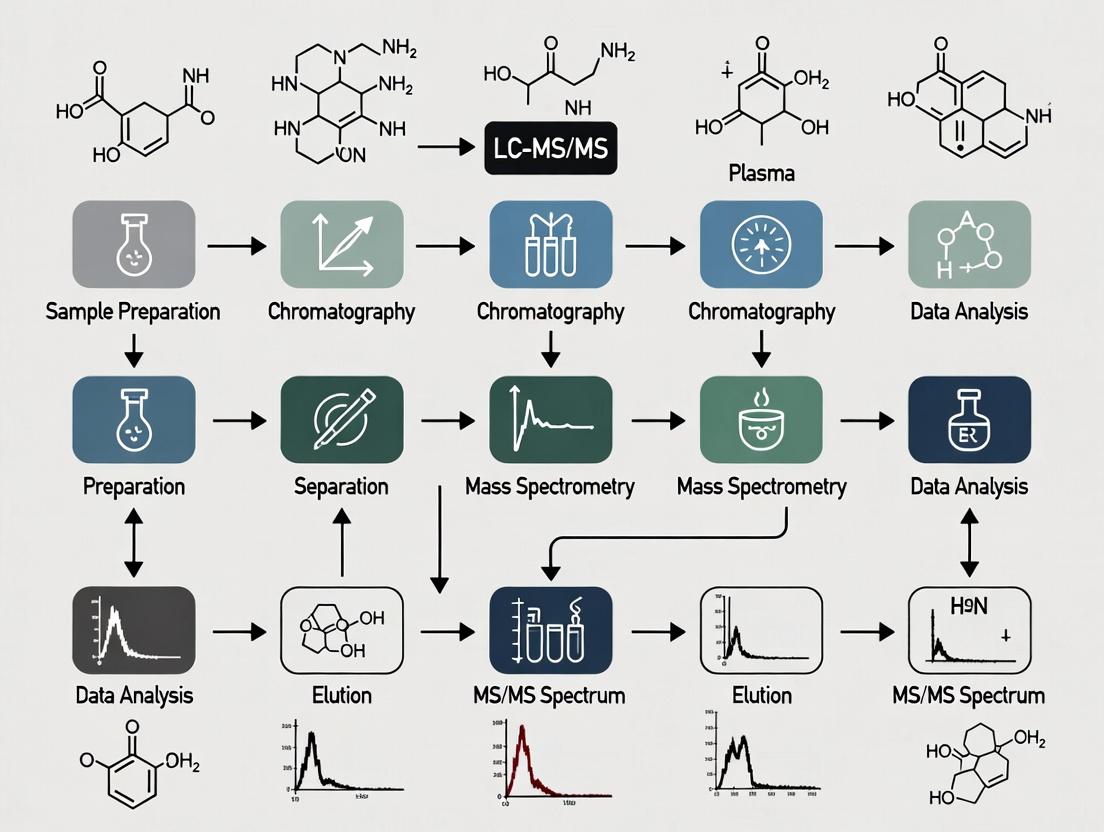
A Comprehensive Guide to Developing and Validating LC-MS/MS Methods for Pharmacokinetic Studies in Plasma
This article provides a complete roadmap for researchers and drug development professionals to establish robust LC-MS/MS methods for pharmacokinetic analysis in plasma.
A Comprehensive Guide to Developing and Validating LC-MS/MS Methods for Pharmacokinetic Studies in Plasma
Abstract
This article provides a complete roadmap for researchers and drug development professionals to establish robust LC-MS/MS methods for pharmacokinetic analysis in plasma. It covers foundational principles, from the critical role of PK studies in drug development to the specific advantages of LC-MS/MS. We detail step-by-step methodological development, including sample preparation, chromatography optimization, and mass spectrometry parameter tuning. The guide addresses common analytical challenges and optimization strategies to enhance sensitivity and specificity. Finally, it outlines the rigorous validation process per regulatory guidelines (ICH M10, FDA) and compares LC-MS/MS with other bioanalytical techniques. This resource aims to empower scientists to generate reliable, high-quality data essential for informed decision-making in preclinical and clinical research.
Understanding LC-MS/MS in Pharmacokinetics: Why It's the Gold Standard for Plasma Analysis
The Critical Role of Pharmacokinetic (PK) Studies in Drug Development
Within the broader thesis on developing and validating a robust LC-MS/MS method for pharmacokinetic studies in plasma, this document outlines the foundational application notes and protocols. The thesis posits that a highly sensitive, selective, and validated LC-MS/MS method is non-negotiable for generating the high-quality PK data required to inform critical decisions in drug development, from lead optimization to regulatory submission.
Key PK Parameters & Quantitative Data
The following table summarizes the core pharmacokinetic parameters derived from plasma concentration-time data, which are critical for assessing the absorption, distribution, metabolism, and excretion (ADME) of a drug candidate.
Table 1: Core Pharmacokinetic Parameters and Their Significance
| Parameter | Symbol | Typical Units | Definition & Significance in Drug Development |
|---|---|---|---|
| Maximum Plasma Concentration | C~max~ | ng/mL or µM | Peak drug concentration post-dose. Indicates absorption rate and extent; critical for efficacy and safety (exposure-toxicity relationship). |
| Time to C~max~ | T~max~ | hours | Time to reach peak concentration. Reflects absorption rate; important for dosing regimen design. |
| Area Under the Curve | AUC~0-t~, AUC~0-∞~ | h·ng/mL | Total drug exposure over time. Primary metric for bioavailability and bioequivalence; correlates with pharmacological effect. |
| Elimination Half-Life | t~1/2~ | hours | Time for plasma concentration to reduce by 50%. Determines dosing frequency and predicts accumulation. |
| Clearance | CL | L/h/kg | Volume of plasma cleared of drug per unit time. Key for dose adjustment in organ impairment. |
| Volume of Distribution | V~d~ | L/kg | Apparent volume into which the drug distributes. Predicts drug penetration into tissues and potential for extravascular distribution. |
| Bioavailability | F | % | Fraction of administered dose that reaches systemic circulation unchanged. Critical for transitioning from IV to oral dosing. |
Experimental Protocols
Protocol: LC-MS/MS Method Development for PK Analysis in Plasma
Objective: To establish a sensitive and specific LC-MS/MS method for the quantification of a small-molecule drug candidate (Compound X) and its major metabolite (M1) in K2EDTA human plasma.
I. Materials & Reagents
- Analytes: Compound X, Metabolite M1 (reference standards).
- Internal Standard (IS): Stable isotope-labeled Compound X-d6.
- Matrix: Blank (drug-free) human plasma with K2EDTA anticoagulant.
- Precipitation Solvent: Acetonitrile (HPLC grade).
- Mobile Phase A: 0.1% Formic acid in water.
- Mobile Phase B: 0.1% Formic acid in acetonitrile.
- Equipment: Triple quadrupole LC-MS/MS system, UHPLC with C18 column (e.g., 2.1 x 50 mm, 1.7 µm), centrifuge, vortex mixer.
II. Procedure
- Sample Preparation (Protein Precipitation): a. Thaw plasma samples on ice. b. Aliquot 50 µL of plasma into a microcentrifuge tube. c. Add 10 µL of working IS solution. d. Vortex for 10 seconds. e. Add 200 µL of ice-cold acetonitrile. f. Vortex vigorously for 2 minutes. g. Centrifuge at 14,000 x g for 10 minutes at 4°C. h. Transfer 150 µL of the supernatant to an autosampler vial with insert. i. Dilute with 50 µL of water, mix gently, and inject 5-10 µL onto the LC-MS/MS.
LC Conditions:
- Column Temperature: 40°C
- Flow Rate: 0.4 mL/min
- Gradient: 5% B (0-0.5 min), 5% → 95% B (0.5-2.5 min), hold 95% B (2.5-3.5 min), 95% → 5% B (3.5-3.6 min), re-equilibrate at 5% B (3.6-5.0 min).
- Injection Volume: 5 µL
MS/MS Conditions (ESI Positive Mode):
- Source Temp: 150°C
- Desolvation Temp: 500°C
- Cone Gas Flow: 150 L/hr
- Desolvation Gas Flow: 1000 L/hr
- Capillary Voltage: 1.0 kV
- MRM Transitions (optimized via direct infusion):
- Compound X: 405.2 → 243.1 (Collision Energy: 20 eV)
- Metabolite M1: 421.2 → 259.1 (Collision Energy: 18 eV)
- IS (X-d6): 411.2 → 249.1 (Collision Energy: 20 eV)
Calibration Curve & QC: Prepare calibration standards (e.g., 1-1000 ng/mL) and quality control samples (Low, Mid, High QCs) in blank plasma. Analyze alongside study samples.
Protocol: In Vivo Rat Pharmacokinetic Study (Single IV & PO Dose)
Objective: To characterize the basic PK profile of Compound X in Sprague-Dawley rats following intravenous (IV) and oral (PO) administration.
I. Materials
- Animals: Male Sprague-Dawley rats (n=6 per route, 250-300g), cannulated (jugular vein).
- Formulations: IV solution (e.g., 5% DMSO in saline), PO suspension (0.5% methylcellulose).
- Equipment: LC-MS/MS system, centrifuge, -80°C freezer.
II. Procedure
- Dosing & Sampling: a. Dose rats (IV: 1 mg/kg via tail vein; PO: 5 mg/kg via oral gavage). b. Collect serial blood samples (∼200 µL) from the jugular cannula at pre-dose, 0.083, 0.25, 0.5, 1, 2, 4, 6, 8, and 24 hours post-dose. c. Immediately transfer blood to K2EDTA tubes, centrifuge at 2000 x g for 10 min at 4°C. d. Harvest plasma and store at -80°C until LC-MS/MS analysis.
- Bioanalysis & PK Analysis: a. Analyze all plasma samples using the validated LC-MS/MS method from Protocol 3.1. b. Plot mean plasma concentration vs. time for each route. c. Use a non-compartmental analysis (NCA) tool (e.g., Phoenix WinNonlin) to calculate PK parameters in Table 1, including absolute bioavailability (F%).
Diagrams
Diagram Title: PK Study Workflow from Bioanalysis to Decision
Diagram Title: Link Between PK Parameters, ADME, and Development Decisions
The Scientist's Toolkit: Research Reagent Solutions
Table 2: Essential Materials for LC-MS/MS-based PK Studies
| Item | Function & Importance in PK Research |
|---|---|
| Stable Isotope-Labeled Internal Standards (SIL-IS) | Compensates for matrix effects and variability in sample preparation/ionization, ensuring assay accuracy and precision. Essential for bioanalytical method validation. |
| Certified Drug-Free Biological Matrices | Blank plasma/serum/tissue homogenates from relevant species for preparing calibration standards and QCs. Critical for establishing a valid analytical range. |
| High-Purity Analytical Reference Standards | Characterized compounds (parent drug and metabolites) for method development, specificity testing, and QC preparation. Purity directly impacts result accuracy. |
| LC-MS/MS Grade Solvents & Additives | Minimize background noise and ion suppression. Essential for consistent mobile phase performance and system cleanliness. |
| Specialized Sample Collection Tubes | Tubes containing appropriate anticoagulants (e.g., K2EDTA) and stabilizers (e.g., esterase inhibitors) to ensure analyte stability from the moment of collection. |
| Validated Bioanalytical Software | Software for instrument control, data acquisition (e.g., MassLynx, Analyst), and processing (e.g., MultiQuant, Skyline). Required for GLP-compliant data integrity. |
Within the framework of a broader thesis on developing a robust LC-MS/MS method for pharmacokinetic (PK) studies in plasma, understanding the core principles of the technology is foundational. LC-MS/MS is the cornerstone of modern bioanalysis due to its unparalleled selectivity, sensitivity, and speed, enabling the precise quantification of drugs and metabolites in complex biological matrices like plasma. This document details the principles, application notes, and protocols essential for implementing LC-MS/MS in PK research.
Core Principles
Liquid Chromatography (LC) Principle
Liquid Chromatography separates compounds in a sample based on their differential partitioning between a mobile phase (liquid solvent) and a stationary phase (packed bed inside a column). Key parameters include:
- Stationary Phase: Typically C18 (octadecylsilane) bonded silica for reversed-phase chromatography, which separates based on hydrophobicity.
- Mobile Phase: A gradient of water (aqueous) and organic solvent (e.g., methanol, acetonitrile) moves analytes through the column.
- Retention Time (tR): The time an analyte spends in the column, unique under set conditions, used for identification.
Tandem Mass Spectrometry (MS/MS) Principle
MS/MS detects and quantifies compounds based on their mass-to-charge ratio (m/z) with high specificity. It involves three core stages:
- Ionization: Analyte molecules are ionized (e.g., by Electrospray Ionization - ESI).
- Mass Selection (Q1): The first quadrupole (Q1) selects the precursor ion of a specific m/z.
- Fragmentation & Detection (Q2, Q3): The selected ion is fragmented in a collision cell (Q2) using inert gas (CID). The resulting product ions are analyzed by the second mass analyzer (Q3). Monitoring a specific precursor→product ion transition (Multiple Reaction Monitoring - MRM) provides exceptional selectivity.
Application Note: Development and Validation of a Plasma PK Method
Objective
To develop and validate a sensitive and specific LC-MS/MS method for the quantification of a small molecule drug (Compound X) in human plasma for a pharmacokinetic study.
Key Quantitative Data from Method Validation
Table 1: Summary of FDA-Guided Method Validation Parameters for Compound X in Plasma
| Validation Parameter | Acceptance Criteria | Result for Compound X |
|---|---|---|
| Linearity Range | R² ≥ 0.995 | 1.0 – 1000 ng/mL |
| Accuracy (% Nominal) | 85-115% (LLOQ: 80-120%) | 92.5 – 105.3% |
| Precision (% RSD) | ≤15% (LLOQ: ≤20%) | 1.2 – 8.7% |
| Lower Limit of Quantification (LLOQ) | S/N ≥ 10, Precision & Accuracy met | 1.0 ng/mL |
| Extraction Recovery | Consistent & High | 85.2% (Mean) |
| Matrix Effect | IS-Normalized MF: 85-115% | 95.4% (CV ≤ 5%) |
| Stability (Bench-top, Processed) | Within ±15% of nominal | Confirmed (24h & 72h) |
Experimental Protocol: Sample Preparation & Analysis
Protocol 1: Solid-Phase Extraction (SPE) of Plasma Samples for LC-MS/MS Title: Plasma Sample Clean-up via SPE Objective: To isolate and concentrate Compound X and its Internal Standard (ISTD) from human plasma while removing interfering matrix components.
Materials:
- Blank human plasma, heparinized.
- Stock solutions of Compound X and deuterated internal standard (Compound X-d6).
- Oasis HLB SPE cartridges (30 mg, 1 mL).
- Solvents: HPLC-grade methanol, acetonitrile, water, and formic acid.
- Vacuum manifold, centrifugal evaporator.
Procedure:
- Pre-SPE: Condition SPE cartridge with 1 mL methanol, then equilibrate with 1 mL water.
- Sample Load: Thaw plasma samples on ice. Pipette 100 µL of plasma into a tube. Add 25 µL of ISTD working solution (in methanol) and 200 µL of 1% formic acid in water. Vortex mix.
- Loading: Load the diluted plasma sample onto the conditioned SPE cartridge.
- Wash: Wash cartridge with 1 mL of 5% methanol in water to remove polar impurities.
- Elution: Elute analytes into a clean collection tube with 1 mL of methanol.
- Evaporation & Reconstitution: Evaporate the eluent to dryness under a gentle stream of nitrogen at 40°C. Reconstitute the dry residue in 150 µL of initial mobile phase (e.g., 30% acetonitrile in 0.1% formic acid). Vortex thoroughly and centrifuge at 13,000 rpm for 5 min. Transfer supernatant to an LC vial for analysis.
Protocol 2: LC-MS/MS Analysis in MRM Mode Title: LC-MS/MS Analysis for PK Quantification Objective: To chromatographically separate and selectively detect Compound X and its ISTD.
Materials:
- Reconstituted sample extracts.
- HPLC system (e.g., Vanquish) with C18 column (e.g., 2.1 x 50 mm, 1.7 µm).
- Tandem mass spectrometer (e.g., Triple Quadrupole, SCIEX or Waters).
LC Conditions:
- Column Temp: 40°C
- Flow Rate: 0.4 mL/min
- Mobile Phase A: 0.1% Formic Acid in Water
- Mobile Phase B: 0.1% Formic Acid in Acetonitrile
- Gradient:
- 0-1.0 min: 30% B
- 1.0-3.0 min: Ramp to 95% B
- 3.0-4.0 min: Hold at 95% B
- 4.0-4.1 min: Ramp to 30% B
- 4.1-5.5 min: Re-equilibrate at 30% B
- Injection Volume: 5 µL
MS/MS Conditions (ESI Positive Mode):
- Ion Source Temp: 150°C
- Desolvation Temp: 500°C
- Capillary Voltage: 3.0 kV
- MRM Transitions (Collision Energy):
- Compound X: 455.2 → 238.1 (25 eV)
- Compound X-d6 (ISTD): 461.2 → 244.1 (25 eV)
- Dwell Time: 50 ms per transition
Data Analysis: Use instrument software to integrate peak areas. Construct a calibration curve by plotting the peak area ratio (Analyte/ISTD) vs. nominal concentration using a weighted (1/x²) linear regression model. Use this curve to calculate unknown sample concentrations.
Visualized Workflows and Relationships
Title: LC-MS/MS Bioanalytical Workflow for Plasma PK
Title: MRM Principle: Precursor Selection and Fragmentation
The Scientist's Toolkit: Key Research Reagent Solutions
Table 2: Essential Materials for LC-MS/MS Plasma PK Studies
| Item / Reagent | Function / Role in Experiment |
|---|---|
| Blank Human Plasma (Matrix) | The biological matrix for calibration standards (spiked) and quality controls. Serves as the baseline for method development and validation. |
| Stable Isotope-Labeled Internal Standard (e.g., Deuterated, ¹³C) | Corrects for variability in sample preparation, ionization efficiency, and matrix effects. Essential for achieving high accuracy and precision. |
| Solid-Phase Extraction (SPE) Cartridges (e.g., Oasis HLB) | Selectively bind and clean up analytes from plasma, removing proteins, salts, and phospholipids to reduce matrix effects and ion suppression. |
| HPLC-Grade Solvents & Additives (MeCN, MeOH, FA, AA) | Form the mobile phase. High purity is critical to minimize background noise and maintain consistent chromatography and ionization. |
| LC-MS/MS Tuning & Calibration Solutions | Used to calibrate and optimize mass spectrometer parameters (e.g., mass accuracy, resolution, sensitivity) for reliable performance. |
| Authentic Reference Standard (Drug & Metabolites) | Provides the definitive identity and purity for preparing calibration curves, enabling accurate quantification of the target analyte. |
Within pharmacokinetic (PK) studies, the accurate quantification of drugs and their metabolites in plasma is paramount. This application note, framed within a broader thesis on developing robust LC-MS/MS methods for PK research, details the core advantages that make LC-MS/MS the gold standard: exceptional selectivity, superior sensitivity, and high analytical speed.
Core Advantages Elucidated
Selectivity
Selectivity is achieved through two orthogonal separation mechanisms. Liquid chromatography (LC) separates analytes based on hydrophobicity, polarity, or size, while tandem mass spectrometry (MS/MS) provides a second dimension of separation based on mass-to-charge ratio (m/z) and fragmentation patterns. This dual separation drastically reduces background chemical noise from the complex plasma matrix.
Sensitivity
LC-MS/MS achieves sensitivity in the low picogram-per-milliliter (pg/mL) range, crucial for quantifying drugs at trace levels during late elimination phases. This is enabled by efficient ionization (e.g., Electrospray Ionization - ESI), advanced detector design, and the noise reduction afforded by Selected/Multiple Reaction Monitoring (SRM/MRM).
Speed
Modern ultra-high-performance liquid chromatography (UHPLC) coupled with fast MS/MS detectors enables run times of 3-7 minutes per sample. High speed facilitates high-throughput analysis, allowing for the rapid processing of hundreds of samples from large PK studies, which is essential for timely decision-making in drug development.
Table 1: Comparative Performance Metrics of a Model LC-MS/MS PK Assay vs. Traditional Techniques
| Analytical Parameter | LC-MS/MS (Model Assay) | HPLC-UV | ELISA |
|---|---|---|---|
| Lower Limit of Quantification (LLOQ) | 1.0 pg/mL | 1.0 ng/mL | 0.1 ng/mL |
| Linear Dynamic Range | 1.0 pg/mL – 1000 ng/mL (6 orders) | 1-1000 ng/mL (3 orders) | 0.1-100 ng/mL (3 orders) |
| Run Time per Sample | 5.5 minutes | 25 minutes | 2-4 hours (plate-based) |
| Selectivity (Matrix Interference) | < 20% (via MRM) | Potential co-elution | High cross-reactivity risk |
| Typical Sample Volume Required | 50 µL | 500 µL | 100 µL |
Table 2: Key MS/MS Parameters for a Model Drug and its Metabolite in a PK Panel
| Compound | Precursor Ion (m/z) | Product Ion (m/z) (Quantifier) | Collision Energy (eV) | Retention Time (min) |
|---|---|---|---|---|
| Drug X | 409.2 | 237.1 | 22 | 4.2 |
| Metabolite M1 | 425.2 | 253.1 | 18 | 3.8 |
| Internal Standard (IS) | 414.2 | 242.1 | 22 | 4.2 |
Experimental Protocols
Protocol 1: Plasma Sample Preparation (Protein Precipitation)
Objective: To efficiently extract the analyte from plasma proteins and prepare a clean sample for LC-MS/MS injection.
- Aliquoting: Pipette 50 µL of thawed, homogenized plasma into a clean 1.5 mL microcentrifuge tube.
- Internal Standard Addition: Add 10 µL of the appropriate internal standard working solution (e.g., 100 ng/mL in methanol).
- Precipitation: Add 200 µL of cold acetonitrile (containing 1% formic acid) to the tube.
- Vortex and Centrifuge: Vortex mix vigorously for 1 minute. Centrifuge at 14,000 x g for 10 minutes at 4°C.
- Supernatant Collection: Carefully transfer 150 µL of the clear supernatant to a clean autosampler vial containing a 250 µL insert.
- Injection: Seal the vial and place it in the autosampler tray maintained at 10°C. A typical injection volume is 5-10 µL.
Protocol 2: LC-MS/MS Method for PK Analysis
Objective: To establish a chromatographic and mass spectrometric method for the simultaneous quantification of a drug and its metabolite. A. Liquid Chromatography Conditions:
- Column: C18, 2.1 x 50 mm, 1.7 µm particle size.
- Mobile Phase A: 0.1% Formic acid in water.
- Mobile Phase B: 0.1% Formic acid in acetonitrile.
- Gradient: 5% B (0-0.5 min), 5% → 95% B (0.5-4.0 min), 95% B (4.0-4.5 min), 95% → 5% B (4.5-4.6 min), 5% B (4.6-5.5 min).
- Flow Rate: 0.4 mL/min.
- Column Oven Temperature: 40°C.
- Autosampler Temperature: 10°C.
B. Tandem Mass Spectrometry Conditions:
- Ion Source: Electrospray Ionization (ESI), positive mode.
- Source Temperature: 150°C.
- Desolvation Gas Flow: 800 L/hr.
- Cone Gas Flow: 50 L/hr.
- Capillary Voltage: 3.0 kV.
- Operation Mode: Multiple Reaction Monitoring (MRM).
- MRM Transitions: As defined in Table 2.
- Dwell Time: 50 ms per transition.
Visualizations
LC-MS/MS Plasma Analysis Workflow
Orthogonal Selectivity in LC-MS/MS
The Scientist's Toolkit
Table 3: Essential Research Reagent Solutions for LC-MS/MS Plasma PK Studies
| Item | Function & Specification | Critical Notes |
|---|---|---|
| Stable Isotope-Labeled Internal Standards (SIL-IS) | Deuterated or 13C-labeled analogs of the analyte. Corrects for variability in extraction, ionization, and matrix effects. | Must be added at the beginning of sample preparation. |
| Mass Spectrometry-Grade Solvents | Acetonitrile, Methanol, Water (with 0.1% Formic Acid or Ammonium Acetate). | Minimizes background ions and suppresses source contamination. |
| Blank Matrix | Drug-free human or species-specific plasma. | Used for preparing calibration standards and quality control samples. |
| Protein Precipitation Reagent | Typically cold acidified organic solvent (e.g., ACN with 1% FA). | Removes proteins, precipitates analytes, and denatures enzymes. |
| LC Column | Reverse-phase (e.g., C18), small particle size (≤ 2µm), 50-100 mm length. | Provides high-resolution, fast separation of analytes from matrix. |
| Calibrators & QCs | Prepared in blank matrix at known concentrations across the expected range. | Used to construct the calibration curve and monitor assay performance. |
Application Notes: Quantification of Core PK Parameters Using LC-MS/MS in Plasma Pharmacokinetic Studies
In the context of developing a robust LC-MS/MS method for pharmacokinetic studies, the accurate measurement of five core pharmacokinetic (PK) parameters is paramount. These parameters provide the foundation for understanding the absorption, distribution, metabolism, and excretion (ADME) of a drug candidate. The sensitivity, specificity, and high-throughput capability of LC-MS/MS make it the industry-standard technique for generating the concentration-time data required for their calculation.
Cmax (Maximum Plasma Concentration): This is the peak concentration of the drug observed in plasma following administration. It is a direct measure of the drug's bioavailability and systemic exposure. In LC-MS/MS assays, Cmax is derived from the highest observed data point in the validated concentration-time profile.
Tmax (Time to Reach Cmax): This parameter indicates the time post-dose at which Cmax occurs. It is a key indicator of the rate of drug absorption. Tmax is directly observed from the concentration-time data generated by serial plasma sample analysis.
AUC (Area Under the Curve): AUC measures the total systemic exposure to the drug over time. The area under the plasma concentration-time curve from zero to the last measurable time point (AUC0-t) and extrapolated to infinity (AUC0-∞) are critical for assessing bioavailability and calculating other parameters like clearance. LC-MS/MS data provides the precise concentration values for trapezoidal rule calculation of AUC.
Half-life (t1/2): The elimination half-life is the time required for the plasma concentration to reduce by 50% during the terminal elimination phase. It is calculated from the elimination rate constant (λz), which is derived from the slope of the log-linear terminal phase of the concentration-time curve generated by LC-MS/MS.
Clearance (CL): Systemic clearance is the volume of plasma cleared of the drug per unit time. It is a fundamental parameter describing the body's efficiency in eliminating the drug. Clearance is calculated as Dose / AUC0-∞ following intravenous administration, or (Dose * F) / AUC0-∞ for extravascular routes, where F is bioavailability. Accurate AUC from LC-MS/MS is essential.
The following table summarizes the core PK parameters, their definition, and their significance in drug development.
Table 1: Core Pharmacokinetic Parameters and Their Significance
| Parameter | Definition | PK Phase | Significance in Drug Development |
|---|---|---|---|
| Cmax | Maximum observed plasma concentration. | Absorption | Indicates bioavailability & potential for efficacy/toxicity. |
| Tmax | Time to reach Cmax. | Absorption | Reflects the rate of drug absorption. |
| AUC0-∞ | Area under the plasma concentration-time curve from zero to infinity. | All (Exposure) | Primary measure of total systemic drug exposure. |
| Half-life (t1/2) | Time for plasma concentration to decrease by 50% in terminal phase. | Elimination | Informs dosing interval and time to steady-state. |
| Clearance (CL) | Volume of plasma cleared of drug per unit time. | Elimination | Describes the body's efficiency in eliminating the drug. |
Experimental Protocols
Protocol 1: LC-MS/MS Method Development and Validation for Small Molecule Quantification in Plasma
Objective: To develop and validate a selective, sensitive, and reproducible LC-MS/MS method for the quantification of a drug candidate in K2EDTA human plasma for support of pharmacokinetic studies.
Materials:
- Analytical Standards: Drug candidate (analyte) and stable isotope-labeled internal standard (IS).
- Biological Matrix: Blank human plasma with K2EDTA anticoagulant.
- Solvents: HPLC-MS grade methanol, acetonitrile, water, and formic acid.
- Equipment: Triple quadrupole LC-MS/MS system, UHPLC system, analytical balance, centrifuge, vortex mixer, calibrated pipettes.
- Software: Analyst or equivalent for data acquisition, Watson LIMS or equivalent for PK analysis.
Procedure:
- Stock Solution Preparation: Independently prepare primary stock solutions of analyte and IS in appropriate solvent (e.g., DMSO/methanol). Dilute to working solutions.
- Calibration Standards & QCs: Spike blank plasma with working solutions to generate a calibration curve (e.g., 1.00–1000 ng/mL) and quality control (QC) samples at Low, Mid, and High concentrations.
- Sample Preparation (Protein Precipitation): a. Aliquot 50 µL of plasma sample (standard, QC, or study sample) into a 96-well plate. b. Add 25 µL of IS working solution in extraction solvent to all samples except double blanks. c. Add 200 µL of ice-cold acetonitrile containing 0.1% formic acid. d. Seal plate, vortex mix for 5 minutes, then centrifuge at 4000 rpm for 15 minutes at 4°C. e. Transfer 150 µL of supernatant to a new 96-well plate, dilute with 100 µL water, and seal for LC-MS/MS analysis.
- LC-MS/MS Analysis: a. Chromatography: Use a C18 reversed-phase column (2.1 x 50 mm, 1.7 µm). Mobile Phase A: 0.1% Formic Acid in Water. Mobile Phase B: 0.1% Formic Acid in Acetonitrile. Employ a gradient elution from 5% B to 95% B over 3.5 minutes. Flow rate: 0.4 mL/min. Column temperature: 40°C. b. Mass Spectrometry: Operate in positive electrospray ionization (ESI+) mode. Optimize source parameters (GS1, GS2, TEM, CUR). Use Multiple Reaction Monitoring (MRM). Define precursor → product ion transitions for analyte and IS. Example: Analyte: 405.2 → 243.1 m/z; IS: 410.2 → 248.1 m/z.
- Data Analysis: Plot peak area ratio (Analyte/IS) vs. nominal concentration. Fit using weighted (1/x²) linear regression. Calculate concentrations of QC and study samples from the calibration curve. Apply acceptance criteria (e.g., ±15% accuracy for QCs).
Protocol 2: Pharmacokinetic Study in Rats and Sample Analysis
Objective: To administer a drug candidate to rats, collect serial blood samples, process to plasma, and analyze via the validated LC-MS/MS method to generate concentration-time data for PK parameter calculation.
Materials:
- Animals: Male Sprague-Dawley rats (n=6 per group), cannulated.
- Formulation: Drug candidate in saline or vehicle for intravenous (IV) or oral (PO) dosing.
- Supplies: Microcentrifuge tubes (K2EDTA coated), centrifuge, wet ice, pipettes.
- LC-MS/MS System: As per Protocol 1.
Procedure:
- Dosing & Sample Collection: Administer a single dose (e.g., 1 mg/kg IV or 5 mg/kg PO) to each rat. Collect blood samples (e.g., ~0.2 mL) via cannula at pre-dose, 0.083, 0.25, 0.5, 1, 2, 4, 8, 12, and 24 hours post-dose.
- Plasma Processing: Immediately place blood tubes on wet ice. Centrifuge at 3000 x g for 10 minutes at 4°C within 30 minutes of collection. Aliquot plasma into labeled tubes and store at ≤ -70°C until analysis.
- Bioanalysis: Thaw study samples. Prepare alongside a fresh calibration curve and QC samples as per Protocol 1. Analyze all samples via the validated LC-MS/MS method.
- PK Analysis: Input the concentration-time data for each animal into a non-compartmental analysis (NCA) tool within Watson LIMS or Phoenix WinNonlin. a. Cmax & Tmax: Directly observed from the data. b. AUC0-t: Calculate using the linear-up/log-down trapezoidal method. c. AUC0-∞: Calculate as AUC0-t + Ct/λz, where Ct is the last measurable concentration. d. Half-life (t1/2): Calculate as 0.693 / λz, where λz is the terminal elimination rate constant from log-linear regression. e. Clearance (CL): For IV, CL = Dose / AUC0-∞. For PO, CL/F = Dose / AUC0-∞.
Table 2: Representative PK Parameters from a Rat Study (Mean ± SD, n=6)
| Dose Route | Dose (mg/kg) | Cmax (ng/mL) | Tmax (h) | AUC0-∞ (h*ng/mL) | t1/2 (h) | CL (mL/min/kg) |
|---|---|---|---|---|---|---|
| Intravenous | 1.0 | 452.3 ± 85.7 | 0.08 (fixed) | 1254.5 ± 210.3 | 3.2 ± 0.5 | 13.6 ± 2.3 |
| Oral | 5.0 | 188.7 ± 45.2 | 0.5 ± 0.2 | 1120.8 ± 189.4 | 3.5 ± 0.6 | - (CL/F: 75.4 ± 12.1) |
The Scientist's Toolkit: Key Research Reagent Solutions
Table 3: Essential Materials for LC-MS/MS based PK Studies
| Item | Function & Rationale |
|---|---|
| Stable Isotope-Labeled Internal Standard (SIL-IS) | Corrects for variability in sample preparation and ionization efficiency in the MS source, improving accuracy and precision. |
| Mass Spectrometry Grade Solvents | Minimize background noise and ion suppression in LC-MS/MS, ensuring optimal sensitivity and consistent performance. |
| SPE or Protein Precipitation Plates | Enable high-throughput, automated sample clean-up to remove phospholipids and proteins that cause matrix effects. |
| Certified Drug-Free Human Plasma | Used as the biological matrix for preparing calibration standards and QCs to match study samples and ensure accurate quantification. |
| Robust UHPLC Column (e.g., C18, 1.7-2.7µm) | Provides high-resolution separation of the analyte from matrix interferences and isobaric compounds, reducing chemical noise. |
| Validated PK Analysis Software | Performs non-compartmental analysis (NCA) to accurately calculate core PK parameters from concentration-time data. |
Visualizations
LC-MS/MS PK Study Workflow
From Data to PK Parameters
This document outlines the critical pre-development considerations for a robust LC-MS/MS method, framed within a thesis focused on pharmacokinetic (PK) studies in human plasma. Success hinges on a deep understanding of the analyte's physicochemical properties and the complexity of the biological matrix, which directly informs sample preparation, chromatographic separation, and mass spectrometric detection choices.
Comprehensive Analysis of Analyte Properties
A systematic evaluation of the target analyte's properties is the first and most crucial step. This data directly dictates every subsequent methodological parameter.
Table 1: Key Analyte Properties and Their Methodological Impact
| Property | Analytical Technique for Assessment | Impact on LC-MS/MS Method | Typical Target Range for Oral Drugs |
|---|---|---|---|
| Molecular Weight | MS calibration | MRM transition selection, Q1 resolution | 150-700 Da |
| pKa | Potentiometric titration, UV-Vis spectroscopy | Mobile phase pH choice for retention/separation | - |
| Log P/D | Shake-flask, HPLC (chromatographic) | Reversed-phase column selection, extraction solvent | Log P 1-5 |
| Solubility | Equilibrium solubility assay | Sample solvent & injection volume | >50 µg/mL |
| Chemical Stability | Forced degradation studies (pH, temp, light) | Sample handling, storage, mobile phase conditions | Stable in matrix ≥24h at 4°C |
| Protein Binding | Equilibrium dialysis, ultrafiltration | Extraction efficiency, required sensitivity | Often >90% |
| Ionization Efficiency | Direct infusion MS in ESI+/ESI- | Ionization mode selection, sensitivity | - |
Protocol 1: Determination of Log D (pH 7.4) via Shake-Flask Method
Objective: To measure the distribution coefficient of the analyte between 1-octanol and phosphate buffer at physiological pH. Reagents: Analyte standard, 1-octanol (saturated with buffer), 0.01M Phosphate buffer pH 7.4 (saturated with 1-octanol). Procedure:
- Prepare a stock solution of analyte in a co-solvent (e.g., DMSO) at 10 mM.
- Add 1.5 mL of each phase (octanol and buffer) to a glass vial.
- Spike 15 µL of stock solution into the vial to achieve a final concentration of ~100 µM.
- Cap tightly and mix on a rotary mixer for 1 hour at 25°C.
- Centrifuge at 3000 x g for 10 minutes to achieve complete phase separation.
- Carefully separate the two layers.
- Quantify the analyte concentration in each phase using a validated UV or LC-MS method.
- Calculation: Log D~7.4~ = Log10 ( [Analyte]~octanol~ / [Analyte]~buffer~ ).
Characterization of the Biological Matrix (Plasma)
Plasma is a complex, variable mixture of proteins, lipids, salts, and endogenous compounds that cause ion suppression/enhancement (matrix effects).
Table 2: Major Plasma Components and Their Interference Potential
| Matrix Component | Typical Concentration | Primary Interference in LC-MS/MS | Mitigation Strategy |
|---|---|---|---|
| Albumin | 35-50 g/L | Non-specific binding, matrix effect | Protein precipitation, stable-isotope internal standard (SIS) |
| Immunoglobulins | 10-20 g/L | Matrix effect | Efficient chromatographic separation |
| Phospholipids | 1-2 mg/mL (total) | Severe ion suppression, column fouling | Phospholipid removal SPE, HILIC chromatography |
| Na+/K+ Salts | ~150 mM | Source contamination, adduct formation | Dilution, solid-phase extraction (SPE) |
| Urea/ Creatinine | 3-8 mM / 50-100 µM | Minor matrix effect | Chromatographic separation |
Protocol 2: Assessment of Phospholipid Removal and Matrix Effect
Objective: To evaluate the efficiency of sample preparation in removing phospholipids and quantify the resulting matrix effect (ME). Reagents: Blank plasma from ≥6 individual donors, analyte, SIS, LC-MS grade solvents. Procedure – Post-Extraction Addition for ME:
- Prepare six individual lots of blank plasma. Process each through the intended sample prep protocol (e.g., Protein Precipitation, SPE).
- Reconstitute the dried extracts from Step 1 with mobile phase containing a known concentration of analyte and SIS (Post-extracted Spike).
- Prepare the same concentration of analyte and SIS in pure mobile phase (Neat Solution).
- Inject all samples (6 individual post-extracted spikes + neat solutions) in triplicate.
- Calculate ME: ME (%) = (Peak Area of Post-extracted Spike / Peak Area of Neat Solution) x 100. An ME of 85-115% is generally acceptable.
- Calculate IS-Normalized ME: Use the SIS response to normalize, indicating reproducibility. Procedure – Monitoring Phospholipids:
- Use a precursor ion scan of m/z 184 (positive mode) or m/z 153 (negative mode) to detect phosphatidylcholines/serines.
- Inject processed blank samples and monitor the chromatographic region where phospholipids elute (typically 1-3 min before analyte in RP).
- Compare the total ion current for m/z 184 in the analyte's retention time window across different prep methods.
Diagram 1: Pre-Method Development Decision Workflow
Title: Decision Pathway for LC-MS/MS Method Planning
Diagram 2: Key Plasma Matrix Effects on LC-MS/MS Signal
Title: Plasma Matrix Effects on Quantification Accuracy
The Scientist's Toolkit: Key Reagent Solutions
| Item | Function in Pre-Development |
|---|---|
| Stable Isotope-Labeled Internal Standard (SIS) | Corrects for losses during sample prep and matrix effects; essential for accuracy. |
| Phospholipid Removal SPE Cartridges (e.g., HybridSPE) | Selectively removes phospholipids from plasma extracts, reducing ion suppression. |
| Multiple Lots of Blank Matrix | Assesses matrix variability and ensures method robustness across populations. |
| Mobile Phase Additives (Ammonium Formate/Acetate) | Provides consistent buffering for stable analyte ionization; volatile for MS compatibility. |
| Protein Precipitation Solvents (ACN, MeOH, acetone) | Rapidly denatures and removes proteins; choice affects phospholipid co-precipitation. |
| In-source CID Standards (e.g., reserpine) | Used for tuning and monitoring source fragmentation stability over time. |
| SPE Wash/Elution Solvent Suite | Allows optimization of selectivity (water, buffers, MeOH, ACN, ethyl acetate). |
Step-by-Step Method Development: From Sample Prep to Data Acquisition
In the context of LC-MS/MS method development for pharmacokinetic (PK) studies, the selection of an appropriate plasma sample preparation technique is a critical determinant of success. The primary objectives are to remove phospholipids, proteins, and other endogenous interferences while efficiently extracting the drug analyte and its metabolites. This ensures assay selectivity, sensitivity, and reproducibility. This application note provides a detailed comparison and protocols for three core techniques: Protein Precipitation (PPT), Liquid-Liquid Extraction (LLE), and Solid-Phase Extraction (SPE), as applied within a thesis focusing on robust LC-MS/MS PK bioanalysis.
Table 1: Comparison of Plasma Sample Preparation Techniques for LC-MS/MS PK Studies
| Feature | Protein Precipitation (PPT) | Liquid-Liquid Extraction (LLE) | Solid-Phase Extraction (SPE) |
|---|---|---|---|
| Principle | Denaturation of proteins using organic solvent or acid. | Partitioning of analyte between immiscible aqueous (plasma) and organic phases. | Selective adsorption and elution of analyte from a solid sorbent. |
| Complexity | Low (Simple) | Moderate | High |
| Throughput | Very High (Amenable to 96-well plate format) | Moderate to High | High (with automation) |
| Cost per Sample | Low | Low | Moderate to High |
| Selectivity | Low (co-precipitation of analytes possible) | Moderate (depends on solvent choice) | High (sorbent and solvent choice) |
| Phospholipid Removal | Poor | Good (with appropriate solvent) | Excellent (with specific sorbents) |
| Matrix Effect (Ion Suppression) | Often High | Moderate | Typically Low (with optimized protocol) |
| Recovery (%) | Variable, often >80% | High, often >70-90% | High and consistent, often >85% |
| Ideal For | High-throughput screening, stable analytes. | Lipophilic to moderately polar analytes. | Complex matrices, low concentration analytes, demanding regulatory assays. |
Detailed Protocols
Protocol 1: Protein Precipitation (PPT)
Objective: To rapidly remove proteins from plasma prior to LC-MS/MS analysis of a small molecule drug. Materials: Plasma samples, internal standard (IS) solution, precipitating solvent (e.g., acetonitrile or methanol), vortex mixer, microcentrifuge, 1.5 mL polypropylene microtubes.
- Aliquot: Transfer 50 µL of plasma into a 1.5 mL microcentrifuge tube.
- Add IS: Add 10 µL of the appropriate internal standard working solution.
- Precipitate: Add 200 µL of ice-cold acetonitrile (a 4:1 solvent-to-plasma ratio).
- Vortex and Incubate: Vortex mix vigorously for 1 minute. Allow to stand on ice or at 4°C for 10 minutes.
- Centrifuge: Centrifuge at 14,000 x g for 10 minutes at 4°C.
- Recover Supernatant: Carefully transfer 150-200 µL of the clear supernatant to a clean autosampler vial or 96-well plate.
- Evaporate & Reconstitute (Optional): Evaporate to dryness under a gentle stream of nitrogen at 40°C. Reconstitute the dry residue in 100 µL of mobile phase (initial LC conditions) and vortex mix.
- Analyze: Inject an aliquot (e.g., 5-10 µL) into the LC-MS/MS system.
Protocol 2: Liquid-Liquid Extraction (LLE)
Objective: To selectively extract a lipophilic analyte from plasma using organic solvents. Materials: Plasma samples, IS solution, extraction solvent (e.g., ethyl acetate, methyl tert-butyl ether (MTBE)), vortex mixer, centrifuge, evaporator (e.g., nitrogen blow-down system), 2 mL polypropylene microtubes.
- Aliquot: Transfer 100 µL of plasma into a 2 mL microtube.
- Add IS: Add 20 µL of IS working solution.
- Add Solvent: Add 1 mL of extraction solvent (e.g., MTBE).
- Extract: Vortex mix for 5 minutes. For better recovery, rock or shake the samples for 10-15 minutes.
- Phase Separate: Centrifuge at 3,000 x g for 5 minutes to separate the layers.
- Transfer Organic Layer: Transfer the upper (organic) layer to a clean tube. For acidic/basic analytes, the aqueous phase pH can be adjusted prior to extraction to improve recovery.
- Evaporate: Evaporate the organic extract to complete dryness under a gentle stream of nitrogen at 40°C.
- Reconstitute: Reconstitute the dry residue in 100 µL of reconstitution solution (e.g., 50:50 water:acetonitrile). Vortex mix for 1-2 minutes.
- Analyze: Centrifuge briefly and inject an aliquot into the LC-MS/MS system.
Protocol 3: Solid-Phase Extraction (SPE)
Objective: To clean and concentrate an analyte from plasma using mixed-mode cation-exchange sorbent. Materials: Plasma samples, IS solution, SPE cartridge/plate (e.g., 30 mg mixed-mode cation-exchange, MCX), vacuum manifold, positive displacement pipettes, solvents (water, methanol, 2% formic acid in water, 5% ammonium hydroxide in methanol).
- Condition: Condition the sorbent bed with 1 mL of methanol, followed by 1 mL of water. Do not let the bed dry out.
- Load: Acidify 200 µL of plasma with 20 µL of 10% formic acid. Add IS. Load the acidified plasma onto the conditioned cartridge.
- Wash 1: Wash with 1 mL of 2% formic acid in water to remove acids, proteins, and polar interferences.
- Wash 2: Wash with 1 mL of methanol to remove neutral interferences and water.
- Dry: Apply full vacuum for 2-3 minutes to dry the sorbent completely.
- Elute: Elute the analyte with 1 mL of 5% ammonium hydroxide in methanol into a clean collection tube.
- Evaporate & Reconstitute: Evaporate the eluent to dryness under nitrogen. Reconstitute in 100 µL of mobile phase, vortex, and centrifuge.
- Analyze: Inject an aliquot into the LC-MS/MS system.
Visualizations
Workflow for Protein Precipitation
Technique Selection Decision Guide
The Scientist's Toolkit
Table 2: Essential Research Reagent Solutions for Plasma Sample Prep
| Item | Function in PK LC-MS/MS Sample Prep |
|---|---|
| Acetonitrile (HPLC/MS Grade) | Common precipitating agent in PPT; also a component of reconstitution and mobile phases. |
| Methyl tert-Butyl Ether (MTBE) | A low-toxicity, low-density organic solvent favored for LLE due to efficient phospholipid removal. |
| Mixed-Mode SPE Sorbents (e.g., MCX, WAX) | Combine reversed-phase and ion-exchange mechanisms for highly selective extraction of ionizable analytes. |
| Stable Isotope-Labeled Internal Standard (SIL-IS) | Isotopically labeled version of the analyte used to correct for variability in extraction, ionization, and matrix effects. |
| Ammonium Formate/Formic Acid | Buffers and pH modifiers crucial for optimizing analyte retention/elution in SPE and chromatography. |
| Phospholipid Removal Plates (e.g., HybridSPE) | Specialized plates that selectively bind phospholipids prior to PPT, significantly reducing matrix effects. |
| Protein Precipitation Plates (96-well) | Filtration plates that combine PPT with ultrafiltration to automate supernatant collection. |
| Bond Elut Plexa / Horizon SLE Plates | Supported Liquid Extraction (SLE) plates that perform LLE in a high-throughput 96-well format without emulsion issues. |
1. Introduction
Within the context of developing a robust, sensitive, and selective LC-MS/MS method for pharmacokinetic (PK) studies in plasma, chromatographic separation is paramount. Optimal separation mitigates matrix effects (ion suppression/enhancement), resolves analytes from isobaric interferences, and ensures accurate quantification. This application note details the systematic optimization of three critical parameters: column selection, mobile phase composition, and gradient elution profile.
2. Research Reagent Solutions & Essential Materials
| Item | Function in LC-MS/MS PK Analysis |
|---|---|
| Hypercarb Porous Graphitic Carbon Column | Provides unique shape-selectivity for polar analytes lacking chromophores; resistant to extreme pH. |
| Acquity UPLC BEH C18 Column (1.7 µm) | High-pressure stable, hybrid silica particle column for superior efficiency and peak capacity in UPLC separations. |
| Ammonium Acetate (LC-MS Grade) | Provides volatile buffer capacity in mobile phase to control pH and stabilize analyte ionization. |
| Formic Acid (LC-MS Grade) | Common mobile phase additive to promote positive ionization in ESI-MS by providing protons. |
| Acetonitrile (LC-MS Grade) | Organic modifier with low viscosity and high elution strength; preferred for ESI-MS due to low chemical noise. |
| Methanol (LC-MS Grade) | Alternative organic modifier with different selectivity; sometimes used for stronger elution of non-polar compounds. |
| Stable Isotope-Labeled Internal Standards (e.g., d3-, 13C-) | Corrects for variability in sample preparation, injection, and matrix effects; essential for quantitative accuracy. |
| Protein Precipitation Plate (e.g., 96-well) | Enables high-throughput removal of plasma proteins prior to chromatographic analysis. |
| Oasis HLB µElution Plate | Provides efficient solid-phase extraction (SPE) for clean-up and concentration of analytes from complex plasma matrix. |
3. Column Selection: A Comparative Protocol
Objective: To evaluate the impact of stationary phase chemistry on the separation efficiency, peak shape, and retention of target analytes and internal standards in a spiked plasma extract.
Protocol:
- Sample Preparation: Prepare a working solution containing your target drug(s) and metabolite(s) at 100 ng/mL in a processed blank plasma matrix (post-protein precipitation).
- Column Candidates: Install the following columns in sequence:
- Column A: C18 (e.g., 2.1 x 50 mm, 1.7 µm)
- Column B: Phenyl-Hexyl (e.g., 2.1 x 50 mm, 1.7 µm)
- Column C: HILIC (e.g., 2.1 x 50 mm, 1.8 µm)
- Isocratic Screening: Use a generic starting mobile phase (e.g., 60% A: 0.1% Formic Acid in Water / 40% B: 0.1% Formic Acid in Acetonitrile). Flow rate: 0.4 mL/min. Column temperature: 40°C.
- Data Acquisition: Inject 5 µL of the prepared sample. Monitor peak asymmetry (As), theoretical plates (N), and retention factor (k).
- Analysis: Select the column yielding the best compromise of retention (k > 2), peak symmetry (As 0.8-1.2), and resolution from the nearest endogenous interference.
4. Mobile Phase Optimization Protocol
Objective: To determine the optimal pH and buffer concentration for maximizing MS signal intensity and chromatographic peak shape.
Protocol:
- pH Screening: Prepare mobile phase A with different pH modifiers:
- Condition 1: 0.1% Formic Acid (~pH 2.7)
- Condition 2: 10 mM Ammonium Formate, pH 3.0
- Condition 3: 10 mM Ammonium Acetate, pH 5.0
- Condition 4: 10 mM Ammonium Bicarbonate, pH 8.0
- Organic Phase: Keep mobile phase B as acetonitrile with the same additive (e.g., 0.1% Formic Acid).
- Gradient Run: Use a fast linear gradient from 5% to 95% B over 3 minutes.
- Evaluation: Inject plasma extracts spiked at the lower limit of quantification (LLOQ). Compare the signal-to-noise ratio (S/N), peak shape, and reproducibility (n=6) for each condition. The condition yielding the highest S/N and consistent retention time is selected.
5. Gradient Elution Optimization Protocol
Objective: To develop a time-efficient gradient that adequately resolves all analytes from each other and matrix components while minimizing run time.
Protocol:
- Initial Scouting: Using the selected column and mobile phase, run a broad gradient (e.g., 5-95% B in 10 min).
- Identify Critical Pair: Note any co-eluting or poorly resolved analyte pairs.
- Shallow Gradient Optimization: For the segment where the critical pair elutes, reduce the gradient slope (e.g., change from 2%/min to 0.5%/min). Use modeling software (e.g., DryLab) if available.
- Equilibration: Ensure the gradient returns to initial conditions and includes a sufficient column re-equilibration time (typically 3-5 column volumes).
- Final Method Test: Validate the final gradient with a calibration curve (e.g., 1-1000 ng/mL) in plasma. Check that resolution (Rs) > 1.5 for all critical pairs and that the total run time is acceptable for high-throughput.
6. Quantitative Data Summary
Table 1: Column Screening Results for a Model Drug (Warfarin) and its Metabolite (7-OH Warfarin)
| Column Type | Retention Factor (k) Warfarin | Peak Asymmetry (As) Warfarin | Resolution (Rs) from Matrix Interference | Theoretical Plates (N) |
|---|---|---|---|---|
| C18 | 4.2 | 1.05 | 2.5 | 18500 |
| Phenyl-Hexyl | 5.1 | 1.10 | 3.8 | 17500 |
| HILIC | 1.8 | 1.35 | 1.2 | 8200 |
Table 2: Impact of Mobile Phase pH on ESI+ Signal (Peak Area) for a Basic Drug (Propranolol)
| Mobile Phase A (pH) | Analyte Peak Area (Counts) | Internal Standard Norm. Response (CV%) | Signal-to-Noise at LLOQ |
|---|---|---|---|
| 0.1% Formic Acid (~2.7) | 1,850,000 | 1.00 (3.2%) | 45 |
| 10mM Amm. Formate (3.0) | 1,920,000 | 0.98 (2.8%) | 48 |
| 10mM Amm. Acetate (5.0) | 950,000 | 1.05 (5.1%) | 22 |
| 10mM Amm. Bicarb. (8.0) | 110,000 | 1.21 (12.5%) | 5 |
Table 3: Optimized Gradient Profile for a Multi-Analyte PK Panel
| Time (min) | % Mobile Phase B | Purpose / Segment |
|---|---|---|
| 0.0 | 5 | Load and focus analytes at head of column |
| 0.5 | 5 | Cleanse very polar matrix components |
| 4.0 | 40 | Shallow elution of early-eluting polar analytes |
| 6.0 | 60 | Steeper gradient for mid-range analytes |
| 7.0 | 95 | Elute strongly retained compounds, wash column |
| 8.5 | 95 | Column wash |
| 8.6 | 5 | Rapid return to initial conditions |
| 10.0 | 5 | Column re-equilibration |
| Total Run Time: | 10.0 minutes |
7. Visualization of Method Development Workflow
Diagram Title: LC-MS/MS Method Development Workflow for PK Studies
8. Visualization of Critical Parameter Interactions
Diagram Title: Core Parameter Interplay in LC Optimization
This application note details the critical procedures for tuning a triple quadrupole mass spectrometer and optimizing Multiple Reaction Monitoring (MRM) transitions within the context of developing a robust and sensitive LC-MS/MS method for pharmacokinetic (PK) studies in plasma. Precise optimization of precursor/product ion selection and collision energy (CE) is fundamental for achieving the required specificity, sensitivity, and reproducibility to quantify drug candidates and metabolites in complex biological matrices.
Key Concepts and Optimization Targets
Precursor Ion Selection and Optimization
The first step involves identifying the optimal precursor ion form (typically [M+H]⁺ for positive mode or [M-H]⁻ for negative mode) of the analyte. This is achieved via direct infusion and Q1 full scan.
- Goal: Maximize signal for the selected adduct.
- Protocol: A standard solution (e.g., 100 ng/mL in 50/50 methanol/water with 0.1% formic acid or ammonium acetate) is directly infused via syringe pump at 5-10 µL/min. Q1 is scanned over an appropriate m/z range (e.g., m/z 50-1000). Source parameters (Gas Temp, Gas Flow, Nebulizer Pressure, Capillary Voltage) are tuned to maximize the intensity of the target precursor ion.
Product Ion Selection and Collision Energy Optimization
The most abundant and specific product ions are selected from the precursor via collision-induced dissociation (CID).
- Goal: Identify 2-3 dominant product ions; the most intense is used for quantification, and the next most intense is used for qualification.
- Protocol: With the optimized precursor ion selected in Q1, product ion scan mode is used. CE is ramped (e.g., from 5 to 50 eV) to generate a comprehensive product ion spectrum. The CE is then finely tuned for each candidate product ion to maximize its signal.
Experimental Protocols
Protocol 2.1: Instrument Tuning and Precursor Ion Confirmation
Objective: To establish optimal source conditions and confirm the primary precursor ion for the analyte(s) of interest. Materials: See "Research Reagent Solutions" table. Procedure:
- Prepare a tuning solution of the analyte at 100-500 ng/mL in a compatible solvent (e.g., 50/50 methanol/water with 0.1% formic acid).
- Connect a syringe pump to the MS ion source via a tee-union. Infuse the solution at a constant flow rate of 5-10 µL/min.
- Set the mass spectrometer to positive (or negative) ionization mode with a dwell time of 1000 ms.
- Perform a Q1 full scan (m/z range: precursor m/z ± 20 Da). Adjust the following source parameters iteratively to maximize the total ion count and the signal for the target precursor ion ([M+H]⁺):
- Nebulizer Gas Pressure: 20-50 psi
- Dry Gas Flow: 5-15 L/min
- Dry Gas Temperature: 200-350°C
- Capillary Voltage: 2.0-4.0 kV (positive mode)
- Record the optimal m/z and source parameters.
Protocol 2.2: Product Ion Discovery and Collision Energy Ramping
Objective: To identify characteristic product ions and determine the optimal CE for each transition. Procedure:
- Using the tuned source parameters from Protocol 2.1, set Q1 to isolate the optimized precursor ion (isolation width ~1-2 Da).
- Set Q3 to scan over a range from m/z 50 to the precursor m/z.
- Initiate a product ion scan while ramping the collision energy linearly from 5 eV to 50 eV over 1-2 minutes.
- Analyze the composite spectrum to identify 2-3 abundant product ions. Avoid common, non-specific fragment ions (e.g., m/z 175 from HPLC mobile phases in positive ESI).
Protocol 2.3: MRM Transition Optimization via CE Breakdown Curve
Objective: To precisely determine the CE that yields the maximum signal for each specific precursor→product ion transition. Procedure:
- For each candidate product ion, create a new MRM transition.
- Fix all parameters except Collision Energy.
- Perform a series of injections or infusions while incrementing the CE in steps of 1-2 eV across a range (e.g., from 5 to 35 eV).
- Plot the peak area or intensity of the product ion against the CE to generate a "breakdown curve."
- Identify the CE value at the apex of the curve. This is the optimal CE for that MRM transition.
Data Presentation
Table 1: Optimized MRM Parameters for a Model PK Compound (Hypothetical Data)
| Analyte | Precursor Ion (m/z) | Product Ion (m/z) | Dwell Time (ms) | Optimal CE (eV) | Function |
|---|---|---|---|---|---|
| Compound X | 407.2 | 285.1 | 50 | 22 | Quantification |
| Compound X | 407.2 | 112.9 | 50 | 35 | Qualification |
| Internal Std (d4-X) | 411.2 | 289.1 | 50 | 22 | Quantification |
Table 2: Typical Source Parameter Ranges for ESI+ Tuning
| Parameter | Typical Range | Purpose |
|---|---|---|
| Nebulizer Gas Pressure | 20-50 psi | Aids in droplet formation and desolvation. |
| Dry Gas (Heater) Flow | 8-12 L/min | Evaporates solvent from charged droplets. |
| Dry Gas Temperature | 300-350°C | Provides heat for desolvation. |
| Capillary Voltage | 2.5-4.0 kV | Applied potential for ion creation. |
| Nozzle Voltage | 500-1500 V | Can enhance ion transmission and sensitivity. |
The Scientist's Toolkit: Research Reagent Solutions
| Item | Function in Tuning & MRM Optimization |
|---|---|
| Reference Mass Solution | Provides calibrant ions (e.g., m/z 121, 922 for ESI+) for mass axis calibration, ensuring accurate m/z assignment. |
| Syringe Pump & Infusion Kit | Enables direct, continuous introduction of tuning solutions into the ion source for stable signal during parameter adjustment. |
| Volatile LC-MS Grade Solvents (Methanol, Acetonitrile, Water) | Used to prepare tuning and standard solutions. High purity minimizes background noise and ion suppression. |
| Volatile Additives (Formic Acid, Ammonium Acetate/Formate) | Promotes efficient ionization (formic acid for ESI+, ammonium acetate for ESI- or neutral molecules). |
| Analytical Standard | High-purity reference material of the target analyte(s) required for establishing the specific MS/MS response. |
| Stable Isotope-Labeled Internal Standard | (e.g., d4, ¹³C-labeled) Corrects for matrix effects and variability in sample preparation and ionization. |
Visualizations
Title: MRM Optimization Workflow for LC-MS/MS
Title: Finding Optimal Collision Energy via Breakdown Curve
In the development of a robust LC-MS/MS method for pharmacokinetic (PK) studies in plasma, the selection of an appropriate internal standard (IS) is critical for ensuring accuracy, precision, and reproducibility. The IS compensates for variability in sample preparation, matrix effects, and instrument performance. The primary choice lies between stable-labeled analogs (isotopically labeled) and structural (unlabeled) analogues. This document provides application notes and detailed protocols for their evaluation and use within a PK research thesis.
Core Comparison: Stable-Labeled vs. Structural Analogues
Table 1: Quantitative Comparison of Internal Standard Types
| Characteristic | Stable-Labeled Analogs (e.g., d3, 13C6) | Structural Analogues (Chemically Similar) |
|---|---|---|
| Chromatographic Co-elution | Yes (identical retention time) | No (similar, but separate retention time) |
| Ionization Efficiency | Matches analyte nearly identically | Can differ significantly |
| Compensation for Matrix Effects | Excellent | Moderate to Poor |
| Risk of Cross-Talk/Interference | Low (resolved by mass) | High (requires chromatographic separation) |
| Cost | High (custom synthesis) | Low to Moderate (commercially available) |
| Availability | May be limited for novel compounds | Generally good |
| Ideal Use Case | Regulatory bioanalysis (GLP), definitive quantitation | Early discovery, screening, when labeled IS unavailable |
Table 2: Impact on Method Performance Metrics in Plasma PK Assays
| Performance Metric | Effect with Stable-Labeled IS | Effect with Structural Analogue IS |
|---|---|---|
| Accuracy (%) | Typically 85-115% | More variable, 80-120% |
| Precision (%CV) | Often <15% at LLOQ | May exceed 15% at LLOQ |
| Matrix Factor (MF) | Corrected effectively (~1.0) | May not be fully corrected (0.8-1.2) |
| Recovery Correction | Highly effective | Less effective |
| Ion Suppression/Enhancement | Fully compensated | Partially compensated |
Detailed Experimental Protocols
Protocol 1: Selection and Qualification of an Internal Standard
Objective: To systematically select and qualify the most appropriate IS for a novel drug candidate (Analyte X) in human plasma.
Materials:
- Analyte X stock solution (1 mg/mL in methanol)
- Candidate Stable-Labeled IS (Analyte X-13C6)
- Candidate Structural Analogue IS (Struct-X, similar core structure)
- Control human plasma (K2EDTA)
- LC-MS/MS system (QqQ)
Procedure:
- Spiking: Prepare three sets of plasma calibration standards for Analyte X.
- Set A: Spiked with Stable-Labeled IS at a fixed concentration (e.g., 50 ng/mL).
- Set B: Spiked with Structural Analogue IS at the same fixed concentration.
- Set C: No IS (for comparison).
- Sample Processing: Use a standardized protein precipitation (PPT) or solid-phase extraction (SPE) protocol across all sets.
- LC-MS/MS Analysis: Inject all samples. Monitor for:
- Chromatographic separation of IS from analyte and endogenous compounds.
- Signal intensity and noise for each IS channel.
- Evaluation:
- Plot peak area ratio (Analyte/IS) vs. nominal concentration for Sets A & B. Assess linearity (R2).
- Process Set C data using IS responses from A and B post-acquisition to calculate precision and accuracy.
- Matrix Effect Experiment: Post-extract spike samples (n=6 from different donors) at low and high QC levels. Compare the analyte/IS response in post-extract spikes vs. neat solution. Calculate IS-normalized Matrix Factor (MF).
Protocol 2: Assessment of IS Compensation for Matrix Effects
Objective: To quantify the ability of each IS type to correct for ionization suppression/enhancement.
Procedure:
- Prepare Samples:
- Neat Solution: Analyte and IS in mobile phase at low and high concentrations (n=5).
- Post-Extraction Spikes: Extract 6 lots of blank control plasma (including hemolyzed and lipemic). Spike the analyte and IS after extraction into the processed sample.
- Pre-Extraction Spikes (Regular QC): Spike analyte into 6 lots of blank plasma before extraction. Add IS before or after based on method design.
- Calculation: For each lot (i) and concentration:
- MF (Analyte) = Peak Area (Post-Extract Spike)i / Mean Peak Area (Neat Solution)
- MF (IS) = Peak Area (Post-Extract Spike)i / Mean Peak Area (Neat Solution) for the IS
- IS-Normalized MF = MF (Analyte) / MF (IS)
- Acceptance: A stable-labeled IS should yield an IS-normalized MF close to 1.0 with low variability (%CV < 15%). A structural analogue IS will show greater deviation.
Visualizations
Diagram Title: Decision Flowchart for Internal Standard Selection
Diagram Title: LC-MS/MS Workflow with Internal Standard
The Scientist's Toolkit
Table 3: Essential Research Reagent Solutions for IS Evaluation in Plasma PK
| Item | Function & Rationale |
|---|---|
| Stable-Labeled IS (≥98% purity, ≥99% isotopic enrichment) | Gold standard for quantification. Minimizes variability by matching analyte's physicochemical properties exactly, differing only in mass. |
| Structural Analogue IS (High Purity) | Alternative when labeled IS is unavailable. Must be structurally similar and chromatographically separable. |
| Charcoal/Dextran-Stripped Human Plasma | Used to prepare calibration standards, ensuring no endogenous analyte is present. Critical for establishing a true blank. |
| Control Matrices (≥6 individual lots) | Pooled and individual lots of blank plasma from diverse donors. Essential for assessing matrix effects and selectivity. |
| Specialty Matrices (Hemolyzed, Lipemic) | Modified control plasmas. Validate IS performance under realistic, variable sample conditions. |
| LC-MS/MS Mobile Phase Additives (e.g., FA, AA) | High-purity formic or acetic acid. Critical for consistent ionization efficiency in ESI. |
| Solid-Phase Extraction (SPE) Cartridges (e.g., Mixed-Mode) | For selective sample clean-up. Choice of sorbent can influence recovery and co-extraction of matrix components. |
| Protein Precipitation Solvents (MeCN, MeOH with 0.1% FA) | For rapid sample preparation. Acidification can improve recovery of acidic/basic analytes and IS. |
This document provides detailed application notes and protocols for the establishment and implementation of calibration curves and quality controls (QCs) within the framework of an LC-MS/MS method for pharmacokinetic (PK) studies in plasma. The reliability of PK data hinges on the accuracy and precision of the bioanalytical method, which is governed by rigorous calibration and QC practices. These protocols are designed to comply with current regulatory expectations from agencies like the FDA and EMA.
Preparation of Calibration Standards and Quality Controls
Stock and Working Solution Preparation
- Primary Stock Solution: Prepare a stock solution of the analyte and stable-labeled internal standard (IS) in an appropriate solvent (e.g., methanol, acetonitrile) at a concentration of ~1 mg/mL. Determine concentration accurately using a certified reference standard.
- Working Solutions: Perform serial dilutions from the primary stock using a compatible solvent to create working solutions for calibration standards (CAL) and QCs. Use separate weighing and dilution schemes for CAL and QC stocks to ensure independence.
Preparation of Calibrators and QCs in Biological Matrix
- Blank Matrix: Use analyte-free human or species-specific plasma. Screen for interference.
- Spiking Procedure: Spike blank plasma with appropriate volumes of working solutions to generate calibration standards spanning the expected concentration range (e.g., from Lower Limit of Quantification (LLOQ) to Upper Limit of Quantification (ULOQ)).
- QC Levels: Prepare QCs at a minimum of three concentrations: Low QC (LQC, ~3x LLOQ), Mid QC (MQC, mid-range), and High QC (HQC, ~75-85% of ULOQ). An additional Dilution QC (DQC) above the ULOQ is required if sample dilution is anticipated.
- Homogeneity and Aliquoting: Vortex thoroughly, aliquot into single-use vials, and store at validated conditions (typically ≤ -70°C).
Table 1: Example Concentrations for CAL and QC in a PK Study
| Level | Type | Nominal Concentration (ng/mL) | Purpose |
|---|---|---|---|
| 1 | CAL (LLOQ) | 1.00 | Defines lower limit of reliable quantification |
| 2 | CAL | 2.50 | |
| 3 | CAL | 5.00 | |
| 4 | CAL | 25.0 | |
| 5 | CAL | 100 | Calibration Curve Points |
| 6 | CAL | 400 | |
| 7 | CAL (ULOQ) | 800 | Defines upper limit of reliable quantification |
| - | LQC | 3.00 | Monitors low-end performance |
| - | MQC | 200 | Monitors mid-range performance |
| - | HQC | 600 | Monitors high-end performance |
| - | DQC | 2000 | Validates the dilution integrity protocol |
Acceptance Criteria
The following criteria are based on current regulatory guidance for bioanalytical method validation and routine application in PK studies.
Table 2: Acceptance Criteria for Calibration Curves and QCs
| Component | Acceptance Criteria |
|---|---|
| Calibration Curve Fit | A minimum of 75% of calibration standards, including LLOQ and ULOQ, must meet back-calculated criteria. The correlation coefficient (r) should be ≥0.99. Weighting (e.g., 1/x, 1/x²) is applied based on heteroscedasticity. |
| Calibration Standard Accuracy | Back-calculated concentration must be within ±15% of nominal (±20% for LLOQ). |
| Quality Control Accuracy & Precision | ≥67% of all QCs (and ≥50% at each concentration) must be within ±15% of nominal. The mean calculated concentration must be within ±15% of nominal. |
| Batch Acceptance | A batch (run) is accepted only if both the calibration curve and QCs meet the above criteria. Samples from a batch that fails QC cannot be reported. |
| IS Response Variability | The IS response in study samples should not deviate significantly from the mean IS response in CAL standards (e.g., not more than a 3-5 fold difference). It is a qualitative monitor for extraction or ionization issues. |
Batch Design for a Pharmacokinetic Study Run
A well-designed analytical batch ensures integrity and efficiency. A batch typically includes:
- Blank sample (matrix without analyte and IS).
- Zero sample (matrix with IS only).
- A set of calibration standards (in duplicate or single) at the beginning and sometimes at the end of the batch.
- QC samples (in replicates of at least 3-5 per level) distributed throughout the batch (start, middle, end).
- Study subject samples (unknowns).
- Reinjection: Possibly a subset of samples for reproducibility check.
Diagram Title: LC-MS/MS Batch Design Workflow for PK Analysis
Detailed Experimental Protocols
Protocol: Preparation of Calibration Curve and QC Samples from Stock
Objective: To prepare calibration standards and QCs in plasma matrix. Materials: See Scientist's Toolkit. Procedure:
- Thaw blank plasma at room temperature or in a refrigerator. Mix gently.
- Label tubes/vials for CAL levels (LLOQ-ULOQ) and QC levels (LQC, MQC, HQC, DQC).
- Calculate the required volume of each working solution to spike into a defined volume of plasma (e.g., 10 µL of spiking solution into 990 µL plasma for a 1:100 dilution).
- Add the appropriate volume of blank plasma to each tube.
- Using calibrated pipettes, add the calculated volume of the respective CAL or QC working solution to the plasma. For the blank and zero samples, add only solvent.
- Vortex each tube vigorously for at least 1 minute.
- Aliquot the spiked plasma into predetermined numbers of polypropylene vials (e.g., 50 µL/vial for micro-sampling methods).
- Clearly label all vials with content, concentration, date, and initials.
- Store all aliquots at ≤ -70°C until use.
Protocol: Processing and Analysis of an Analytical Batch
Objective: To execute an LC-MS/MS batch for the quantification of analyte in study samples. Procedure:
- Thawing: Thaw CALs, QCs, and unknown samples in a refrigerator or at room temperature. Vortex to mix.
- Sample Extraction: Transfer a precise aliquot (e.g., 50 µL) of each sample to a clean tube/plate. Add a fixed volume of the Internal Standard Working Solution (e.g., 25 µL). Add precipitation/extraction solvent (e.g., 200 µL of acetonitrile/methanol containing 0.1% formic acid). Vortex mix for 5 minutes, then centrifuge at >4000xg for 10 minutes at 4°C.
- Transfer: Transfer the clean supernatant to an autosampler vial/plate. Seal.
- LC-MS/MS Analysis: Inject the recommended volume (e.g., 5-10 µL) onto the LC-MS/MS system following the sequence outlined in Section 4 (Batch Design).
- Data Processing: Integrate chromatographic peaks for the analyte and IS. Calculate the peak area ratio (Analyte/IS) for each sample.
- Calibration Curve Construction: Using the CAL standards, plot the peak area ratio (y) against the nominal concentration (x). Apply the appropriate regression model (linear, quadratic) and weighting. Do not force the curve through the origin.
- QC and Sample Calculation: Use the regression equation from the calibration curve to back-calculate the concentrations of the QCs and unknown samples.
- Acceptance Decision: Apply the criteria from Table 2. If the batch is accepted, report unknown sample concentrations. If failed, investigate root cause and repeat the batch.
The Scientist's Toolkit: Key Research Reagent Solutions
Table 3: Essential Materials for LC-MS/MS PK Method Development
| Item | Function & Rationale |
|---|---|
| Certified Reference Standard (Analyte) | Provides the known, high-purity substance for preparing calibration standards. Essential for establishing traceability and accuracy. |
| Stable-Labeled Internal Standard (IS) | (e.g., ¹³C, ²H-labeled). Compensates for variability in sample preparation, injection, and ionization efficiency in MS, improving precision and accuracy. |
| Mass Spectrometry-Grade Solvents | (Acetonitrile, Methanol, Water). Minimize background noise and ion suppression in the MS source, ensuring optimal sensitivity and specificity. |
| LC-MS Grade Additives | (Formic Acid, Ammonium Acetate/Formate). Used in mobile phases to promote analyte protonation/deprotonation and improve chromatographic peak shape. |
| Analyte-Free Biological Matrix | (Human/animal plasma). Serves as the blank matrix for preparing calibrators and QCs, and for assessing selectivity and specificity of the assay. |
| Protein Precipitation Plates/Tubes | Facilitate high-throughput sample preparation by allowing parallel processing, centrifugation, and filtration of samples in a 96-well format. |
| Certified Volumetric Glassware & Calibrated Pipettes | Ensure accurate and precise measurement of liquids during stock solution preparation and sample aliquoting, a foundational requirement for a valid method. |
| Low-Binding Polypropylene Tubes & Vials | Prevent adsorption of the analyte to container surfaces, which is critical for achieving accurate results, especially at low concentrations (ng/mL-pg/mL). |
Solving Common LC-MS/MS Challenges: Matrix Effects, Sensitivity, and Reproducibility
In the development and validation of LC-MS/MS methods for pharmacokinetic (PK) studies in plasma, matrix effects represent a critical source of analytical bias. Ion suppression or enhancement caused by co-eluting endogenous plasma constituents (e.g., phospholipids, salts, metabolites) or administered drug formulation excipients can significantly alter the ionization efficiency of the target analyte and its internal standard. This compromises accuracy, precision, and the reliable quantification of drug concentrations over time, which is foundational to deriving key PK parameters like AUC, C~max~, and t~1/2~. This application note details protocols for identifying, quantifying, and mitigating these effects to ensure robust bioanalytical method performance.
Quantitative Assessment of Matrix Effects
The magnitude of matrix effects (ME) is quantitatively assessed using the post-extraction addition method and the post-column infusion method. Key metrics are summarized below.
Table 1: Quantitative Metrics for Matrix Effect Assessment
| Metric | Calculation Formula | Acceptance Criteria | Interpretation |
|---|---|---|---|
| Matrix Factor (MF) | MF = (Peak Area in Post−Extract Spiked Sample) / (Peak Area in Neat Solution) | CV of MF ≤ 15% | MF = 1: No effect; MF < 1: Suppression; MF > 1: Enhancement |
| IS-Normalized MF | MFIS-Norm = MFAnalyte / MF_IS | CV ≤ 15% | Corrects for variability when stable isotope-labeled IS is used. |
| % Matrix Effect | % ME = (MF − 1) × 100% | Ideally within ±15% | Direct percentage expression of suppression/enhancement. |
Table 2: Common Sources & Magnitude of Plasma Matrix Effects
| Source | Typical LC-MS Zone | Potential % Ion Suppression | Comment |
|---|---|---|---|
| Phospholipids | ~1-4 min (RP-LC, C18) | Up to 70-90% | Major cause; lysophosphatidylcholines are highly suppressive. |
| Non-Volatile Salts | Early elution, dead time | Up to 50% | e.g., Na+, K+ from plasma. |
| Formulation Excipients | Varies with compound | Can exceed 80% | e.g., PEG, Tween 80, propylene glycol in discovery PK. |
| Endogenous Metabolites | Throughout chromatogram | Variable (5-40%) | e.g., urea, bile acids, fatty acids. |
Experimental Protocols
Protocol 1: Post-Extraction Addition for Matrix Factor Determination
Objective: To quantify the absolute matrix effect for an analyte in different lots of matrix. Materials: See "Scientist's Toolkit" section. Procedure:
- Prepare six different lots of control (blank) plasma from individual donors.
- Process each lot through the entire sample preparation procedure (e.g., protein precipitation, SPE, LLE).
- After evaporation and reconstitution, spike the processed blank extracts with a known concentration of analyte and internal standard (IS). Label these "Post-Extract Spiked" samples.
- Prepare equivalent concentrations of analyte and IS in pure reconstitution solvent (e.g., mobile phase A/B). Label these "Neat Solutions."
- Analyze all samples by LC-MS/MS in a single batch.
- Calculate: For each lot, MF = (Peak Area of Post-Extract Spike) / (Peak Area of Neat Solution). Calculate the mean and CV% of the MF across the six lots.
- Interpretation: A CV > 15% indicates a significant and variable matrix effect. An MF consistently <0.85 or >1.15 indicates suppression or enhancement, respectively.
Protocol 2: Post-Column Infusion for Temporal Mapping
Objective: To visually identify chromatographic regions of ion suppression/enhancement. Procedure:
- Infuse a constant stream of analyte (at a concentration producing a stable signal) directly into the MS source via a T-union connected post-column.
- Simultaneously, inject a processed blank plasma extract onto the LC column.
- The LC eluent mixes with the post-column infused analyte before entering the MS.
- Monitor the selected MRM transition for the infused analyte. A stable baseline indicates no matrix effect.
- Interpretation: A dip in the baseline indicates ion suppression from co-eluting matrix components. A peak indicates ion enhancement. This map identifies "danger zones" to avoid during method development.
Protocol 3: Mitigation via Enhanced Sample Cleanup (SPE)
Objective: To reduce phospholipid-induced suppression using mixed-mode SPE. Procedure:
- Condition a mixed-mode (e.g., C8/SCX) SPE cartridge with methanol followed by water.
- Load acidified plasma sample (e.g., with 1% formic acid).
- Wash with 5% methanol in water to remove salts and proteins, followed by a wash with methanol to remove neutral interferences.
- Elute the analyte (now charged) with a basic organic solvent (e.g., 5% NH4OH in methanol).
- Evaporate and reconstitute for LC-MS/MS analysis.
- Assessment: Compare the matrix factor and the chromatographic baseline in the early elution region (1-4 min) to results from protein precipitation. A significant reduction in early baseline noise and an MF closer to 1 indicate successful mitigation.
Visualization: Workflows and Relationships
Diagram Title: Matrix Effect Identification and Mitigation Decision Workflow
Diagram Title: Matrix Effect Origin in LC-MS/MS Workflow
The Scientist's Toolkit: Essential Reagents & Materials
Table 3: Key Research Reagent Solutions for Matrix Effect Studies
| Item / Solution | Function & Rationale |
|---|---|
| Control Plasma (≥6 individual lots) | Assess inter-lot variability of matrix effects. Pooled plasma may mask lot-specific effects. |
| Stable Isotope-Labeled Internal Standard (SIL-IS) | Gold standard for correcting matrix effects. Co-elutes with analyte, experiences nearly identical suppression/enhancement. |
| Mixed-Mode Solid Phase Extraction (SPE) Cartridges | Provide selective cleanup (e.g., Oasis MCX, HLB). Remove phospholipids and ionic interferences better than simple PPT. |
| Ammonium Acetate / Formate Buffers | Volatile mobile phase additives compatible with MS. Aid in reproducible chromatography and ionization. |
| Phospholipid Removal Cartridges (e.g., HybridSPE) | Specialized sorbents to selectively bind and remove phospholipids from plasma samples prior to analysis. |
| Post-Column Infusion T-Union | PEEK union to combine LC eluent with a constant infusion of analyte for temporal mapping experiments. |
| Lysophosphatidylcholine Standards | Used as markers to identify the phospholipid elution region during method development and screening. |
Within the development and execution of a Liquid Chromatography-Tandem Mass Spectrometry (LC-MS/MS) method for pharmacokinetic (PK) study in plasma, maintaining optimal sensitivity is paramount. A decline in signal response directly impacts data quality, leading to poor precision, inaccurate quantification of drugs and metabolites, and potential failure to meet regulatory bioanalytical guidelines. This application note systematically addresses two primary corrective actions: source cleaning and instrumental parameter re-optimization, framed within the workflow of a clinical PK study.
Diagnostic Workflow for Sensitivity Loss
The following decision pathway should be followed when a significant loss in signal (>20-30%) is observed during a PK sample batch analysis.
Title: Diagnostic Pathway for LC-MS/MS Signal Loss in PK Studies
Protocol I: Source Cleaning and Maintenance
Contamination of the ion source is the most frequent cause of gradual sensitivity loss. This protocol details a comprehensive cleaning procedure for an electrospray ionization (ESI) source.
Materials & Safety
- Personal Protective Equipment: Nitrile gloves, safety glasses.
- Solvents: HPLC-grade methanol, acetonitrile, water. Isopropanol (IPA).
- Cleaning Tools: Lint-free wipes, cotton swabs, non-metallic sonicators.
- Replacement Parts: New nebulizer, drying gas lines, and O-rings as per manufacturer schedule.
Step-by-Step Protocol
- Vent System & Power Down: Follow the manufacturer's procedure to safely vent the mass spectrometer and disconnect high-voltage power.
- Disassemble Source: Remove the entire ion source assembly. Further disassemble to access key components: the ESI probe (nebulizer), capillary, cone/skimmer (if applicable), and all metal/orifice plates.
- Sonication: Place metal parts (plates, capillary) in separate beakers. Sonicate for 15 minutes in each of the following solvent sequences:
- Beaker 1: 50:50 Methanol:Water
- Beaker 2: 100% Isopropanol
- Beaker 3: 100% Acetonitrile
- Wipe & Dry: After sonication, gently wipe all parts with lint-free wipes soaked in methanol. Use cotton swabs for intricate spaces. Allow all components to air-dry completely in a clean environment.
- Nebulizer/Probe Check: Inspect the nebulizer for clogs. If compromised, replace with a new one. Do not sonicate the piezoelectric nebulizer assembly.
- Reassemble & Reinstall: Carefully reassemble the source with clean O-rings. Reinstall onto the instrument.
- Leak Check & System Start-up: Perform a system leak check. Restart the instrument and allow temperatures and pressures to stabilize.
Protocol II: Parameter Re-optimization
If cleaning does not fully restore signal, key MS/MS parameters may require re-optimization due to component aging or drift.
Key Parameters for Re-optimization
The following table summarizes the critical parameters, their typical influence, and recommended optimization approach.
Table 1: Critical MS/MS Parameters for Re-optimization
| Parameter | Typical Range (ESI+) | Impact on Sensitivity | Optimization Method |
|---|---|---|---|
| Source Temperature | 150°C - 600°C | Evaporation & Desolvation | Flow injection analysis (FIA) of analyte. |
| Nebulizer Gas Pressure | 20 - 80 psi | Spray Stability & Droplet Size | FIA, maximize precursor signal. |
| Drying Gas Flow & Temp | 5 - 20 L/min, 200°C-400°C | Desolvation Efficiency | FIA, co-optimize with source temp. |
| Capillary Voltage | 0.5 - 6.0 kV (instrument dependent) | Ionization Efficiency | FIA, test in 0.5 kV increments. |
| Cone Voltage/Fragmentor | 10 - 200 V | Precursor Ion Transmission | FIA, balance signal and in-source fragmentation. |
| Collision Energy (CE) | 5 - 80 eV | Product Ion Yield | Direct infusion of analyte, optimize for each MRM transition. |
Experimental Protocol for Parameter Tuning
- Prepare Standard Solution: Prepare a mid-level concentration of the target analyte (e.g., 100 ng/mL) in a matrix-matched solvent (e.g., 50:50 mobile phase).
- Initial Direct Infusion (for CE): Dilute standard to ~1 µg/mL. Connect syringe pump directly to MS. Infuse at 5-10 µL/min.
- Optimize Collision Energy: For each MRM transition, ramp the CE (e.g., from 5 to 60 eV in 5 eV steps). Plot response vs. CE. Select the CE value yielding the maximum product ion signal.
- Flow Injection Analysis (for Source Params): Set up LC flow (e.g., 0.4 mL/min, 50:50 A/B) without a column. Inject 5-10 µL of the 100 ng/mL standard.
- Design of Experiment (DoE): Use a univariate or multivariate approach. For example, iteratively adjust Source Temp, Gas Flow, and Capillary Voltage around the original method values, monitoring the precursor ion intensity.
- Validation: After optimal settings are identified, reinject the original calibration standards (e.g., 1-1000 ng/mL) and quality control (QC) samples to verify restoration of accuracy, precision, and sensitivity (Signal-to-Noise ratio >10 for LLOQ).
The Scientist's Toolkit: Key Research Reagent Solutions
Table 2: Essential Materials for LC-MS/MS PK Method Troubleshooting
| Item | Function in Troubleshooting |
|---|---|
| HPLC-grade Methanol & Acetonitrile | Primary solvents for source cleaning and mobile phase preparation. Low UV absorbance and minimal MS background. |
| Isopropanol (IPA) | Effective solvent for removing non-polar and lipid-based contaminants from source components. |
| Ammonium Acetate / Formic Acid | Common volatile buffers and pH modifiers for mobile phase. Critical for reproducible ionization. |
| Stable-Labeled Internal Standards (e.g., d₃, ¹³C) | Corrects for variability in sample prep and ionization efficiency; essential for accurate quantification. |
| Quality Control (QC) Plasma Samples (Low, Mid, High) | Used to monitor method performance and validate system suitability before/after troubleshooting. |
| Commercial ESI Tuning Mix / Reference Standard | Contains compounds with known ionization properties for mass calibration and sensitivity verification. |
| Lint-Free Wipes & Precision Swabs | For physically removing particulate contamination without leaving fibers on sensitive components. |
| Replacement Nebulizer & Capillary | Consumable parts with defined lifespans; direct replacement often resolves spray stability issues. |
| Blank (Drug-Free) Plasma | For preparing calibration standards and assessing matrix effects and background interference. |
This application note is framed within the context of developing and validating a robust LC-MS/MS method for pharmacokinetic studies in plasma. Consistent chromatographic performance is critical for generating accurate, precise, and reproducible bioanalytical data. Peak tailing, carryover, and retention time shift are three prevalent issues that can compromise data integrity, leading to inaccurate quantification of drug candidates and metabolites. This document details protocols for diagnosing, troubleshooting, and resolving these challenges.
Peak Tailing
Thesis Context Impact: In PK studies, peak tailing reduces method sensitivity, impairs accurate integration (affecting AUC and Cmax calculations), and can lead to poor separation from endogenous compounds or metabolites.
Diagnosis & Quantitative Assessment
Peak tailing is quantified using the USP Tailing Factor (T). T = (a + b) / 2a, where a and b are the distances from the peak front and tail to the peak center at 5% of peak height. A symmetric peak has T ≈ 1.0.
Table 1: Common Causes and Diagnostic Data for Peak Tailing
| Root Cause | Typical Tailing Factor Range | Diagnostic Experiment |
|---|---|---|
| Secondary Interactions (e.g., with silanols) | T > 1.5, often across all analytes | Compare tailing on different column chemistries (C18, phenyl, polar-embedded). |
| Column Voiding/Degradation | T increases progressively over time/injections. | Monitor system pressure and T over a column lifetime study. |
| Excessive Extra-Column Volume | T > 1.3, more pronounced for early-eluting peaks. | Replace capillaries with smaller ID (e.g., 0.12 mm vs 0.18 mm). |
| Injection Solvent Too Strong | T elevated for early-eluting peaks only. | Dilute sample in mobile phase or weaker solvent. |
| Inadequate Mobile Phase pH Control | T high for ionizable compounds; varies with pH. | Analyze at pH ± 0.5 units from analyte pKa. |
Experimental Protocol: Systematic Diagnosis of Peak Tailing
Objective: To identify the primary cause of peak tailing for a basic drug candidate in plasma extract.
Materials:
- LC-MS/MS system.
- Test analyte (1 µg/mL in reconstitution solvent).
- Three analytical columns: Standard C18, Polar-embedded C18, and Phenyl-hexyl.
- Mobile Phase A: 0.1% Formic acid in water.
- Mobile Phase B: 0.1% Formic acid in acetonitrile.
- Washed plasma extract (processed blank matrix).
Procedure:
- Initial Assessment: Inject the test analyte using the standard C18 column and the validated gradient. Record peak shape (T) and retention time.
- Column Chemistry Test: Repeat the injection identically on the polar-embedded and phenyl-hexyl columns. Note any significant improvement in T.
- Extra-Column Volume Test: On the standard C18 system, replace the LC-MS capillary (e.g., from 0.18 mm ID to 0.12 mm ID) and re-inject. Compare T.
- Injection Solvent Test: Prepare the test analyte in: a) The standard reconstitution solvent (e.g., 90% organic), b) A 1:1 mix of reconstitution solvent and mobile phase A, c) Primarily mobile phase A. Inject and observe T for the analyte peak.
- pH Adjustment Test: Prepare mobile phases buffered at pH 3.0 and 4.5 (using ammonium formate) instead of formic acid. Re-inject the test analyte and plasma extract. Compare T and selectivity.
Interpretation: Improvement with a polar-embedded column suggests silanol interaction. Improvement with smaller ID capillary indicates system dispersion issues. Improvement with weaker injection solvent confirms solvent mismatch. Change with pH confirms ionization control need.
Diagram Title: Logical Flow for Diagnosing Peak Tailing Causes
Carryover
Thesis Context Impact: Carryover from a high-concentration PK sample (e.g., Cmax) into a subsequent sample (e.g., a later time point or blank) causes falsely elevated concentrations, invalidating PK parameters.
Diagnosis & Quantitative Assessment
Carryover is calculated as: % Carryover = (Peak Area in Post-Blank / Peak Area of High Concentration Standard) × 100. Acceptance criteria is typically ≤20% of LLOQ area or ≤0.1% of the high standard area.
Table 2: Carryover Sources and Mitigation Strategies
| Source | Typical % Carryover | Diagnostic/Mitigation Protocol |
|---|---|---|
| Autosampler Injector (Syringe, Needle, Seat, Seal) | 0.1% - 5% | Perform extensive needle wash with strong/weak solvents. Replace worn seals/vials. |
| Active Sites in Flow Path (Valves, Transfer Lines) | 0.01% - 0.5% | Install wash valve; use divert valve; flush system with strong wash. |
| Column-based Carryover | Can be compound-specific | Use longer wash step at high organic; consider column heating. |
Experimental Protocol: Identifying Source of Carryover
Objective: To isolate the component (autosampler vs column/system) responsible for significant carryover (>0.2%).
Materials:
- LC-MS/MS system with a switching valve installed post-column.
- High concentration standard (e.g., Upper Limit of Quantification in matrix).
- Blank matrix (processed).
- Strong needle wash solvent (e.g., 50:50:0.2 Water:ACN:Formic Acid).
- Strong column wash solvent (e.g., 90:10 ACN:Water).
Procedure:
- System Setup: Configure the divert valve to send flow to waste from 0.0 to 1.0 min (solvent front) and after the analyte elutes.
- High Concentration Injection: Inject the ULOQ standard (n=3). Note the peak area (A_high).
- Post-Injection Blank (Diagnostic):
- a) Full System Test: Inject a processed blank immediately after the ULOQ. Do not alter valve timing. Record any peak area in blank (Ablankfull).
- b) Autosampler-Only Test: Inject the ULOQ. Immediately after injection, manually switch the divert valve to waste before the sample reaches the column. Flush the autosampler flow path to the valve for 2 minutes. Then, inject a blank with the valve in the normal position. Record any peak area (AblankAS).
- Calculate Carryover:
- % CarryoverFull = (Ablankfull / Ahigh) * 100
- % CarryoverAS = (AblankAS / Ahigh) * 100
- Significant difference implies column/system contribution.
Interpretation: If % CarryoverAS is high, focus on autosampler wash optimization and hardware inspection. If % CarryoverFull is high but % Carryover_AS is low, the carryover is occurring in the column or post-column flow path.
Diagram Title: Workflow to Isolate Carryover Source
Retention Time Shift
Thesis Context Impact: RT shifts can cause misidentification of analytes, integration errors, and failure of scheduled MRM windows, leading to inaccurate PK data across a large batch.
Diagnosis & Quantitative Assessment
Monitor RT variability as %RSD over a batch. Acceptance is often ≤±0.05 min or ≤2% RSD. Drifts (>0.1 min over a batch) and sudden jumps are problematic.
Table 3: Common Causes of Retention Time Shifts
| Root Cause | Observed Pattern | Corrective Action Protocol |
|---|---|---|
| Mobile Phase Degradation / Evaporation | Progressive drift over time. | Prepare fresh mobile phases daily; use solvent bottle lids with tubing ports. |
| Column Temperature Fluctuation | Drift correlated with lab temp changes. | Ensure column oven is functioning; allow thermal equilibration. |
| Column Degradation | Progressive shortening of RT for early peaks. | Replace column; implement guard column. |
| Inadequate Mobile Phase Equilibration | RT instability at start of batch. | Increase equilibration volume (5-10 column volumes). |
| pH or Buffer Concentration Variation | Sudden or progressive shift. | Accurately measure buffer salts; monitor pH of final mix. |
Experimental Protocol: Investigating Systematic RT Drift
Objective: To identify the cause of a progressive increase in analyte RT over a 100-injection PK batch.
Materials:
- LC-MS/MS system with active solvent pre-heater/degasser.
- Freshly prepared mobile phases from new solvent bottles.
- New analytical column (same lot).
- Calibration standards.
Procedure:
- Baseline Column Performance: On the old column, replicate the problematic batch sequence using freshly prepared mobile phases from newly opened bottles. Monitor RT of the analyte and an internal standard every 10 injections.
- Temperature Monitoring: Log the column oven temperature and laboratory ambient temperature throughout the run.
- Column Replacement Test: Replace the old column with a new column from the same manufacturer and lot. Repeat an abbreviated sequence (e.g., 20 injections) using the same mobile phase bottles from Step 1. Monitor RT.
- Mobile Phase Stability Test: On the new column, prepare a second set of mobile phases from new bottles. Run another abbreviated sequence. Monitor RT.
- Data Analysis: Plot RT vs Injection Number for all four experiments.
Interpretation: If RT stabilizes with new mobile phases (Step 4), the cause was degradation. If RT stabilizes only with the new column (Step 3), the cause was column degradation. If RT drift correlates with lab temperature, improve temperature control.
Diagram Title: Troubleshooting Retention Time Shift Issues
The Scientist's Toolkit: Research Reagent Solutions
Table 4: Essential Materials for Resolving Chromatographic Issues in LC-MS/MS PK Studies
| Item / Reagent | Function / Rationale |
|---|---|
| Polar-Embedded or Phenyl Alkyl Columns | Reduce secondary silanol interactions for basic compounds, alleviating peak tailing. |
| Low-Volume, Low-Pressure Mixing Chambers | Minimize post-pump, pre-column dwell volume to reduce gradient delay and potential remixing. |
| High-Purity Silanizing Reagents (e.g., TMS) | Passivate glassware and autosampler vials to minimize adsorption of hydrophobic/adsorptive compounds. |
| Pre-mixed Mobile Phase Buffer Salts | Ensure consistent ionic strength and pH for reproducible retention times; reduces preparation error. |
| Pre-slit Silicone/PEEK Septa for Vials | Prevents coring by autosampler needle, a common source of carryover and blockage. |
| Strong Needle Wash Solvents | Custom blends (e.g., water/ACN/isopropanol with acid) to solubilize diverse analytes and reduce carryover. |
| In-Line Filter or Guard Column | Protects analytical column from particulate matter, extending lifetime and stable retention. |
| Liquid Chromatography Data System (LCDS) with Peak Review Algorithms | Automatically flags peaks with abnormal tailing, area, or RT for investigator review. |
Application Notes
In the context of LC-MS/MS method development for pharmacokinetic (PK) studies in plasma, achieving absolute specificity is paramount for accurate quantification of the drug and its metabolites. Two persistent challenges are interference from in-source or in-vivo generated metabolites and the resolution of isobaric compounds. This document outlines strategies, protocols, and solutions to manage these specificity challenges, ensuring data integrity for regulatory submission.
1. Challenge: Metabolite Interference Metabolites, particularly labile phase II conjugates like glucuronides or sulfates, can fragment back to the parent ion in the ion source (in-source fragmentation), leading to falsely elevated parent drug concentrations. Similarly, co-eluting metabolites can undergo cross-talk in the collision cell if precursor/product ion transitions are insufficiently selective.
2. Challenge: Isobaric Compounds Isobaric species, including isomers (e.g., hydroxylated metabolites at different positions), isotopes, or co-administered drugs with identical nominal masses, produce identical precursor ion (m/z) signals. Distinguishing them requires chromatographic or advanced mass spectrometric separation.
Quantitative Data Summary of Common Interferences & Solutions
Table 1: Common Metabolite Interferences in PK LC-MS/MS
| Interference Type | Example | Potential Impact on Parent Quantification | Typical Resolution Strategy |
|---|---|---|---|
| In-Source Fragmentation | Glucuronide conjugate → Parent ion | False positive increase (>20% bias) | Optimize source parameters (CE, Temp); Use MRM of intact conjugate. |
| In-Vivo Conversion | Ester prodrug → Active drug in sample | Overestimation of active drug | Stabilize matrix (esterase inhibitors); Rapid processing. |
| Cross-Talk | Metabolite → same MRM as parent | False positive signal | Optimize MRM transitions (unique product ions); Improve chromatographic separation. |
| Isobaric Interference | Positional isomers (e.g., OH at C4 vs. C5) | Conflation of PK profiles | High-resolution chromatography (UPLC); MS/MS spectral differentiation. |
Table 2: Performance Metrics of Specificity Enhancement Techniques
| Technique | Typical Resolution (Rs) Gain | Impact on Run Time | Approximate Specificity Improvement* |
|---|---|---|---|
| Ultra-High-Performance LC (UPLC) | 1.5 to 2.5 fold over HPLC | Decrease (~30%) | High |
| Hydrophilic Interaction LC (HILIC) | Excellent for polar isomers | Variable | Very High for polar analytes |
| High-Resolution MS (HRMS) | Mass accuracy < 5 ppm | Similar to MS/MS | Definitive (by exact mass) |
| Differential Mobility Spectrometry (DMS) | Separates by ion mobility | Minimal increase | Moderate to High |
*Qualitative assessment based on literature and application data.
Experimental Protocols
Protocol 1: Assessing and Mitigating In-Source Fragmentation Objective: To diagnose and eliminate interference from labile metabolite conjugates. Materials: Blank plasma, spiked plasma samples (parent drug, synthesized glucuronide metabolite), stabilized mobile phases. Procedure:
- Infusion Experiment: Continuously infuse a solution of the pure glucuronide metabolite (e.g., 100 ng/mL) into the MS source via a T-connector while running the LC method with mobile phase only.
- MRM Monitoring: Monitor the MRM transition for the parent drug. The appearance of a signal indicates in-source fragmentation.
- Source Parameter Optimization: Systematically adjust the following parameters to minimize the fragment signal:
- Capillary Voltage/Needle Voltage: Reduce in increments of 0.1 kV.
- Source Temperature: Reduce in increments of 10°C.
- Cone Voltage/Orifice Voltage (Fragmentor): Reduce in increments of 5 V.
- Validation: Re-analyze spiked plasma samples containing only the glucuronide. The signal in the parent MRM channel should be <20% of the LLOQ response.
Protocol 2: Resolving Isobaric Compounds Using Chromatographic Optimization Objective: To achieve baseline separation (Rs > 1.5) for two isobaric drug metabolites. Materials: Mixed standard of isobaric metabolites (M1 and M2), various LC columns (C18, phenyl, HILIC), different pH modifiers. Procedure:
- Column Screening: Inject the mixed standard on different stationary phases (e.g., 2.1 x 100 mm, sub-2µm). Use a generic gradient (5-95% organic in 10 min).
- pH Optimization: For ionizable compounds, prepare mobile phase buffers at pH 3.0 (formic acid), 4.5 (ammonium formate), 6.8 (ammonium acetate), and 8.2 (ammonium bicarbonate). Test each with the best column from step 1.
- Temperature Gradient: On the optimal column/pH combination, vary column temperature (30°C, 40°C, 50°C) to modify selectivity.
- Gradient Slope Optimization: Flatten the gradient around the expected retention window (e.g., change from 1%/min to 0.5%/min) to maximize resolution. Calculate Rs = 2*(tR2 - tR1)/(w1+w2). Target Rs ≥ 1.5.
- Final Method: Apply the optimized conditions to a plasma extract to verify separation in the matrix.
Mandatory Visualizations
Diagram 1: Pathway of In-Source Fragmentation Interference
Diagram 2: Specificity Problem-Solving Workflow
The Scientist's Toolkit: Research Reagent Solutions
Table 3: Essential Materials for Managing Specificity
| Item | Function & Relevance to Specificity |
|---|---|
| Stable-Labeled Internal Standards (IS) | (e.g., d4-, 13C6- analogs) Compensate for matrix effects and ionization variability; critical for accurate quantification when interferences affect ionization efficiency. |
| Synthesized Metabolite Standards | Used as reference materials to confirm retention times, generate unique MRM transitions, and diagnose in-source fragmentation. |
| Phosphatase/Esterase Inhibitors | Added to blood collection tubes to prevent ex-vivo conversion of prodrugs or labile metabolites, preserving the authentic analyte profile. |
| High-Purity LC Columns | Columns with different chemistries (C18, Phenyl, HILIC, Chiral) are essential for resolving isobaric and isomeric compounds. |
| Mass Defect Filters (Software) | HRMS data processing tool that filters spectra based on exact mass shifts typical of metabolites, helping identify and exclude interferences. |
| Differential Mobility Spectrometry (DMS) Cell | An add-on device to the MS source that separates ions by mobility in high/low fields, providing an orthogonal separation dimension for isobars. |
Strategies for Enhancing Method Robustness and Long-Term Performance
Within the context of developing a robust LC-MS/MS method for pharmacokinetic (PK) studies in plasma, ensuring long-term performance is critical for generating reliable data across multi-week or multi-site clinical trials. This document outlines application notes and detailed protocols focused on mitigating common sources of variability and failure.
Key factors impacting LC-MS/MS robustness in PK analyses are summarized below.
Table 1: Key Variability Sources and Mitigation Strategies
| Variability Source | Impact on Method | Recommended Mitigation Strategy |
|---|---|---|
| Matrix Effects (Plasma) | Ion suppression/enhancement, altered calibration curve slope. | Use stable isotope-labeled internal standards (SIL-IS); optimize sample clean-up (SPE, PPT); employ post-column infusion. |
| Column Degradation | Peak broadening, tailing, retention time shift. | Implement column guarding (pre-column filter); establish pressure/backflush protocols; use dedicated columns per project. |
| Autosampler Carryover | Inaccurate quantitation of subsequent samples. | Optimize wash solvent composition (high organic > needle wash); include blank injections after high-concentration samples. |
| Instrument Sensitivity Drift | Reduced signal, higher LLOQ failure rate. | Schedule routine system suitability tests (SST); use quality control (QC) samples bracketing unknowns. |
| Mobile Phase & Solvent Quality | Increased baseline noise, ghost peaks. | Use HPLC/MS-grade solvents; prepare fresh mobile phases daily; implement solvent filtration. |
Detailed Experimental Protocols
Protocol 2.1: Systematic Assessment of Matrix Effects
Objective: To quantify and map matrix effects across different lots of plasma. Materials: Drug analyte, SIL-IS, drug-free plasma from ≥6 individual donors, 1 pooled lot. Procedure:
- Prepare post-extraction spiked samples: Extract 50 µL of each donor's plasma via protein precipitation (PPT) with 200 µL of acetonitrile containing SIL-IS. Spike the extracted supernatant with a known mid-level concentration of analyte.
- Prepare neat solutions: In clean tubes, prepare analyte+SIL-IS in mobile phase at the same concentration.
- Analyze all samples (neat and post-extraction) in a single batch via LC-MS/MS.
- Calculation: Matrix Factor (MF) = (Peak Area of analyte in post-extraction spike) / (Peak Area of analyte in neat solution). Normalize MF using SIL-IS response.
- Acceptance Criteria: CV of normalized MF across all lots should be ≤15%. A normalized MF of 1 indicates no matrix effect; <1 indicates suppression; >1 indicates enhancement.
Protocol 2.2: Establishing a System Suitability Test (SST) and QC Bracket Protocol
Objective: To monitor and control system performance throughout a batch. Materials: Pre-prepared SST sample at mid-concentration, Low/Med/High QC samples. Procedure:
- SST Injection: At the beginning of each batch, perform 6 replicate injections of the SST sample.
- Criteria: Calculate %CV for analyte and IS retention time (RT) (max ±0.1 min) and peak area (≤5%). Ensure signal-to-noise ratio >10 for LLOQ-level check.
- QC Bracket: Analyze clinical samples in batches not exceeding 100 injections. Use the following sequence: Blank → SST → Calibrators → QC Low → Unknowns (max 10) → QC Med → Unknowns (max 10) → QC High → Unknowns → QC Low, Med, High.
- Batch Acceptance: ≥67% of all QCs (and ≥50% at each level) must be within ±15% of nominal value.
Visualizing Key Workflows and Relationships
Diagram Title: Roadmap to LC-MS/MS Method Robustness
Diagram Title: QC-Bracketed Batch Sequence for PK Analysis
The Scientist's Toolkit: Key Research Reagent Solutions
Table 2: Essential Materials for Robust Plasma LC-MS/MS PK Assays
| Item | Function & Rationale |
|---|---|
| Stable Isotope-Labeled Internal Standards (SIL-IS) | Gold standard for correcting for losses during extraction and variable matrix effects. Provides identical chemistry but distinct MS detection. |
| HybridSPE or Similar Phospholipid Removal Plates | Selectively removes phospholipids, a major source of ion suppression and column contamination in plasma extracts. |
| HPLC/MS-Grade Solvents with Stabilizers | High-purity solvents (acetonitrile, methanol, water) minimize background ions. Stabilized solvents (e.g., water with 0.1% formic acid) prevent pH drift. |
| Polypropylene Lo-Bind Microtubes | Minimizes nonspecific adsorption of analytes to tube walls, critical for low-concentration PK samples. |
| In-Line or Guard Column Filter (0.5 µm) | Protects the expensive analytical column from particulates in injected samples, extending column life. |
| Certified Matrix (Plasma) Lots | Well-characterized, single-donor or pooled plasma lots for consistent preparation of calibration standards and QCs. |
| System Suitability Test (SST) Reference Material | A standardized, stable sample used to verify instrument performance meets pre-defined criteria before sample batch analysis. |
Method Validation and Benchmarking: Ensuring Regulatory Compliance and Data Integrity
Within pharmacokinetic (PK) studies using LC-MS/MS, robust and validated bioanalytical methods are critical for generating reliable data to support regulatory submissions. The International Council for Harmonisation (ICH) M10 guideline on bioanalytical method validation and the U.S. Food and Drug Administration (FDA) Bioanalytical Method Validation Guidance provide the definitive frameworks. This article details their application for a plasma-based LC-MS/MS PK assay.
Guideline Comparison and Key Requirements
The following table summarizes the core validation parameters as defined by ICH M10 and the FDA, highlighting their alignment for LC-MS/MS methods.
Table 1: Comparison of Key Validation Parameters (ICH M10 vs. FDA)
| Validation Parameter | ICH M10 Requirement | FDA Guidance Requirement | Common Protocol for LC-MS/MS PK Assay |
|---|---|---|---|
| Selectivity/Specificity | No interference ≥20% of LLOQ for analyte and ≥5% for IS. | No interference ≥20% of LLOQ. | Analyze ≥6 individual blank plasma matrices. Assess interference at LLOQ. |
| Accuracy & Precision | Within-run: ±15% (20% at LLOQ). Between-run: ±15% (20% at LLOQ). | ±15% of nominal, 20% at LLOQ. | Run QC samples (LLOQ, Low, Mid, High) in ≥3 runs, ≥5 replicates per run. |
| Calibration Curve | Minimum of 6 non-zero standards. Use simplest model. | Minimum of 6 concentration levels. | 1/x or 1/x² weighted linear regression typical. R² ≥0.99. |
| Lower Limit of Quantification (LLOQ) | S/N ≥5. Accuracy & Precision ±20%. | S/N typically ≥5. Accuracy & Precision ±20%. | Determine from selectivity/sensitivity experiments. |
| Carryover | ≤20% of LLOQ and ≤5% of IS. | Should not be significant. | Inject blank after high-concentration sample. |
| Matrix Effect | Assess with ≥6 individual lots. IS-normalized MF: CV ≤15%. | Assess with ≥6 individual matrices. | Post-column infusion or post-extraction spike experiment. |
| Dilution Integrity | Demonstrate with ≥2 replicates at ≥2 dilutions. Accuracy & Precision ±15%. | Accuracy & Precision ±15%. | Spike above ULOQ, dilute into range with blank matrix. |
| Stability | Evaluate in matrix under all conditions. | Evaluate in matrix under all conditions. | Bench-top, processed, freeze-thaw, long-term at -70°C. |
| Incurred Sample Reanalysis (ISR) | ≥10% of subjects or 10 samples, whichever is greater. | ≥7% of analyzed subject samples. | Reanalyze incurred samples from PK study; ≥67% within ±20%. |
Detailed Application Notes and Protocols
Protocol: Full Method Validation for a Small Molecule PK Assay
Objective: To validate an LC-MS/MS method for the quantification of "Compound X" in human plasma per ICH M10/FDA guidelines.
Materials & Reagents:
- Analyte: Compound X (reference standard).
- Internal Standard (IS): Stable isotope-labeled Compound X (e.g., ¹³C₃-Compound X).
- Matrix: Blank human plasma (K2EDTA), ≥6 individual lots.
- Solvents: LC-MS grade methanol, acetonitrile, water, formic acid.
- Equipment: Triple quadrupole LC-MS/MS system, UHPLC, positive displacement pipettes.
Procedure:
- Solution Preparation: Prepare separate stock solutions of analyte and IS. Prepare calibration standards (e.g., 0.1, 0.5, 1, 5, 10, 25, 50, 100 ng/mL) and QC samples (LLOQ: 0.1 ng/mL, Low: 0.3 ng/mL, Mid: 10 ng/mL, High: 80 ng/mL) in plasma.
- Sample Extraction (Protein Precipitation): a. Aliquot 50 µL of plasma sample (calibrator, QC, or study sample) into a microcentrifuge tube. b. Add 10 µL of IS working solution. c. Vortex mix for 10 seconds. d. Add 200 µL of cold acetonitrile containing 0.1% formic acid. e. Vortex vigorously for 2 minutes. f. Centrifuge at 13,000 × g for 10 minutes at 4°C. g. Transfer 150 µL of supernatant to an autosampler vial with insert. h. Inject 5 µL onto the LC-MS/MS system.
- LC-MS/MS Conditions:
- Column: C18 (50 x 2.1 mm, 1.7 µm)
- Mobile Phase A: 0.1% Formic acid in water.
- Mobile Phase B: 0.1% Formic acid in acetonitrile.
- Gradient: 5% B to 95% B over 3.0 min, hold 0.5 min, re-equilibrate.
- Flow Rate: 0.4 mL/min.
- MS Detection: ESI+, MRM transitions: Compound X: 345.2 → 128.1; IS: 350.2 → 133.1.
Validation Runs: Execute validation batches as per Table 1 parameters. A typical accuracy/precision batch contains calibration curve in duplicate and QC samples (n=5 per level).
Protocol: Incurred Sample Reanalysis (ISR)
Objective: To demonstrate the reproducibility of the validated method for actual study samples.
Procedure:
- After completing the main bioanalysis for a PK study, select samples per the guideline (≥10% of subjects, covering Cmax, elimination phase near t=24h, and near Cmin).
- Thaw selected incurred samples alongside freshly prepared calibration standards and QC samples.
- Reanalyze the selected incurred samples using the exact validated method.
- Calculate the percentage difference for each reassayed sample: % Difference = [(Original - Repeat) / Mean] * 100.
- Acceptance Criterion: At least 67% of the repeat results should be within ±20% of the original concentration.
Table 2: The Scientist's Toolkit – Essential Reagents & Materials
| Item | Function in LC-MS/MS PK Assay |
|---|---|
| Stable Isotope-Labeled Internal Standard | Corrects for variability in extraction efficiency, matrix effects, and instrument ionization. |
| LC-MS Grade Solvents & Additives | Minimize background noise, ion suppression, and column degradation for robust sensitivity. |
| Charcoal-Stripped Plasma | Used for preparing calibration standards to mimic analyte-free matrix, if needed. |
| Quality Control (QC) Samples | Monitor the performance of the assay during validation and every study sample run. |
| Appropriate Anticoagulant Plasma | Ensures biological relevance (e.g., K2EDTA for most small molecules). |
| Certified Reference Standard | Ensures accuracy of the quantitative result. Must be of known identity and purity. |
Visualized Workflows
Bioanalytical Method Lifecycle from Dev to ISR
Batch Acceptance Criteria Decision Flow
This application note details the experimental protocols for validating key analytical parameters in the development and execution of a robust LC-MS/MS method for pharmacokinetic (PK) studies in human plasma. The validation is framed within a broader thesis on quantifying a novel small-molecule drug candidate (referred to as "Compound X"). Adherence to these parameters—Selectivity, Sensitivity (as defined by the Lower Limit of Quantification, LLOQ), Accuracy, Precision, and Linearity—is critical for generating reliable concentration-time data to support regulatory submissions.
Selectivity and Specificity
Objective: To unequivocally demonstrate that the method can differentiate and quantify the analyte (Compound X and its internal standard, IS) in the presence of endogenous plasma matrix components and other potential interferents.
Protocol:
- Samples Preparation:
- Blank Matrix: Prepare six individual sources of drug-free human plasma (including heparinized, EDTA, and citrate if relevant).
- Zero Sample: Fortify each blank matrix with the IS at the working concentration.
- LLOQ Sample: Fortify each blank matrix with Compound X at the LLOQ concentration and the IS.
- Analysis: Inject and analyze all samples using the proposed LC-MS/MS method.
- Acceptance Criteria: Chromatographic response in blank matrices at the retention time of Compound X and the IS should be ≤ 20% of the LLOQ response for the analyte and ≤ 5% for the IS. No significant interference (≥20% of LLOQ) should be observed at the analyte/IS retention times in the zero samples.
Key Reagents & Materials:
- Individual Donor Plasma: (Six minimum) To assess biological variability.
- Hemolyzed & Lipemic Plasma Lots: To test method robustness against variable matrix.
- Potential Coadministered Drugs: For interference testing.
Sensitivity: The Lower Limit of Quantification (LLOQ)
Objective: To establish the lowest concentration of Compound X that can be measured with acceptable accuracy and precision.
Protocol:
- Sample Preparation: Prepare a minimum of five independent LLOQ samples (e.g., at 1.00 ng/mL) from blank plasma.
- Analysis: Analyze these samples in a single batch alongside a calibration curve.
- Acceptance Criteria:
- Accuracy: Mean calculated concentration must be within ±20% of the nominal concentration.
- Precision: The coefficient of variation (CV%) must be ≤20%.
- Signal-to-Noise Ratio: The analyte response at the LLOQ should typically be ≥5:1 compared to a blank sample.
Accuracy and Precision
Objective: To evaluate the closeness of measured values to the true value (Accuracy) and the degree of scatter between a series of measurements (Precision) at multiple concentration levels.
Protocol (Intra- and Inter-day Validation):
- QC Sample Preparation: Prepare Quality Control (QC) samples at four concentration levels: LLOQ, Low QC (3x LLOQ), Mid QC (~mid-range of calibration curve), and High QC (~75-85% of the upper limit of quantification, ULOQ).
- Intra-day (Within-run): Analyze six replicates of each QC level in a single analytical run. Calculate mean accuracy (% Bias) and precision (CV%).
- Inter-day (Between-run): Analyze six replicates of each QC level across three separate analytical runs on different days. Calculate overall mean accuracy and precision.
- Acceptance Criteria: Accuracy (mean % Bias) should be within ±15% (±20% at LLOQ). Precision (CV%) should be ≤15% (≤20% at LLOQ).
Table 1: Representative Accuracy & Precision Data for Compound X in Plasma
| QC Level (ng/mL) | Intra-day Accuracy (% Bias) | Intra-day Precision (CV%) | Inter-day Accuracy (% Bias) | Inter-day Precision (CV%) |
|---|---|---|---|---|
| LLOQ (1.00) | +3.5 | 5.2 | +4.8 | 6.7 |
| Low (3.00) | -2.1 | 3.8 | -1.5 | 4.5 |
| Mid (40.0) | +0.8 | 2.5 | +1.2 | 3.1 |
| High (80.0) | -1.4 | 2.1 | -0.9 | 2.8 |
Linearity
Objective: To demonstrate that the detector response is directly proportional to the concentration of Compound X over the intended working range.
Protocol:
- Calibration Curve Preparation: Prepare a minimum of six non-zero calibration standards, appropriately spaced (e.g., 1.00, 2.00, 5.00, 20.0, 50.0, 100. ng/mL) to define the range. A blank (matrix without analyte or IS) and a zero (matrix with IS only) sample are also prepared.
- Analysis: Analyze calibration curves in triplicate across three separate runs.
- Data Processing: Plot peak area ratio (Analyte/IS) vs. nominal concentration. Apply a weighted (e.g., 1/x²) least-squares linear regression.
- Acceptance Criteria: The correlation coefficient (r) should be ≥0.99. Each back-calculated standard concentration must be within ±15% of nominal (±20% at LLOQ).
Table 2: Back-calculated Concentrations from a Representative Calibration Curve
| Nominal (ng/mL) | Mean Calculated (ng/mL) | % Deviation | Run 1 CV% | Run 2 CV% |
|---|---|---|---|---|
| 1.00 | 1.03 | +3.0 | 4.5 | 5.8 |
| 2.00 | 1.94 | -3.0 | 3.1 | 2.9 |
| 5.00 | 5.15 | +3.0 | 2.5 | 2.0 |
| 20.0 | 19.7 | -1.5 | 1.8 | 2.2 |
| 50.0 | 49.5 | -1.0 | 1.5 | 1.3 |
| 100.0 | 101.2 | +1.2 | 1.2 | 1.7 |
The Scientist's Toolkit: Essential Research Reagents & Materials
| Item | Function in LC-MS/MS PK Method |
|---|---|
| Stable Isotope-Labeled Internal Standard (e.g., [¹³C₃, ²H₃]-Compound X) | Corrects for variability in sample preparation, ionization efficiency, and matrix effects. |
| Charcoal-Stripped Human Plasma | Provides an "interference-free" matrix for preparing calibration standards, ensuring accurate baseline measurements. |
| Phosphate-Buffered Saline (PBS) | Used for dilution of samples, standards, and as a component of mobile phases. |
| Protein Precipitation Solvents (e.g., Acetonitrile, Methanol with 0.1% Formic Acid) | Denatures and precipitates plasma proteins to extract the analyte, simplifying sample cleanup. |
| LC-MS Grade Solvents & Additives | High-purity water, acetonitrile, methanol, and formic acid minimize background noise and system contamination. |
| Reverse-Phase LC Column (e.g., C18, 2.1 x 50 mm, 1.7-2.6 µm) | Provides chromatographic separation of the analyte from matrix isobars and ion suppressants. |
| Quality Control (QC) Pooled Plasma | In-house prepared pools at low, mid, and high concentrations used to monitor method performance in every analytical run. |
Visualization: LC-MS/MS Method Validation Workflow
Diagram Title: Method Validation Parameter Sequence
Visualization: Key Experimental Protocol for QC Analysis
Diagram Title: Accuracy & Precision Validation Protocol
Within the context of developing and validating a robust LC-MS/MS method for pharmacokinetic (PK) studies in plasma, comprehensive stability assessment is a critical component of method validation. It ensures the integrity of analyte measurements from the moment of sample collection through final analysis, which is fundamental for generating reliable PK parameters. This document outlines application notes and detailed protocols for evaluating key stability types as per regulatory guidance (FDA, EMA).
Stability Types & Experimental Design
Stability experiments must be conducted using QC samples (low, medium, and high concentrations) prepared in the appropriate biological matrix. The following table summarizes the core stability parameters to be assessed.
Table 1: Stability Assessment Parameters and Acceptance Criteria
| Stability Type | Experimental Condition | Typical Duration | Acceptance Criteria |
|---|---|---|---|
| Bench-Top | Ambient temperature, exposed to light | ~4-24 hours | Mean concentration within ±15% of nominal; RSD ≤15% |
| Freeze-Thaw | Multiple cycles (e.g., -70°C to ambient) | Minimum 3 cycles | Mean concentration within ±15% of nominal; RSD ≤15% |
| Long-Term | At storage temperature (e.g., -70°C) | Study duration (weeks/months) | Mean concentration within ±15% of nominal; RSD ≤15% |
| Processed Sample (Autosampler) | Post-preparation in autosampler (e.g., 4-10°C) | Up to 72 hours | Mean concentration within ±15% of nominal; RSD ≤15% |
Detailed Experimental Protocols
Protocol 1: Bench-Top Stability
Objective: To evaluate analyte stability in matrix at room temperature over the typical sample handling period.
- Preparation: Prepare six replicates of low and high QC (LQC, HQC) samples from spiked plasma.
- Processing: Aliquot and keep these samples at room temperature (e.g., 25°C) on the laboratory bench, unprotected from light for a predetermined period (e.g., 4, 8, 24h).
- Control: Concurrently, prepare and immediately extract six replicates of freshly spiked LQC and HQC samples (time-zero control).
- Analysis: After the bench-top period, extract all samples (stability and control) alongside a freshly prepared calibration curve.
- Calculation: Calculate the mean concentration of the stability samples. The percent deviation from the nominal concentration and from the time-zero control must be within ±15%.
Protocol 2: Freeze-Thaw Stability
Objective: To assess analyte stability through multiple freeze and thaw cycles.
- Preparation: Prepare eighteen replicates each of LQC and HQC samples. Divide into three sets (A, B, C) of six replicates.
- Cycle Initiation: Freeze all samples at the intended storage temperature (e.g., -70°C) for a minimum of 12 hours.
- Thawing: Thaw Set A at room temperature completely (≈2 hours). Once thawed, refreeze for 12-24 hours.
- Repetition: Repeat the thaw/refreeze process for two more cycles. After the third complete cycle, thaw and extract Set A.
- Staggered Analysis: Repeat steps 3-4 for Set B (starting 1-2 days later) and Set C.
- Control: Analyze alongside freshly spiked QC samples. Stability is demonstrated if the mean concentration after three cycles is within ±15% of nominal.
Protocol 3: Long-Term Stability
Objective: To determine the maximum allowable storage time for samples under study conditions.
- Preparation: Prepare a large batch of LQC and HQC samples (minimum n=6 per level per time point).
- Storage: Store all samples at the intended long-term storage temperature (e.g., -70°C or -80°C).
- Time Points: Remove and analyze replicates at predetermined intervals (e.g., 1, 3, 6, 9, 12 months). At each time point, extract and analyze stability samples with a freshly prepared calibration curve and freshly spiked QC samples.
- Evaluation: Compare the mean measured concentration at each time point to the nominal concentration. Samples are considered stable for the duration where results remain within ±15% of nominal.
Protocol 4: Processed Sample (Autosampler) Stability
Objective: To ensure extracted samples are stable in the autosampler for the duration of an analytical batch.
- Preparation: Prepare and extract six replicates of LQC and HQC samples. Also, prepare a calibration curve.
- Initial Injection: Immediately inject a portion of the processed samples to establish the "time-zero" concentration.
- Storage: Leave the remaining processed vials in the autosampler under set conditions (typically 4-10°C).
- Delayed Injection: Re-inject the same processed vials after a period exceeding the longest expected batch runtime (e.g., 24, 48, 72h).
- Analysis: Compare the mean analyte response (peak area) or back-calculated concentration of the delayed injections to the initial injections. The deviation should be within ±15%.
Visualization of Stability Assessment Workflow
Title: Workflow for Key Stability Assessments in Bioanalysis
The Scientist's Toolkit: Essential Research Reagents & Materials
Table 2: Key Research Reagent Solutions for LC-MS/MS Stability Studies
| Item | Function in Stability Assessment |
|---|---|
| Stable Isotope-Labeled Internal Standard (SIL-IS) | Corrects for variability during sample processing, extraction, and ionization; critical for accurate stability assessment. |
| Control (Blank) Plasma | The biological matrix used to prepare calibration standards and QC samples; must be from the appropriate species (e.g., human, rat). |
| Analyte Stock & Working Solutions | Prepared in appropriate solvent (e.g., methanol, DMSO) at high concentration for precise spiking into matrix for QC sample creation. |
| Protein Precipitation Solvents (e.g., ACN, MeOH) | Used for rapid sample cleanup, denaturing proteins, and precipitating potential degrading enzymes in plasma. |
| LC-MS/MS Mobile Phases (A: Aq. Buffer, B: Organic) | Typically, volatile buffers (e.g., ammonium formate) and organic solvents (ACN/MeOH) for chromatographic separation and MS detection. |
| Calibration Standards | A series of samples (typically 6-8) spiked with known analyte concentrations to construct the standard curve for quantification. |
| Quality Control (QC) Samples | Independently prepared samples at low, mid, and high concentrations used to monitor assay performance and conduct stability tests. |
| Ultra-Low Temperature Freezer (-70°C to -80°C) | For reliable long-term and freeze-thaw stability studies, ensuring consistent and stable storage conditions. |
| Temperature-Controlled Autosampler (4-10°C) | Essential for conducting processed sample stability experiments under controlled conditions. |
Cross-Validation and Partial Validation for Method Modifications
Within the framework of a thesis developing and applying a Liquid Chromatography-Tandem Mass Spectrometry (LC-MS/MS) method for pharmacokinetic (PK) studies in plasma, the validation of the analytical method is paramount. The core thesis investigates the bioanalytical quantification of a novel small-molecule drug candidate and its major metabolite to support non-compartmental PK analysis. As method modifications are inevitable during long-term studies—due to instrument upgrades, column availability, or internal standard lot changes—a rigorous strategy for demonstrating continued method suitability is required. This document outlines application notes and protocols for two key approaches: Full Cross-Validation and Partial Validation, ensuring data integrity and regulatory compliance (e.g., FDA/EMA bioanalytical method validation guidelines) throughout the research lifecycle.
Definitions and Regulatory Rationale
- Cross-Validation: A full, comparative validation performed when a modified method is considered a new method. It is required for significant changes, such as transferring the method to a different laboratory or instrument, changing the detection principle, or altering the sample processing procedure fundamentally. All key validation parameters are re-assessed between the original and modified methods using the same sample sets.
- Partial Validation: A focused, abbreviated validation to demonstrate that a minor modification does not adversely affect the method's performance. It is appropriate for changes such as a new analyst, a new lot of internal standard, a new column of the same type, or minor mobile phase pH adjustments. Only the validation parameters likely to be impacted are re-evaluated.
Table 1: Decision Matrix for Validation Type Based on Method Modification
| Modification Type | Examples | Recommended Validation Approach | Key Parameters to Assess |
|---|---|---|---|
| Significant Change | Method transfer to a different lab; Change in sample preparation (e.g., PPT vs. LLE); Change in mass analyzer type (e.g., QqQ to Q-TOF). | Full Cross-Validation | Accuracy, Precision, Selectivity, Sensitivity (LLOQ), Linearity, Matrix Effects, Stability (all). |
| Moderate Change | New LC-MS/MS instrument of same model/manufacturer; New analyst; Change in source or desolvation temperature. | Partial Validation | Accuracy, Precision, Sensitivity, Carryover. |
| Minor Change | New lot of internal standard; New column from same supplier/specification; Minor (±0.2) mobile phase pH adjustment. | Partial Validation | Accuracy at LLOQ and QC levels, Selectivity (for new IS lot). |
Table 2: Example Cross-Validation Results for an LC-MS/MS PK Method Transfer
| Validation Parameter | Original Method (Lab A) | Modified Method (Lab B) | Acceptance Criteria | Result (Pass/Fail) |
|---|---|---|---|---|
| Accuracy (LLOQ, % Bias) | -4.2% | +5.1% | ±20% | Pass |
| Precision (LLOQ, %CV) | 6.8% | 7.5% | ≤20% | Pass |
| Accuracy (Mid-QC, % Bias) | +2.1% | -1.8% | ±15% | Pass |
| Precision (Mid-QC, %CV) | 4.5% | 5.2% | ≤15% | Pass |
| Calibration Curve R² | 0.998 | 0.997 | ≥0.995 | Pass |
| Matrix Effect (IS-Norm MF, %CV) | 3.2% | 4.8% | ≤15% | Pass |
| Processed Sample Stability (24h, %Change) | -2.5% | -3.1% | ±15% | Pass |
Experimental Protocols
Protocol 1: Full Cross-Validation for LC-MS/MS Method Transfer
Objective: To demonstrate equivalence between the original (Reference) and modified (Transferred) bioanalytical methods. Materials: Blank plasma from at least 6 individual sources, spiked calibration standards, quality control (QC) samples at LLOQ, Low, Mid, and High concentrations, stable isotope-labeled internal standard (IS), reagents for sample processing. Procedure:
- Sample Preparation: Prepare a single, large batch of spiked calibration standards (n=6 replicates per level) and QC samples (n=18 replicates per level) from the same stock solutions.
- Split-Sample Analysis: Divide each replicate evenly. Analyze one half using the Original Method (Lab A) and the other half using the Modified Method (Lab B) within the same analytical run window (e.g., 24-48 hours).
- Sequence Design: Use an interspersed sequence to avoid bias. Include calibration curves, QCs, and double-blank samples for both methods.
- Data Analysis: Calculate the concentration for each sample from its respective calibration curve. For the modified method, establish accuracy, precision, and linearity per standard validation protocols.
- Statistical Comparison: Perform a statistical comparison (e.g., Bland-Altman plot, paired t-test at each QC level, or assessment of whether the 90% confidence interval for the ratio of means falls within 80-125%) of the reported concentrations from the two methods.
- Acceptance: The modified method must meet all standard validation criteria, and no statistically significant difference (p>0.05) or a mean bias within ±15% should be observed between methods for the QC samples.
Protocol 2: Partial Validation for a New Internal Standard Lot
Objective: To confirm that a new lot of stable isotope-labeled Internal Standard (IS) does not affect method accuracy, precision, and selectivity. Materials: New and old IS lots, blank plasma from at least 6 individual sources, spiked QCs at LLOQ, Low, Mid, High concentrations. Procedure:
- Selectivity Check: Process and analyze 6 individual blank plasma samples with the new IS lot added. Verify the absence of significant interfering peaks at the retention times of the analyte and IS.
- QC Sample Analysis: Prepare and process three analytical runs (n=6 replicates per QC level per run) using the new IS lot. Use the existing calibration curve prepared with the old IS lot (to test cross-compatibility) or a new curve with the new IS (as per final intent).
- Data Analysis: Assess intra-run and inter-run accuracy and precision for QC samples. Compare the IS response (area) and IS-normalized matrix factor between the old and new lots in a subset of samples.
- Acceptance: QC accuracy and precision must meet standard criteria (±15%, ≤15% CV). No significant drift in IS response or matrix effect should be observed that adversely impacts data quality.
Diagrams
Title: Decision Workflow for Method Modification Validation
Title: Full Cross-Validation Experimental Workflow
The Scientist's Toolkit: Key Research Reagent Solutions
Table 3: Essential Materials for LC-MS/MS PK Method Validation
| Item | Function & Specification | Example/Note |
|---|---|---|
| Stable Isotope-Labeled Internal Standard (IS) | Corrects for variability in sample preparation, ionization efficiency, and matrix effects. Ideally ¹³C or ¹⁵N labeled (>99% purity) to co-elute with the analyte. | d₃-Metformin, ¹³C₆-Phenytoin. Critical for reliable quantification. |
| Blank Biological Matrix | Used to prepare calibration standards and QCs. Must be analyte-free from relevant donors/species. Pooled and individual lots are needed for selectivity tests. | Human, rat, or monkey plasma. Verify absence of interfering endogenous compounds. |
| Analyte Reference Standard | The highest purity chemical standard of the drug and metabolite for preparing stock solutions. Certificate of Analysis (CoA) with purity and storage conditions is required. | Typically >98% purity. Stored at -20°C or -80°C as recommended. |
| Mass Spectrometry Grade Solvents | Used for mobile phases and sample reconstitution. Low volatility, low UV absorbance, and minimal chemical background to reduce noise and contamination. | Acetonitrile, Methanol, Water (with 0.1% Formic Acid). |
| Protein Precipitation / Extraction Reagents | For sample clean-up. Choice depends on method. PPT: Cold organic solvents (Acetonitrile). LLE: MTBE, Ethyl Acetate. SPE: Specific sorbent cartridges. | Acetonitrile with 0.1% Formic Acid is common for PPT. Ensures high analyte recovery. |
| Calibration & QC Stock Solutions | Prepared gravimetrically in suitable solvent (e.g., DMSO, methanol). Separate weighing events for calibration and QC stocks are mandatory to introduce independent error. | Stored in aliquots at -80°C to avoid freeze-thaw cycles. |
Within the context of developing a robust LC-MS/MS method for pharmacokinetic (PK) studies in plasma, selecting the appropriate bioanalytical platform is critical. The choice impacts method sensitivity, specificity, throughput, cost, and regulatory compliance. This application note provides a comparative analysis of common platforms, focusing on their application in quantifying small-molecule drugs and metabolites in biological matrices.
Table 1: Comparative Overview of Bioanalytical Platforms for Plasma PK Studies
| Feature | LC-MS/MS | ELISA | HPLC-UV | GC-MS |
|---|---|---|---|---|
| Typical Sensitivity | 0.1-1 pg/mL (high sensitivity) | 1-100 pg/mL | 1-10 ng/mL | 0.1-1 ng/mL |
| Dynamic Range | 3-4 orders of magnitude | 2-3 orders of magnitude | 2-3 orders of magnitude | 3-4 orders of magnitude |
| Specificity | Very High (mass/charge & retention time) | High (antibody-dependent) | Moderate (retention time only) | High (mass/charge & retention time) |
| Multiplexing Capability | Moderate (MRM) | High (multiple antibodies/plates) | Low | Moderate |
| Sample Throughput | Moderate-High (5-10 min/sample) | Very High (batch processing) | Low-Moderate (15-30 min/sample) | Moderate |
| Sample Volume Required | Low (50-100 µL) | Low (25-100 µL) | High (500-1000 µL) | Medium (100-500 µL) |
| Development Time/Cost | High initial, lower per-sample | High initial, very low per-sample | Moderate initial, moderate per-sample | High initial, moderate per-sample |
| Ideal Analytes | Small molecules, metabolites | Proteins, peptides, large molecules | Small molecules with chromophores | Volatile/small molecules, steroids, fatty acids |
| Susceptibility to Matrix Effects | High (requires careful mitigation) | Low-Medium | Low-Medium | Medium (requires derivatization) |
Detailed Experimental Protocols
Protocol: Generic LC-MS/MS Method for Small Molecule PK in Plasma
Objective: To quantify a proprietary small-molecule drug candidate (Compound X) and its major metabolite in human plasma for a pharmacokinetic study.
Materials: See "The Scientist's Toolkit" below.
Procedure:
- Sample Collection & Storage: Collect venous blood into K₂EDTA tubes. Centrifuge at 1500 × g for 10 minutes at 4°C. Aliquot plasma into polypropylene tubes and store at -80°C until analysis.
- Internal Standard (IS) Addition: Thaw samples on ice. Aliquot 50 µL of calibration standard, QC, or study sample into a 96-well plate. Add 10 µL of IS working solution (stable isotope-labeled analog of Compound X).
- Protein Precipitation (PPT): Add 200 µL of cold acetonitrile (containing 0.1% formic acid) to each well. Seal the plate and vortex mix vigorously for 5 minutes.
- Centrifugation & Dilution: Centrifuge the plate at 4000 × g for 15 minutes at 10°C. Transfer 150 µL of the supernatant to a new 96-well plate containing 50 µL of 10% aqueous methanol. Seal and vortex briefly.
- LC-MS/MS Analysis:
- Chromatography: Inject 5-10 µL onto the UHPLC system.
- Column: C18 (2.1 x 50 mm, 1.7 µm).
- Mobile Phase A: 0.1% Formic acid in water.
- Mobile Phase B: 0.1% Formic acid in acetonitrile.
- Gradient: 5% B to 95% B over 3.0 min, hold for 1.0 min, re-equilibrate for 1.5 min.
- Flow Rate: 0.4 mL/min. Column Temp: 40°C.
- Mass Spectrometry: Operate in positive electrospray ionization (ESI+) mode with multiple reaction monitoring (MRM).
- Compound X: Q1 → Q3: 405.2 → 289.1 (Collision Energy: 22 eV).
- Metabolite M1: 421.1 → 305.1 (Collision Energy: 18 eV).
- Internal Standard: 410.2 → 294.1 (Collision Energy: 22 eV).
- Source Conditions: Gas Temp: 300°C, Gas Flow: 10 L/min, Nebulizer: 45 psi, Capillary Voltage: 3500 V.
- Chromatography: Inject 5-10 µL onto the UHPLC system.
- Data Analysis: Using the instrument software, plot the peak area ratio (analyte/IS) vs. nominal concentration of calibration standards. Apply a weighted (1/x²) linear regression model. Back-calculate QC and study sample concentrations from the calibration curve.
Protocol: Competitive ELISA for Therapeutic Antibody PK in Plasma
Objective: To quantify a humanized monoclonal antibody (mAb Y) in plasma.
Procedure:
- Coating: Coat a 96-well microplate with 100 µL/well of target antigen (2 µg/mL in PBS). Incubate overnight at 4°C.
- Blocking: Wash plate 3x with PBS-T (0.05% Tween-20). Add 300 µL/well of blocking buffer (5% BSA in PBS). Incubate for 2 hours at room temperature (RT).
- Sample/Standard Incubation: Wash plate 3x. Add 50 µL/well of diluted plasma samples, standards (mAb Y in assay buffer), and controls. Immediately add 50 µL/well of biotinylated detection antibody (specific to mAb Y). Incubate for 2 hours at RT with shaking.
- Detection Antibody Incubation: Wash plate 5x. Add 100 µL/well of streptavidin-HRP conjugate (1:5000 dilution). Incubate for 1 hour at RT.
- Signal Development: Wash plate 5x. Add 100 µL/well of TMB substrate solution. Incubate in the dark for 15-30 minutes.
- Reaction Stop & Readout: Add 50 µL/well of 2N H₂SO₄ to stop the reaction. Read the absorbance immediately at 450 nm (reference 650 nm) using a plate reader.
- Data Analysis: Generate a 4-parameter logistic (4PL) curve fit of absorbance vs. standard concentration. Interpolate unknown sample concentrations from the curve.
Visualizations
Title: Bioanalytical Platform Selection Workflow for PK Studies
Title: LC-MS/MS Bioanalysis Workflow from Plasma to Data
The Scientist's Toolkit: Essential Research Reagent Solutions
Table 2: Key Materials for LC-MS/MS PK Method Development
| Item / Reagent Solution | Function in Protocol | Critical Notes |
|---|---|---|
| Stable Isotope-Labeled Internal Standard (IS) | Corrects for analyte loss during sample prep and matrix effects during ionization. | Ideally deuterated or ¹³C-labeled analog of the analyte. Essential for reliable quantification. |
| Mass Spectrometry-Grade Solvents (Acetonitrile, Methanol, Water) | Used in mobile phases and protein precipitation. Minimizes background chemical noise. | Low volatile organic impurity levels are crucial for baseline stability and sensitivity. |
| Acid/Base Additives (Formic Acid, Ammonium Acetate, etc.) | Modifies pH to optimize analyte ionization (ESI+) and improve chromatographic peak shape. | Concentration (typically 0.1%) is critical for reproducibility. |
| Solid-Phase Extraction (SPE) Plates or µElution Plates | Alternative to PPT for cleaner extracts. Provides sample cleanup and analyte concentration. | Various chemistries (C18, mixed-mode) available. Increases sensitivity and reduces matrix effects. |
| Matrix (Plasma) from Appropriate Species | Used for preparing calibration standards and quality controls (QCs). | Should be analyte-free. Use same species as study samples (e.g., human, rat, monkey). |
| UHPLC Column (e.g., C18, 1.7-2.7 µm particle size) | Provides high-resolution chromatographic separation of analytes from matrix components. | Column chemistry and dimensions are optimized for analyte polarity and retention. |
Conclusion
Developing and validating a reliable LC-MS/MS method for pharmacokinetic studies in plasma is a multifaceted but critical process in modern drug development. By understanding the foundational principles, meticulously executing method development, proactively troubleshooting issues, and rigorously validating the assay per regulatory standards, researchers can generate high-quality, defensible PK data. This data forms the backbone of critical decisions regarding drug dosing, safety, and efficacy. Future directions in the field point toward increased automation, higher throughput via multiplexed assays, the integration of microsampling techniques, and the application of high-resolution mass spectrometry for even greater specificity and metabolite identification. Mastering LC-MS/MS methodology empowers scientists to accelerate the translation of promising drug candidates from the lab to the clinic, ultimately contributing to improved therapeutic outcomes.